Abstract
Hypophosphatemic rickets and short stature are observed in nephropathic cystinosis, an orphan autosomal recessive lysosomal storage disease due to a deficiency of cystinosin (CTNS gene). Although bone impairment is not common, it nevertheless appears to be more and more discussed by experts, even though the exact underlying pathophysiology is unclear. Four hypotheses are currently discussed to explain such impairment: copper deficiency, bone consequences of severe hypophosphatemic rickets during infancy, cysteamine toxicity and abnormal thyroid metabolism. In murine models, the invalidation of the CTNS gene is associated neither with renal phosphate wasting nor with renal failure, but causes severe osteopenia and growth retardation, thus raising the hypothesis of a specific underlying bone defect in cystinosis. Moreover, the in vitro ability of mesenchymal stromal cells isolated from bone marrow to differentiate along the osteoblastic lineage is reduced in patients with cystinosis as compared with cells obtained from healthy controls, this cellular abnormality being reverted after cysteamine treatment. From our experience of three pediatric patients with cystinosis and severe bone deformations having undergone a thorough biochemical evaluation, as well as a bone biopsy, we conclude that even though copper deficiency, high-doses cysteamine regimens and abnormal thyroid metabolism may worsen the bone picture in cystinosis patients, the exact pathophysiology of such impairment remains to be defined. The role of chronic hypoparathyroidism due to chronic phosphate wasting could also be discussed. In the future, larger and prospective studies should focus on this topic because of the potential major impact on patients' quality of life.
Introduction
Nephropathic cystinosis (1/100 000–200 000 living births) is an orphan autosomal recessive lysosomal storage disease characterized by a deficiency of the cystine lysosomal transport protein (that is, cystinosin encoded by the CTNS gene), resulting in systemic accumulation of cystine crystals, thus leading to tissue damage, primarily in the kidney and the cornea.1 At early stages, patients suffer from complete proximal tubulopathy; the natural evolution is a progressive chronic interstitial nephritis, leading to end-stage renal disease (ESRD) within the first decade of life. The use of cysteamine therapy since the 1980s has postponed ESRD and other extra-renal morbidities to the second decade of life.1,2
There are very few data in the literature describing bone impairment in patients with nephropathic cystinosis;3,4,5 however, more and more physicians discuss likely bone consequences of this orphan disease, four main hypotheses being raised to explain such an impairment: copper deficiency, bone consequences of severe hypophosphatemic rickets during infancy due to the severe proximal tubulopathy, cysteamine toxicity and abnormal thyroid metabolism.
From our experience of three pediatric patients with cystinosis and severe bone deformations having undergone a thorough biochemical evaluation, as well as a bone biopsy (and reported below), we believe that even though copper deficiency, high-doses cysteamine regimens and abnormal thyroid metabolism may worsen the bone picture in cystinosis patients, the exact pathophysiology of such impairment remains to be defined. The aim of this manuscript is therefore to report our experience and to propose a review on the current available data on bone impairment in nephropathic cystinosis.
Patients and Methods
The three cases reported below correspond to retrospective case reports of bone impairment observed in two European pediatric nephrology reference centers. We obtained the approval from local ethical committees (French IRB, CPP Lyon Sud-Est II and Italian IRB, Bambino Gesu Children's Hospital internal Ethical committee).
The three bone biopsies were performed to improve clinical decision making in accordance to the international guidelines of CKD-MBD management,6 and thus did not require a specific ethical committee approval. However, they were performed after parents received information and gave consent; the biopsies were performed at the time of orthopedic surgery at the tibia in patients 1 and 2, and 3 years after orthopedic surgery of the right leg in patient 3; moreover, they were obtained with a Jamshidi core needle, therefore preventing us to perform complete histomorphometry analysis. The biopsies were performed at the tibia that was the site of surgery for patients 1 and 2.
Description of the three clinical cases
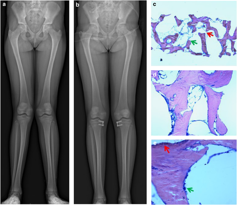
Patient 1 is a girl who was diagnosed with nephropathic cystinosis lately at 5 years of age (clinical picture of renal Fanconi syndrome but delay in diagnosis); from 7 years of age, she developed progressive genu valgum with important bone pains and decreased walking ability, thus requiring surgical correction and bone biopsy at 13 years of age. At the time of onset of leg deformations, her glomerular filtration rate (GFR) measured with inulin clearance was 37 ml min−1 1.73 m−2; thyroid hormones were normal, while serum calcium, phosphate and PTH were in the lower normal range (2.17–2.39 mmol l−1, 1.10–1.42 mmol l−1 and 24–34 ng l−1, respectively, during the year following the beginning of leg deformations). At the time of the surgical procedure, she was receiving growth hormone (from the age of 6), alfacalcidol and calcium carbonate; she was on the waiting list for renal transplantation but not undergoing renal replacement therapy, with an estimated GFR (eGFR) as estimated with the Schwartz 2009 equation of 13 ml min−1 per 1.73 m2. Thyroid hormone, thyroid stimulating hormone, copper, ceruloplasmin, phosphate, bicarbonate and calcium concentrations were normal; PTH level was 141 ng l−1 with an average PTH level during the year before surgery of 119 ng l−1 (Elsa PTH CisBio, Bioassays, Codolet, France, reference values 14–66 ng l−1). Average 25 D level during the year before surgery was 71 nmol l−1, ranging from 43 to 131 nmol l−1 (DIASORIN radioimmunology, Diasorin SA, Antony, France); 1–25 D levels were measured once at 127 pmol l−1 (reference local values between 72 and 192, RIA, Diasorin). Under cysteamine bitartrate, leukocyte hemi-cystin levels were within the target range, ranging between 0.3 and 0.9 nmol of leukocyte cystin per mg of proteins during the year before surgery (local normal values <0.4); at the time of biopsy, she was receiving cysteamine bitartrate four times a day, at a daily dose of 44 mg kg−1. Compliance had always been satisfying in this patient. The bone biopsy showed normal mineralization with active areas for both bone resorption and formation. Figure 1 illustrates the radiographs obtained before surgery at the time of bone biopsy (Figure 1a), the radiographs obtained after the surgical correction (Figure 1b) and the bone histology (Figures 1c–e).
Figure 1.
Bone data in the first patient. (a) Radiographs of the legs at the time of bone biopsy: there is no evidence for abnormal mineralization; the bones are slightly bowed. (b) Radiographs of the legs after surgery: there is a complete correction of the deformations. (c) Bone biopsy at the tibia, May–Grünwald–Giemsa × 2.5: under low power, slightly impaired trabecular structure with focal trabecular separations (red arrow) and loose trabecular endings (green arrow) were seen. (d) Bone biopsy at the tibia, May–Grünwald–Giemsa × 10: mineralization appears normal. (e) Bone biopsy at the tibia, May–Grünwald–Giemsa × 20: mineralization appears normal, with active areas for both bone resorption (multinucleated osteoclasts) within resorption lacunae (green arrow) and bone formation (osteoblasts; red arrow).
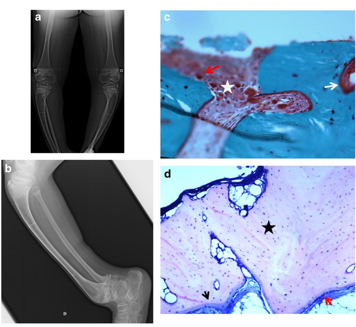
Patient 2 is a boy diagnosed with cystinosis at 2 years of age; in addition to a neurological developmental delay of unknown etiology, he developed severe bone deformation with a progressive and complete impairment of walking ability from 12 years of age. He received five pamidronate infusions because of decreased bone density, the last infusion occurring 3 years before bone biopsy. He never received growth hormone therapy. He also developed cutaneous side effects of cysteamine bitartrate at the age of 7 years. Cutaneous symptoms improved as daily dose of cysteamine decreased, as reported previously;3 however, at the time of cutaneous impairment, the daily doses adjusted on body surface area were usual (1.3 mg m−2 per day), and leukocyte hemi-cystin levels were in the lower range (ranging from 0.01 to 1 nmol of leukocyte cystin per mg of proteins during the follow-up, local normal values <0.4). A kinetic study of hemi-cystin levels was therefore performed to better understand the clinical phenotype; an important drop was observed after oral intake of cysteamine, reflecting likely cysteamine overdose. Copper deficiency was also found but 2 years of oral copper supplementation did not improve bone deformations; in the same time, copper levers remained either low, or in the normal lower range. A surgical correction and bone biopsy were therefore performed at 14 years of age as he was given alfacalcidol, calcium carbonate and phosphate supplementation. At that time, eGFR was 72 ml min−1 1.73 m−2; thyroid hormone, phosphate, bicarbonate and calcium concentrations were normal; PTH was 22 ng l−1. Bone biopsy showed active areas of mineralization and bone formation, with extended and unusual areas of bone resorption. Figure 2 illustrates the radiographs obtained 2 years before surgery (Figure 2a), the radiographs obtained at the time of bone biopsy (Figure 2b), and the bone histology (Figures 2c and d).
Figure 2.
Bone data in the second patient. (a) Radiographs of the legs 2 years before bone biopsy: severe deformations and sequelae of rickets. (b) Radiographs of the legs at the time of bone biopsy: severe deformations, sequelae of rickets and fractures. (c) Bone biopsy at the tibia, Goldner's trichrome, × 10: bone tissue with a partly fibrotic bone marrow (white star) and unusual resorption areas, with many multinucleated osteoclasts (red arrow). Mineralization appears normal (even though some part of the trabecular surface shows non mineralized osteoid, therefore leading to discuss a moderate surface osteoidosis; white arrow). (d) Bone biopsy at the tibia, May–Grünwald–Giemsa, × 10: elongated thin osteoid seams are colored in blue (black arrow) but mineralization appears normal. Numerous osteocytes (blue dots; marked with black star) are embedded in the mineralized matrix (pink), and some empty osteocytic lacuna (white dots) are observed.
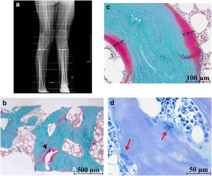
Patient 3 is a boy from a consanguineous family who was treated with cysteamine starting at 11 months of age. He was born at 35 weeks gestation with a neonatal course complicated by birth asphyxia, respiratory distress syndrome, pneumothorax and seizures. Surgery was performed at 1 month of age to treat cricoid cartilage stenosis; he was diagnosed with hypothyroidism at 10 months of age. He received growth hormone therapy from age 5 to 11. At 10 years of age, he developed leg and joint pains, bilateral knee valgus, severe scoliosis, muscle weakness, neuromotor regression and dark red skin lesions on both elbows without any history of trauma. At that time, his daily dose of cysteamine chlorohydrate was 90 mg kg−1 per day. Brain magnetic resonance imaging showed moderate reduction of white matter thickness, which was attributed to the perinatal asphyxia. Analysis of skin biopsy specimens from the elbow lesions revealed benign vascular proliferation on light microscopy and clear abnormalities of elastin fibers and some collagen fibers, with increased variability of collagen fiber caliber on electron microscopy. Since the exact etiology of the elbow lesions was unknown, they were surgically removed; however, they relapsed, and 18 months after the patient's first presentation, he also developed red striae on both thighs. One month later, he was hospitalized with asthenia, vomiting, behavioral disturbances and severe muscle weakness, impairing completely his walking ability. Electroencephalography and brain magnetic resonance imaging were stable. Bone densitometry showed a Z score of −1.3. The patient was then switched from cysteamine chlorohydrate to cysteamine bitartrate and the total daily dose of cysteamine was decreased to 40 mg kg−1 per day. He demonstrated clinical improvement in the ensuing months including waning of the neurological symptoms and increased muscular strength; however, his abnormal walking pattern persisted because of severe knee deformities, despite maximized phosphate and vitamin D supplementation, requiring orthopedic surgery to realign his lower limbs at 16 years of age. Remarkably a bone callus did not develop until several months after surgery which prevented removal of the external pins and fixator for >1 year. At the age of 17 years, he underwent complete thyroid removal because of a follicular adenoma. Eventually, because of a rapid bowing of long bone, bone pains and severe osteopenia, but no evidence for bone fracture, he underwent a bone biopsy at 20 years of age. Bone biopsy showed diffuse defective mineralization with excess of osteoid matrix. There were active osteoclasts and little osteoblastic activity. Figure 3 illustrates the radiographs obtained 8 months after the bone biopsy (Figure 3a) and the bone histology (Figures 3b–d). Table 1 summarizes the main data for the three patients.
Figure 3.
Bone data in the third patient. (a) Radiographs of the legs 8 months after bone biopsy: severe deformations, but no evidence of abnormal mineralization. (b) Goldner's trichrome, original magnification × 20: bone trabeculae showed focal deep defective mineralized matrix (red-stained bone regions; black arrow). (c) Goldner's trichrome, original magnification × 20: up to 50-μm-thick osteoid seams (red-stained bone regions) are observed on the surface of mineralized (green-stained portions of bone matrix) bone trabeculae. (d) Toluidine blue, original magnification × 40: enlarged osteoclasts contained up to eight isomorphic nuclei (red arrows). No marrow fibrosis was apparent in this specimen.
Table 1. Summary of the main clinical and biochemical findings in the three pediatric patients with severe bone deformations and nephropathic cystinosis.
| Usual values | Patient 1 | Patient 2 | Patient 3 | |
|---|---|---|---|---|
| Age at diagnosis (years) | 5 | 2 | 0.9 | |
| Age at onset of bone symptoms (years) | 7 | 10 | 10 | |
| Age at BB (years) | 13 | 14 | 20 | |
| Renal function at the time of BB (ml min−1 1.73 m−2) | >90 | 13 (conservative management) | 72 (conservative management) | 22 (conservative management) |
| Thyroid status | Normal | Normal | Hypothyroidism treated with L-thyroxine | |
| Copper status | Normal | Copper deficiency | Copper deficiency | |
| Treatment with growth hormone | Yes | No | Yes | |
| Bicarbonates (average during the year before BB, mmol l−1) | 19–24 | 22 | 24 | 23 |
| Total calcium (average during the year before BB, mmol l−1) | 2.20–2.55 | 2.19 | 2.39 | 2.35 |
| Phosphorus (average during the year before BB, mmol l−1) | 0.74–1.45 | 1.39 | 1.37 | 1.19 |
| PTH levels (average during the year before BB, pg ml−1) | 15–65 | 119 | 15 | <2.5 |
| 25 OH vitamin D levels (average during the year before BB, mmol l−1) | 75–150 | 71 | 31 | Not performed |
| Hemi-cystine levels (leukocyte cystin in nmol per mg of protein) | <0.4 | Between 0.3 and 0.9 during the year before surgery | 0.01–1 | 0.7 |
| Daily dose of cysteamine bitartrate (mg kg−1) | 44 | 40 | 27 | |
| X-ray analysis | No evidence for abnormal mineralization | Sequelae of rickets | No evidence for abnormal mineralization | |
| BB results | Normal mineralization with active areas for both bone resorption and formation | Active areas of mineralization and bone formation, with extended and unusual areas of bone resorption | Diffuse defective mineralization. Active osteoclasts and little osteoblastic activity | |
| Cystine crystals (BB) | No | No | No |
Abbreviation: BB, bone biopsy.
Discussion
There are very few data in the literature describing bone impairment in patients with cystinosis;3,4,5 one case of co-occurrence of Osteogenesis Imperfecta type VI and cystinosis as a contiguous gene syndrome was described.7 However, international experts treating patients worldwide currently discuss unexplained bone impairment such as stress fractures and bone pains/deformations in teenagers and young adults with cystinosis undergoing conservative management of CKD. This occurrence of ‘late novel symptoms' in cystinosis may be explained by the improved global prognosis of the disease observed in developed countries.2 In animal models, the invalidation of the CTNS gene in certain strains of mice is associated neither with renal phosphate wasting nor with renal failure, but causes severe osteopenia and growth retardation,8 thus raising the hypothesis of a specific underlying bone defect in cystinosis. It has been shown recently that the in vitro ability of mesenchymal stromal cells isolated from bone marrow to differentiate along the osteoblastic lineage is reduced in patients with cystinosis as compared with cells obtained from healthy controls; this cellular abnormality can be reverted after cysteamine treatment.9
Cysteamine (HSCH2CH2NH2) is a cystin-depleting drug used in cystinosis: indeed, it cleaves the disulfide bond with cysteine and depletes lysosomal cysteine in most tissues, but also decreases apoptotic cell death and cell oxidation, nevertheless without any significant effect on the renal Fanconi syndrome.10 Although it does not cure cystinosis, it dramatically improves the overall prognosis of this severe orphan disease. Cysteamine was approved for clinical use in the 1990s, and current evidence obtained in cystinosis patients born between 1970 and 2000 demonstrates that its early initiation delays the onset of end-stage renal failure by almost a decade,2 but also the onset of non-renal comorbidities. In animals, cysteamine has been shown to stimulate growth hormone secretion in yaks,11 and to stimulate growth rates in different models, notably in fish.12 In patients, it has been reported from clinical data that cysteamine could promote growth and bone maturation, allowing several patients to reach normal adult height without GH therapy;13 however, it remains debatable whether it is a cysteamine effect per se, or an effect of a better global management of patients. Conversely, the maximum daily dose of cysteamine should not exceed 1.95 g m−2, because of possible side effects of the drug.10
Indeed, some patients develop symmetric angioendotheliomatosis lesions on the elbows, and less frequently long-bone deformations, striae rubrae, muscular and neurological symptoms. These side effects, probably secondary to excessive doses of cysteamine (at least partly), were reported by Besouw3, affecting ∼1.5% of treated patients. In this series of six patients (among them Patient 2 and 3 from the present report), skin lesions and neurologic symptoms improved with decreased daily doses of cysteamine. The trend was similar for bone impairment and muscular pain; however, in our two patients, with 2 additional years of follow-up, the improvement was only transient and incomplete. With cysteamine overdose, wound healing issues were described, and one can hypothesize that cysteamine overdose may also delay the healing of stress fractures. Indeed, with the high peak levels observed after cysteamine bitartrate intake, there can be a considerable effect of collagen cross-linkings and in copper chelation, by analogy with D-penicillamine effects on collagen.3 Besouw3 also discussed a likely interference of cysteamine with interstitial matrix proteins, thus leading to a stimulation of angiogenesis and the development of reactive angioendotheliomatosis in skin. However, we did not observe apparent abnormal vascular structures in these three bone biopsies, and our patients received relatively low doses of cysteamine, as illustrated in Table 1. It is nevertheless important to stress out that normal Wistar rats receiving cysteamine for 6 months develop severe skeletal deformities (severe kyphosis), as well as cardiovascular abnormalities (dissecting aneurysm of the thoracic aorta).14
Interestingly, in two out of our three patients, a developmental delay was also noted whereas cystinosis was genetically confirmed. Although not uncommon in cystinosis, neurobehavioral abnormalities are not a classical feature of the disease during adolescence;15 in our two cases, they may have worsened the bone phenotype. The main clinical and biochemical findings in our three pediatric patients with severe bone deformations and nephropathic cystinosis (Table 1) allow to conclude that even though copper deficiency, high-dose cysteamine regimens and abnormal thyroid metabolism may worsen the clinical bone picture, the exact pathophysiology of such an impairment remains to be completely defined in this orphan disease since Patient 1 did not present any of these confounding factors. Moreover, severe rickets/osteomalacia to explain such bone impairment can be ruled out from the biopsies.
Renal Fanconi syndrome is usually thought to be the most important factor to explain bone deformations and rickets in patients with cystinosis; however, in these three patients, once the diagnosis of cystinosis was made, they received an adapted calcium and phosphate oral supplementation, with electrolytes within the normal range, and there was no evidence of abnormal mineralization on the X-rays performed at the time of the biopsy (Table 1). We nevertheless cannot completely rule out a ‘washing out' of calcium and phosphate from bone to maintain normal circulating levels. In that setting, the low PTH levels observed in patients appear interesting: one may wonder whether chronic renal phosphate wasting can induce chronic hypoparathyroidism (after ‘adjustment' on renal function), and therefore slightly decrease ionized calcium and therefore favor the onset of bone deformations/impairment. Unfortunately, neither ionized calcium levels nor biomarkers of bone formation (for example, osteocalcin), resorption (for example, CTX) and osteocytic activity (for example, FGF23 or sclerostin) were available in our patients. With such a hypothesis, it would be interesting in clinical practice to assess ionized calcium and to provide patients with oral calcium supplementation in addition to vitamin D analogs and phosphate supplementation.
In this specific setting of cystinosis, the distinction between bone lesions due to CKD and renal osteodystrophy on one hand, and bone lesions due to cystinosis itself (whatever the exact underlying pathophysiology) on the other hand, remains challenging. We can nevertheless rule out in these three patients the role of corticosteroids after renal transplantation on bone. Primary hypogonadism may also worsen the clinical bone picture, but in these three prepubertal patients this factor was probably a minor confounding factor.1 It would also have been interesting to perform EM imaging of the bone, but this technique was unfortunately unavailable.
Conclusion
Bone impairment in nephropathic cystinosis appears to be more and more described. From our experience on three pediatric patients with nephropathic cystinosis who developed severe bone disease, we believe that we can rule out the main hypotheses usually raised to explain completely such a bone impairment (for example, copper deficiency, bone consequences of severe hypophosphatemic rickets during infancy, cysteamine toxicity and abnormal thyroid metabolism): as such, we discuss the role of chronic hypoparathyroidism due to chronic phosphate wasting to explain at least partly the clinical picture.
We hope that this description of three cases of bone impairment in cystinosis will open a new field of investigation in the future. In addition to experimental data, larger and prospective studies evaluating this specific topic are required, since this comorbidity can have a major impact on the patients' quality of life.
Acknowledgments
We would like to acknowledge Pr Georges Deschenes (Hopital Robert Debré, Paris, France) for his skillful scientific help. We would also like to acknowledge Dr Kariman Abelois-Genevois (Hôpital Femme Mère Enfant, Bron, France) who performed bone biopsies in the French patients, and Brigitte Burt-Pichat (INSERM-UMR 1033, Lyon, France) for her skillful technical help in preparing bone biopsies. They also would like to acknowledge Dr Cécile Acquaviva-Bourdain (Département de Biologie, Hospices Civils de Lyon, France) for biochemical measurements.
Author Contributions: JB collected the data and wrote the manuscript. MG provided the clinical data from the third patient and approved the final version of the manuscript. FN provided the clinical data from the second patient and approved the final version of the manuscript. JZ and GB interpreted the bone biopsies, edited the manuscript and approved the final version of the manuscript. AB-T provided the clinical data from the first patient, edited the manuscript and approved the final version of the manuscript. FE and PC edited the manuscript and approved the final version of the manuscript.
Footnotes
JB has received a research grant from Raptor pharmaceuticals for a clinical prospective research project on bone impairment in nephropathic cystinosis. MG is principal investigator for the RP 103 study in Italy. PC is a medical expert for Raptor pharmaceuticals and co-investigator for the RP 103-3, 103-4 and 103-7 studies. FE is a medical expert for Raptor pharmaceuticals and co-investigator for the RP 103-3, 103-4 and 103-7 studies. AB-T is a medical expert for Raptor pharmaceuticals and co-investigator for the RP 103-3, 103-4 and 103-7 studies. The remaining authors declare no conflict of interest.
References
- Nesterova G, Gahl WA. Cystinosis: the evolution of a treatable disease. Pediatr Nephrol 2013; 28: 51–59. [DOI] [PMC free article] [PubMed] [Google Scholar]
- Bertholet-Thomas A, Bacchetta J, Tasic V, Cochat P. Nephropathic cystinosis--a gap between developing and developed nations. N Engl J Med 2014; 370: 1366–1367. [DOI] [PubMed] [Google Scholar]
- Besouw MT, Bowker R, Dutertre JP, Emma F, Gahl WA, Greco M et al. Cysteamine toxicity in patients with cystinosis. J Pediatr 2011; 159: 1004–1011. [DOI] [PubMed] [Google Scholar]
- Klusmann M, Van't Hoff W, Monsell F, Offiah AC. Progressive destructive bone changes in patients with cystinosis. Skeletal Radiol (e-pub ahead of print 28 September 2013; doi: 10.1007/s00256-013-1735-z. [DOI] [PubMed] [Google Scholar]
- Sirrs S, Munk P, Mallinson PI, Ouellette H, Horvath G, Cooper S et al. Cystinosis with sclerotic bone lesions. JIMD Rep 2014; 13: 27–31. [DOI] [PMC free article] [PubMed] [Google Scholar]
- Kidney Disease: Improving Global Outcomes CKDMBDWG. KDIGO clinical practice guideline for the diagnosis, evaluation, prevention, and treatment of Chronic Kidney Disease-Mineral and Bone Disorder (CKD-MBD). Kidney Int 2009; 113: S1–S130. [DOI] [PubMed] [Google Scholar]
- Tucker T, Nelson T, Sirrs S, Roughley P, Glorieux FH, Moffatt P et al. A co-occurrence of osteogenesis imperfecta type VI and cystinosis. Am J Med Gen 2012; 158: 1422–1426. [DOI] [PubMed] [Google Scholar]
- Cherqui S, Sevin C, Hamard G, Kalatzis V, Sich M, Pequignot MO et al. Intralysosomal cystine accumulation in mice lacking cystinosin, the protein defective in cystinosis. Mol Cell Biol 2002; 22: 7622–7632. [DOI] [PMC free article] [PubMed] [Google Scholar]
- Conforti A, Taranta A, Biagini S, Starc N, Pitisci A, Bellomo F et al. Cysteamine treatment restores the in vitro ability to differentiate along the osteoblastic lineage of mesenchymal stromal cells isolated from bone marrow of a cystinotic patient. J Transl Med 2015; 13: 143. [DOI] [PMC free article] [PubMed] [Google Scholar]
- Emma F, Nesterova G, Langman C, Labbe A, Cherqui S, Goodyer P et al. Nephropathic cystinosis: an international consensus document. Nephrol Dial Transplant 2014; 29: 87–94. [DOI] [PMC free article] [PubMed] [Google Scholar]
- Hu R, Wang Z, Peng Q, Zou H, Wang H, Yu X et al. Effects of GHRP-2 and cysteamine administration on growth performance, somatotropic axis hormone and muscle protein deposition in yaks (bos grunniens) with growth retardation. PLoS ONE 2016; 11: e0149461. [DOI] [PMC free article] [PubMed] [Google Scholar]
- Tse MC, Cheng CH, Chan KM. Effects of chronic cysteamine treatment on growth enhancement and insulin-like growth factor I and II mRNA levels in common carp tissues. Br J Nutr 2006; 96: 650–659. [PubMed] [Google Scholar]
- Markello TC, Bernardini IM, Gahl WA. Improved renal function in children with cystinosis treated with cysteamine. N Engl J Med 1993; 328: 1157–1162. [DOI] [PubMed] [Google Scholar]
- Jayaraj AP. Dissecting aneurysm of aorta in rats fed with cysteamine. Br J Exp Pathol 1983; 64: 548–552. [PMC free article] [PubMed] [Google Scholar]
- Broyer M, Tete MJ, Guest G, Bertheleme JP, Labrousse F, Poisson M. Clinical polymorphism of cystinosis encephalopathy. Results of treatment with cysteamine. J Inherit Metab Dis 1996; 19: 65–75. [DOI] [PubMed] [Google Scholar]


