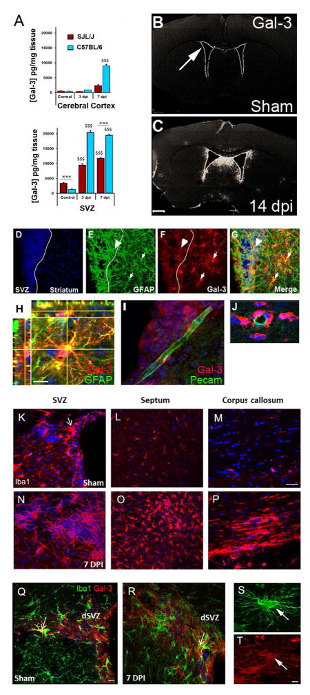
Fig. 2. TMEV infection in mice increased periventricular Gal-3 expression.
(A) Gal-3 protein levels in the cerebral cortex and the SVZ measured by ELISA. $$$ P<0.001 compared to controls, ***P<0.001 SJL/J compared to C57BL/6. (B–C) Gal-3 immunofluorescence was detected in the SVZ (arrow) in sham mice and was increased after TMEV. (D–G) Arrowhead points to a GFAP+ (green) and Gal-3+ (red) double-positive cell in the SVZ and the small arrows to double-positive cells in the striatum, 7 dpi (SJL/J mice). (H) Confocal image of striatal astrocyte expressing GFAP (green) and Gal-3 (red) (SJL/J mice). (I–J) Gal-3 was associated with blood vessels but seldom expressed by PECAM+ endothelial cells. (K–M) Iba1+ immunoreactivity in sham controls in the SVZ (ex. arrow), septum and corpus callosum (SJL/J mice). (N–P) Iba1+ immunoreactivity in the SVZ, septum and corpus callosum 7 dpi, (SJL/J mice). (Q–R) Iba1 (green) and Gal-3 immunoreactivity (red) in a sham mouse SVZ and 7 dpi. Arrows show rare double-positive cells (C57BL/6 mice). (S–T) Iba1+/Gal-3+ cell in the corpus callosum 7 dpi. Scale bars: C=500 μm; H=5 μm; M=15 μm; Q=10 μm; T=2 μm.

