Abstract
Background:
Many plants growing in Saudi Arabia are used in folk medicine for treatment of several diseases.
Objective:
Information of the chemical constituents and biological activities of plants is desirable for the discovery of therapeutic agents and discovering the actual value of folkloric remedies.
Materials and Methods:
The compounds were isolated and purified using silica gel column chromatography and preparative high-performance liquid chromatography-diode array detector (HPLC-DAD) Method. The alcoholic extracts of these plants were evaluated for biological activities.
Results:
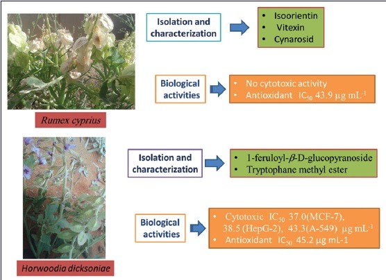
Isolation and characterization of 1-feruloyl-β-D-glucopyranoside (1) as well as new secondary metabolite tryptophan methyl ester (2) were isolated for the 1st time from the Horwoodia dicksoniae. The three flavones were isolated from Rumex cyprius identified as isoorientin (3), vitexin (4), and Cynarosid (5). The structures of these compounds were characterized by nuclear magnetic resonance and mass spectrometry analysis and comparing with literature. The compounds were isolated and purified using silica gel column chromatography and preparative HPLC-DAD Method. The alcoholic extracts of these plants were evaluated for antimicrobial activities against two Gram-positive bacteria, two Gram-negative bacteria, and four pathogenic fungi. Both plants showed good activities against Syncephalastrum racemosum and Streptococcus pneumoniae with minimal inhibitory concentrations (MICs) 0.98 and 1.95 μg/mL, respectively. H. dicksoniae showed good activity against Aspergillus fumigates with an MIC 1.95 μg/mL. The two extracts showed also effective free radical scavenging activities in the 1,1-diphenyl-2-picrylhydrazyl assay. H. dicksoniae exhibited remarkable cytotoxic activity against Human breast cancer mammary cancer cells-7, Human liver cancer human hepatoma carcinoma cells-2, and human lung carcinoma (A-549) cell lines.
Conclusions:
It was suggested that further work should be carried out to isolate, purify, and characterize the active constituents responsible for the activity of these plants.
SUMMARY
New secondary metabolite Tryptophane methyl ester as well as 1-feruloyl-β-D-glucopyranoside were isolated for the first time from the HD.
Isoorientin, vitexin and Cynarosid were isolated from RC.
HD exhibited good activity against Aspergillus fumigates with an MIC 1.95 µg mL-1.
HD showed significant cytotoxic activity against Human breast cancer (MCF-7), Human liver cancer (HepG-2) and Human lung carcinoma (A-549) cell lines.
Keywords: Antimicrobial, antioxidant, cytotoxicity, horwoodia, Rumex
INTRODUCTION
Plant-derived substances have recently become of great interest owing to their versatile applications. Medicinal plants are the richest bioresource of drugs of traditional systems of medicine, modern medicine, and chemical precursors for synthetic drugs. Information of the chemical constituents of plants is desirable for the discovery of therapeutic agents and discovering the actual value of folkloric remedies.[1] Many plants growing in Saudi Arabia are used in folk medicine for treatment of several diseases.[2,3,4] Horwoodia dicksoniae Turrill is known in Arabic as Khuzama. Rumex genus belongs to polygonaceae family, and several species of this genus have indicated noteworthy therapeutic potentials. There are some reports in literature about evaluation of a few species of Rumex for various medicinal potential[5] Rumex cyprius revealed antibacterial activity against methicillin resistant Staphylococcus aureus, which has shown relatively high resistant when exposed to reference antibiotics.[6] In the present work, phytochemical analyses as well as antimicrobial, antioxidant, and cytotoxic activities of the alcoholic extracts of these plants were carried out.
MATERIALS AND METHODS
Experimental
General
Ultraviolet (UV) spectra were determined with a Shimadzu UV-1650PC spectrophotometer; infrared (IR) spectra were carried out on a Nicolet 205 Fourier transform IR spectrometer connected to a Hewlett-Packard ColorPro Plotter. ESIMS (positive ion acquisition mode) was carried out on an XEVO TQD triple quadruple instrument (Waters Corporation, Milford, MA 01757, USA) mass spectrometer. Proton nuclear magnetic resonance (1HNMR) and 13CNMR spectra were obtained in dimethyl sulfoxide (DMSO)-d6 on a Varian Gemini 400 MHz spectrometer using tetramethylsilane as internal standard; chemical shifts are reported as (ppm) at the Department of Chemistry, Faculty of Science, King Abdul-Aziz University, Jeddah 21589, KSA. Preparative high-performance liquid chromatography (HPLC) was performed on a Waters® 2545 Quaternary Gradient Module (150 mm × 19 mm, ID) column, prepacked with Prep C18 (OBD) with flow rate 24 mL/min, Waters® fraction collector III and Waters® 2707 auto-sampler. The entire system was controlled using Empower 3 Software. Detection was achieved with a Waters® 2998 diode array detector, and chromatograms were recorded at 254–400 nm. Vacuum liquid chromatography (VLC) was carried out using Silica Gel 60, 0.04–0.063 mm mesh size (Merck). Solid phase extraction (SPE) was performed on SPE-C18 cartridges (A Strata column). Thin layer chromatography was carried out on precoated silica gel 60 F254 (Merck) plates. Developed chromatograms were visualized by spraying with 1% vanillin-H2SO4, followed by heating at 100°C for 5 min.
Plant material
H. dicksoniae and R. cyprius herbs were collected from Rafha region, Kingdom of Saudi Arabia, in March, 2014, and were kindly identified by Dr. Abdelrahman Talhah Abdelwahab Abdelrahman, Head of biology department, Faculty of Science, Northern Border University, Kingdom of Saudi Arabia. A voucher specimen has been deposited in the Department of Pharmacognosy, Faculty of Pharmacy, Northern Border University, Kingdom of Saudi Arabia.
Preparation of plant extracts
The air-dried powdered herbs of H. dicksoniae (200 g) and R. cyprius (250 g) were subjected to exhaustive extraction with 70% methanol (2 L × 3 L) at room temperature. The combined methanolic extracts were concentrated under reduced pressure to dryness which gives 15 and 20 g, respectively. The concentrated methanolic extracts were suspended separately in distilled water (100 mL) and partitioned successively with n-hexane, chloroform, ethyl acetate and n-butanol to give n-hexane, chloroform, ethyl acetate, n-butanol and water fractions for each plant.
Preliminary phytochemical screening
The chemical tests were carried out on the alcoholic extract or the powdered plant material using the procedures outlined by Harborne,[7] Trease and Evans.[8] The results are presented in Table 3.
Table 3.
Phytochemical screening of the Horwoodia dicksoniae and Rumex cyprius

Isolation of the compounds
The MeOH fraction (15 g) of H. dicksoniae was subjected to VLC on silica gel (300 g) using 0.5 L each of n-hexane, n-hexane/EtOAc (50:50) and (25:75), EtOAc, EtOAc/MeOH (1:1), and MeOH to give seven fractions (A1-A7). Fraction F5 (1.2 g) was applied to a sephadex LH-20 column to deliver eight subfractions (1–8). Subfraction 2 and 3 were subjected to subsequent purification on semi-preparative RP-C18 HPLC eluting with MeOH/H2O (75:25) using XBridge _ RP-18 (150 mm, 10 mm, 5 µ) at a flow rate of 6.6 mL/min and detection at 254 nm to give compounds 1 (30 mg, 20 min) and 2 (20 mg, 33 min), respectively.
1-Feruloyl-β-D-glucopyranoside (1)
Yellow amorphous powder; λ nm: 237, 329; IR (KBr) ν/cm; 1HNMR (400 MHz, CD3 OD) δ 3.43 (m, 1H, H-4').,45 (m, 1H, H-2'), 3.50 (t, 1H, J 8.2, H-3'), 3.55 (m, 1H, H-5'), 3.61 (dd, 1H, J 4.7, 11.2, H-6'b), 3.81 (m, 1H, H-6'a), 3.88 (s, OMe), 5.62 (d, 1H, J 7.6, H-1'),6.39 (d, 1H, J 15.8, α), 6.82, (d, 1H, J 8.2, H-5), 7.08 (brd, 1H, J 8.1, H-6), 7.17 (d, 1H, J 1.3, H-2), 7.71 (d, 1H, J 15.8, β), d,; 13CNMR (100 MHz, CD3 OD) δ 55.14, 60.98, 69.72, 72.67, 76.62, 77.37, 94.41,110.49, 113.33, 115.18, 123.04, 126.16,146.95, 147.96, 149.47, 166.43; ESIMS m/z 355 [M-H]−.
Tryptophan methyl ester (2)
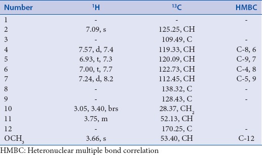
Light brown amorphous powder; λ nm: 237, 329; IR (KBr) ν/cm: 1HNMR, 13CNMR [Table 1], ESIMS m/z 241 [M+Na] +, 226 [M-Me+Na].
Table 1.
Nuclear magnetic resonance data of compound 2 (CD3OD, 400 and 100 MHz)
About 10 g of the MeOH extract of R. cyprius were subjected to VLC on silica gel (250 g) using successively n-hexane, n-hexane/EtOAc (50:50–25:75), EtOAc, EtOAc/MeOH (1:1) and MeOH 1 L each, to give six fractions (F1-F6). Fraction F3 (2.1 g) was subjected to SPE-C18 cartridges to deliver eight subfractions (1–8). Subfraction 3 (100 mg) was injected (200 µL) into the reverse phase preparative HPLC-diode array detector (DAD) using a linear gradient elution with a mixture of two solvents. Solvent A consisted of methanol and solvent B consisted of water. The solvent gradient consisted in a series of linear gradients, starting from 5% to 100% of solvent B over 20 min at a flow rate of 24 mL/min and detection at 210–400 nm to give compounds 1 (10 mg), 2 (13 mg) and 3 (10 mg), respectively.
High-performance liquid chromatography-diode array detector characterization of the isolated flavones
Through comparison of different RP-HPLC retention times of compounds subfraction 3 (13.87, 14.82, and 15.94 min respectively) [Figure 1], it was clear that no quite difference in their polarities. The UV spectra of all compounds show the same lambda maxima because the responsible chromophoric group in all compounds is the same one. The importance is the difference in the UV-absorption intensities Figure 2 between the derivatives, where it originated from the difference in the concentration of the samples.
Figure 1.

High-performance liquid chromatography-diode array detector chromatogram of subfraction (F-3) of Rumex cyprius
Figure 2.
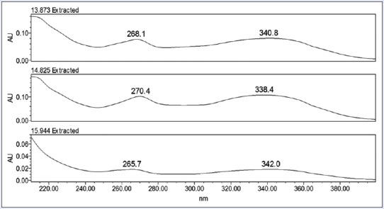
High-performance liquid chromatography-diode array detector chromatogram of compounds 1, 2, and 3, respectively
Vitexin (apigenin-8-C-glucoside) (3)
Yellow amorphous powder; λ nm: 268.1, 340.8; IR (KBr) ν/ν/cm: 3430, 1660, 1610, 1595, 1510; 1HNMR (400 MHz, DMSO-d6) δ 3.41 (m, 1H, H-6”b), 3.30–3.55 (m, 3H, H-3”, 4”, 5”), 3.76 (d, 1H, J 12.0, H-6”a), 4.08 (m, 1H, H-2”),4.72 (d, 1H, J 9.2, H-1”), 6.25 (s, 1H, H-3), 6.68 (s, 1H, H-6), 7.92 (d, 2H, J 8.0, H-3', 5'), 7.94 (d, 2H, J = 8.0 Hz, H-2', 6'), 13.10 (brs, OH); 13CNMR (100 MHz, DMSO-d6,) δ 61.11, 70.3, 70.87, 73.20, 78.32, 81.26, 98.35, 102.26, 103.73, 104.10, 115.9, 121.54, 128.87, 155.99, 160.27,160.88, 163.50, 164.16, 182.04; ESIMS m/z 431 [M-H]−, 430 [M-2H]−, 269 (M-H-162) = (aglycone-H).
Isoorientin (luteolin-6-C-glucoside) (4)
Yellow amorphous powder; λ/nm: 270.4, 338.4; IR (KBr) ν/ν/cm: 3400, 1662, 1615, 1590, 1510; 1HNMR (400 MHz, DMSO-d6) δ 3.19 (1H, m, H-5”), 3.22 (t, 1H, J 8.4, H-3”), 3.25 (d, 1H, J 8.4, H-4”), 3.45 (d, 1H, J 11.6, H-6”b), 3.68 (d, 1H, J 11.6, H-6”a), 4.10 (m, 1H, H-2”), 4.59 (d, 1H, J 9.6, H-1”), 6.91 (d, 1H, J 8.4, H-5'), 6.51 (s, 1H, H-3), 6.64 (s, 1H, H-8), 7.37 (brs, 1H, H-2'), 7.39 (d, 1H, J 8.8, H-6'), 13.46 (brs, OH); 13CNMR (100 MHz, DMSO-d6) δ 61.27, 70.11, 70.31, 72.93, 78.67, 81.17, 93.65, 102.63, 103.24, 108.50, 113.08, 116.08, 119.07, 121.35, 145.58, 149.65, 156.33, 160.43, 163.40, 163.81, 181.8; ESIMS m/z 447 [M-H]−, 493 [M + HCOOH-H]−.
Cynarosid (luteolin-7-O-β-D-glucopyranoside) (5)
Yellow powder; λ nm: 265.7, 342; IR (KBr) ν/ν/cm: 3423, 1637, 1612, 1562, 1459; 1HNMR (400 MHz, DMSO-d6) δ 3.22 (t, 1H, J 8.4, H-2”), 3.41 (d, 1H, J 11.2, H-6”b), 3.47 (t, 1H, J 9.2, H-3”),3.47 (t, 1H, J 10.4, H-4”), 3.73 (d, 1H, J 11.2, H-6”b), 3.76 (d, 1H, J 12.0, H-6”a), 5.02 (d, 1H, J 7.6, H-1”), 6.45 (brs, 1H, H-6), 6.70 (s, 1H, H-3), 6.82 (brs, 1H, H-8), 6.91 (d, J 8.4, H-5'), 7.39 (brs, H-2'), 7.44 (d, J 8.4, H-6'), 12.77 (brs, OH); 13CNMR (100 MHz, DMSO-d6) δ 60.70, 69.41, 72.81, 75.81, 76.71, 94.88, 99.55, 99.60, 102.88, 105.27, 113.19, 119.42, 116.07, 121.26, 145.74, 149.74, 156.90, 160.71, 162.69, 164.64, 181.98; ESIMS m/z 449 [M + H]+, 471 [M + Na]+.
Antimicrobial assays
Antimicrobial activities of plant extracts were investigated in vitro against different bacteria and fungi. Two standard strains of Gram-positive bacteria (Streptococcus pneumonia Regional Center for Mycology and Biotechnology antimicrobial unit [RCMB 010010], Bacillus subtilis [RCMB 010067]) and two standard strains of Gram-negative bacteria (Pseudomonas aeruginosa [RCMB 010043], Escherichia coli [RCMB 010052]) were used for antibacterial assay. Four clinical pathogenic fungi (Aspergillus fumigates [RCMB 02568], Syncephalastrum racemosum [RCMB 05922], Geotricum candidum [RCMB 05097], and Candida albicans [RCMB 05036]) were used for antifungal assay. The microbial species are environmental and clinically pathogenic microorganisms obtained from RCMB, Al-Azhar University. Antimicrobial tests were carried out by the agar well-diffusion method,[9,10] using 100 μL of suspension containing 1 × 10⁸ colony forming units (CFU)/mL for tested bacteria and 1 × 10⁴ spore/mL fungi spread on nutrient agar and malt extract agar, respectively. After the media had cooled and solidified, wells (6 mm in diameter) were made in the solidified agar and loaded with 100 μL of tested sample solutions in 1 mL DMSO with concentrations of 10 mg/mL. Negative controls were prepared using DMSO employed for dissolving the tested samples while ampicillin, gentamycin, and amphotericin B were used as positive controls for Gram-positive bacteria, Gram-negative bacteria, and fungi, respectively. The inoculated plates were then incubated for 24 h at 37°C for bacteria and 48 h at 28°C for fungi and the diameter of any resulting zones of inhibition of growth was measured in millimeter (mm). Each experiment was performed in triplicates and the data were expressed as mean ± standard deviation. The minimal inhibitory concentrations (MICs) were determined using the two-fold serial dilution technique.[11,12] The two-fold serial dilutions of the tested sample solutions were prepared. The final concentrations of the solutions were 500–0.007 μg/mL. The tubes were then inoculated with the test organisms, grown in their suitable broth for tested pathogenic bacteria (1 × 10⁸ CFU/mL for bacteria and 1 × 10⁴ spore/mL for fungi); each 0.5 mL received 100 μL of the above inoculum and was incubated at 37°C for 24 h for bacteria and after 48 h of incubation at 28°C for fungi. MIC values were taken as the lowest sample concentration that prevents visible bacterial growth. Each experiment was made 3 times.
Antioxidant assay
The antioxidant activity of the plant extracts was determined by the 1, 1-diphenyl-2-picrylhydrazyl (DPPH) free radical scavenging assay.[13] Freshly prepared (0.004% w/v) methanol solution of DPPH radical was prepared and stored at 10°C in the dark. A methanol solution of the test sample was prepared. A 40 µL aliquot of the methanol solution was added to 3 mL of DPPH solution. Absorbance measurements were recorded immediately with a UV-visible spectrophotometer (Milton Roy, Spectronic 1201). The decrease in absorbance at 515 nm was determined continuously, with data being recorded at 1 min intervals until the absorbance stabilized (16 min). The absorbance of the DPPH radical without antioxidant (control) and the reference compound ascorbic acid were also measured. All the determinations were performed in three replicates and averaged. The percentage inhibition (PI) of the DPPH radical was calculated according to the formula:
PI = ([(AC − AT)/AC] × 100)
Where AC = absorbance of the control at t = 0 min and AT = absorbance of the sample + DPPH at t = 16 min.
Cytotoxicity assays
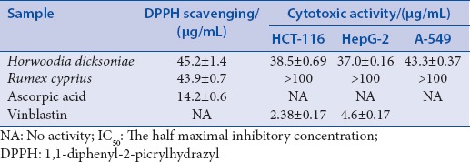
The plant extracts were tested for cytotoxicity against three human tumor cell lines: Human breast cancer mammary cancer cells-7 (MCF-7), Human liver cancer hepatoma carcinoma cells (HepG-2), and Human lung carcinoma (A-549) cell lines. The cells were obtained from the American Type Culture Collection (ATCC, Rockville, MD, USA). The cells were grown on Roswell Park Memorial Institute 1640 medium (Nissui Pharm. Co., Ltd., Tokyo, Japan) supplemented with 10% inactivated fetal calf serum and 50 μg/mL gentamycin. The cells were maintained at 37°C in a humidified atmosphere with 5% CO2 and were sub cultured 2–3 times a week. The cytotoxic activity was determined using cell viability assay method as described previously.[14,15] Percentage cell viability was calculated as the mean absorbance of control cells/mean absorbance of treated cells. Dose-response curves were prepared and the IC50 value was determined. The results are presented in Table 2.
Table 2.
1,1-diphenyl-2-picrylhydrazyl scavenging/(half maximal inhibitory concentration) (μg/mL) and cytotoxic activity/(half maximal inhibitory concentration) (μg/mL) of Horwoodia dicksoniae and Rumex cyprius extracts
Statistical analysis
All data were expressed as mean ± standard error of the mean. Student's t-test was applied for detecting the significance of difference between each sample; P < 0.05 was considered statistically significant.[16]
RESULTS AND DISCUSSION
The preliminary phytochemical screening of two medicinal plants, H. dicksoniae and R. cyprius, is summarized in Table 3. The results revealed the presence of medicinally active compounds in the two plants studied. Flavonoids and tannins were present in both plants. Alkaloids and saponins are present only in R. cyprius. Anthraquinones are absent from the two plants. Several studies confirmed that the presence of these important constituents contributes medicinal as well as physiological activities to the studied plants in the treatment of different diseases. Therefore, extracts from these plants can be used as a good source for valuable drugs.
The H. dicksoniae and R. cyprius extract of was subjected to a succession of chromatographic procedures, including silica gel chromatography, gel permeation chromatography using Sephadex LH-20 and preparative HPLC to afford two pure isolates 1-feruloyl-β-D-glucopyranoside (1), tryptophan methyl ester (2), isoorientin (3), vitexin (4), and cynarosid (5) [Figure 3]. The structures of the isolated compounds were established using spectroscopic analysis, especially, NMR spectra and direct comparison with published data.[17,18]
Figure 3.
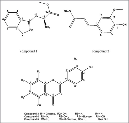
The structures of compounds 1, 2, 3, 4, and 5
The antimicrobial activities of the plant extracts were investigated in vitro against different bacteria and fungi. H. dicksoniae and R. cyprius extracts demonstrated growth inhibitory effect and variable antimicrobial activity against most of the specific organisms tested [Table 4 and Figure 4]. They demonstrate a good activity against Aspergillus fumigatus, Streptococcus pneumoniae and E. coli. They appear to be more active against S. racemosum than the standard. They showed week antimicrobial activity against Geotricum candidum and B. subtilis. Both plant extracts did not show antimicrobial activity against the Gram-negative bacteria, P. aeruginosa and the fungus C. albicans.
Table 4.
Antimicrobial activity as diameter of inhibition zone/mm of plant extracts against selected microorganisms
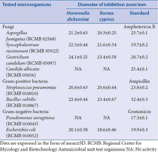
Figure 4.
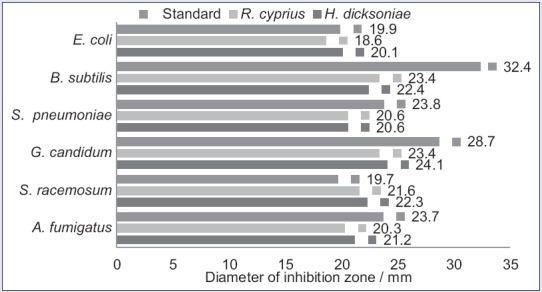
Antimicrobial activity as diameter of inhibition zone/mm of plant extracts
The results of the MIC determinations [Table 5 and Figure 5] showed noticeable MIC values for the tested plant extracts against the entire set of the tested organisms except for P. aeruginosa and C. albicans. All the tested plant extracts exhibited both antibacterial and antifungal activities. The obtained MICs varied from 0.98 to 3.9 µg/mL for the tested plant extracts. The lowest MIC values (0.98 µg/mL) observed with the plant extracts against S. racemosum, G. candidum and B. subtilis. When regarding the activity of the positive standard against the tested microbial species, the two plant extracts showed the highest level of activity against S. racemosum with an MIC 0.98 µg/mL (nearly fourfold lower than that of amphotericin B), followed by H. dicksoniae extract against the fungus A. fumigatus and the two plant extracts against the Gram-positive bacteria S. pneumoniae with MICs 1.95 µg/mL (nearly only twofold greater than that of amphotericin B and ampicillin). H. dicksoniae extract is active against E. coli as the same as the standard gentamycin with an MIC 3.9 µg/mL. R. cyprius extract showed the lowest level of activity against A. fumigatus and E. coli.
Table 5.
Antimicrobial activity as minimum inhibitory concentration/(μg/mL) of plant extracts
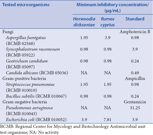
Figure 5.
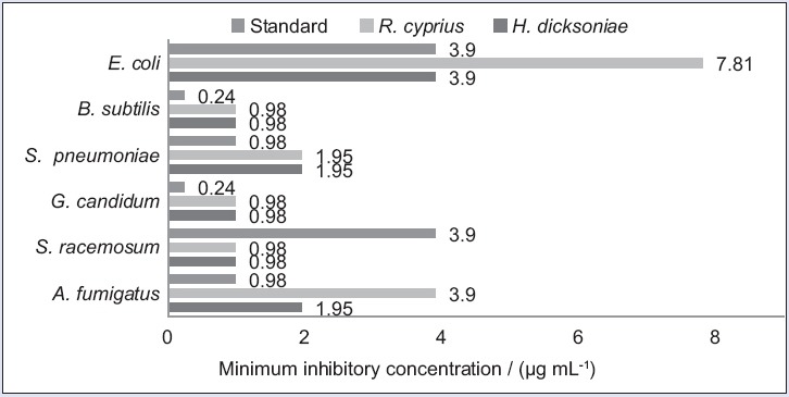
Minimal inhibitory concentrations (μg/mL) of plant extracts
The antioxidant activity
There are significant variations in the capacity of the plant extracts to scavenge the DPPH radical with IC50 ranging from 43 to 45 µg/mL [Table 2, Figures 6 and 7]. From the estimated IC50 values, the order of potency is R. cyprius extract with IC50 43.9 µg/mL followed by H. dicksoniae extract with IC50 45.2 µg/mL. The IC50 of these extracts are higher than the IC50 of the positive control (ascorbic acid 14.2 µg/mL).
Figure 6.
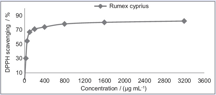
Antioxidant activity of Rumex cyprius extract using 1, 1-diphenyl-2-picrylhydrazyl scavenging
Figure 7.
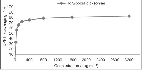
Aantioxidant activity of Horwoodia dicksoniae extract using 1, 1-diphenyl-2-picrylhydrazyl scavenging
The cytotoxic activity
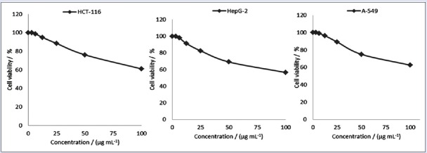
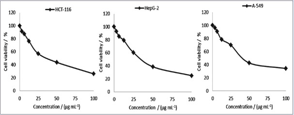
Cytotoxic activities of the H. dicksoniae and R. cyprius extracts were tested against three cancer cell lines Human breast cancer (MCF-7), Human liver cancer (HepG-2), and Human lung carcinoma (A-549) [Table 2, Figures 8 and 9]. As a result, H. dicksoniae exhibited a remarkable cytotoxic activity against the three cell lines with values of IC50 37.0, 38.5, and 43.3 µg/mL, respectively. On the other hand, R. cyprius extract showed no cytotoxic activity against the tested cell lines.
Figure 8.
Cytotoxic activity of Horwoodia dicksoniae extract against cancer cell lines HCT-116, hepatoma carcinoma cells-2 and A-549 respectively
Figure 9.
Cytotoxic activity of Rumex cyprius extract against cancer cell lines HCT-116, hepatoma carcinoma cells-2 and A-549 respectively
CONCLUSIONS
It was suggested that further work should be carried out to isolate, purify, and characterize the active constituents responsible for the activity of these plants. Also, additional work is encouraged to elucidate the possible mechanism of action of these constituents.
Financial support and sponsorship
The authors would like to express their deep gratitude to the Northern Border University, Kingdom Saudi Arabia for providing financial support of that research with the Grant No (435-016-3)
Conflicts of interest
There are no conflicts of interest.
ABOUT AUTHOR

Mohammed F. Abdelwahab
Mohammed F. Abdelwahab, is an Associate Professor at the Department of Natural product and complementary medicine, Faculty of pharmacy, NBU university, Rafha, Saudia. He has projects in collaboration with national and international institutions. Has experience in the area of Pharmacognosy and Chemistry of Natural Products, working mainly in: Medicinal plants, New Research Drugs, HPLC-DAD and NMR.
Acknowledgment
The authors would like to specific their appreciation to the Northern Border University Kingdom Saudi Arabia and director of the Mycology and biotechnology, University of Al-Azhar, for antimicrobial, antioxidant and cytotoxic activities.
REFERENCES
- 1.RNS Yadav, Munin A. Phytochemical analysis of some medicinal plants. J Phytology. 2011;3:10–4. [Google Scholar]
- 2.Chaudhary SA. Flora of the Kingdom of Saudi Arabia Illustrated. I. Riyadh, Saudi Arabia: Ministry of Agriculture and Water, National Agriculture and Water Research Center, Cruciferae (Brassicaceae); 2001. pp. 464–533. [Google Scholar]
- 3.Ruberto G, Baratta MT, Deans SG, Dorman HJ. Antioxidant and antimicrobial activity of Foeniculum vulgare and Crithmum maritimum essential oils. Planta Med. 2000;66:687–93. doi: 10.1055/s-2000-9773. [DOI] [PubMed] [Google Scholar]
- 4.Shinwari MI, Khan MA. Folk use of medicinal herbs of Margalla Hills National Park, Islamabad. J Ethnopharmacol. 2000;69:45–56. doi: 10.1016/s0378-8741(99)00135-x. [DOI] [PubMed] [Google Scholar]
- 5.Nighat F, Muhammad Z, Riaz R, Zarrin FR, Safia A, Bushra M, et al. Biological activities of Rumex dentatus L. evaluation of methanol and hexane extracts. Afr J Biotechnol. 2009;8:6945–51. [Google Scholar]
- 6.Wangchuk P, Keller PA, Pyne SG, Taweechotipatr M, Tonsomboon A, Rattanajak R, et al. Evaluation of an ethnopharmacologically selected Bhutanese medicinal plants for their major classes of phytochemicals and biological activities. J Ethnopharmacol. 2011;137:730–42. doi: 10.1016/j.jep.2011.06.032. [DOI] [PubMed] [Google Scholar]
- 7.Harborne JB. Phytochemical Methods. New Delhi: Springer (India) Pvt. Ltd; 2005. p. 17. [Google Scholar]
- 8.William Charles Evans, Trease and Evans Pharmacognosy. 15th ed. Edinburgh, UK: W.B. Saunders; 2002. [Google Scholar]
- 9.Jorgensen JH, Turnidge JD. Susceptibility test methods: Dilution and disk diffusion methods. In: Murray PR, Baron EJ, Jorgensen JH, Landry ML, Pfaller MA, editors. Manual of Clinical Microbiology. 9th ed. Washington, D.C: ASM Press; 2007. pp. 1152–72. [Google Scholar]
- 10.Brown D, Macgowan A. Harmonization of antimicrobial susceptibility testing breakpoints in Europe: Implications for reporting intermediate susceptibility. J Antimicrob Chemother. 2010;65:183–5. doi: 10.1093/jac/dkp432. [DOI] [PubMed] [Google Scholar]
- 11.Cleidson V, Simone M, Elza FA, Artur SJ. Screening methods to determine antibacterial activity of natural products. Braz J Microbiol. 2007;38:369–80. [Google Scholar]
- 12.Rajbhandari M, Schöpke T. Antimicrobial activity of some Nepalese medicinal plants. Pharmazie. 1999;54:232–4. [PubMed] [Google Scholar]
- 13.Tailor CS, Goyal A. Antioxidant activity by DPPH radical scavenging method of Ageratum conyzoides Linn. leaves. Am J Ethnomed. 2014;1:244–9. [Google Scholar]
- 14.Gangadevi V, Muthumary J. Preliminary studies on cytotoxic effect of fungal taxol on cancer cell lines. Afr J Biotechnol. 2007;6:1382–6. [Google Scholar]
- 15.Saravanan BC, Sreekumar C, Bansal GC, Ray D, Rao JR, Mishra AK. A rapid MTT colorimetric assay to assess the proliferative index of two Indian strains of Theileria annulata. Vet Parasitol. 2003;113:211–6. doi: 10.1016/s0304-4017(03)00062-1. [DOI] [PubMed] [Google Scholar]
- 16.Lyman R. Ott. An Introduction to Statistical Methods and Data Analysis. 6th ed. Canada: Brooks Cole; 2010. pp. 140–205. [Google Scholar]
- 17.Lin YL, Kuo YH, Shiao MS, Chen CC, Ou JC. Flavonoid glycosides from Terminalia catappa L. J Chin Chem Soc. 2000;47:253–6. [Google Scholar]
- 18.Wen P, Han H, Wang R, Wang N, Yao X. C-glycosylfavones and aromatic glycosides from Campylotropis hirtella (Franch.) Schindl. Asian J Tradit Med. 2007;2:149–53. [Google Scholar]


