Dear Sir,
Previous reports have illustrated reversible structural changes in optic disc cupping, the lamina cribrosa, and the retinal nerve fiber layer (RNFL) following glaucoma surgery[1]–[3]. However, few studies have assessed changes in macular thickness following such intervention. Our aim was to investigate the impact of intraocular pressure (IOP) reduction after glaucoma surgery on total macular thickness utilizing spectral domain-optical coherence tomography(SD-OCT) (Spectralis, Heidelberg Engineering, Carlsbad, CA, USA). The Wills Eye Glaucoma Research Center retrospectively reviewed the charts of glaucoma patients who underwent trabeculectomy or tube shunt surgery between January 2012 and February 2013. The institutional review board of Wills Eye Hospital approved the study. Patients were included if they had macular SD-OCT scans within one year prior to and up to two years following surgery. Patients with maculopathy, trauma to the eye, previous glaucoma surgeries prior to 2012, or scans with image quality score (Q) less than 16 were then excluded to determine the final sample size[4]. The main outcome measures were the mean IOP and the total macular thickness. A 2-sided paired t-test was used to analyze the changes in these outcome measures. Ten patients were included in the study. Baseline characteristics and changes in mean IOP and total macular thickness following surgery are shown in Table 1. The majority of patients were diagnosed with primary open angle glaucoma and underwent trabeculectomy. Prior to surgery, mean IOP was 25.9 mm Hg (SD: 8.7 mm Hg). Mean total macular thickness increased from 261 µm to 271 µm (SD: 17.3 µm, P=0.096) following a significant reduction in IOP (SD: 6.2 mm Hg, P<0.001). Following surgery, mean IOP was 16.2 mm Hg (SD: 7.3 mm Hg). Seven out of 10 (70%) patients showed increases in total macular thickness. There was no correlation between the amount of IOP reduction and change in macular thickness (P>0.20). Cystoid macular edema was not noted in any of the scans.
Table 1. Baseline characteristics and changes in mean IOP and total macular thickness following glaucoma surgery.
| Variables | Value (n=12) | P |
| Age (x±s), a | 72.9±8.8 | N/A |
| Sex, n (%) | N/A | |
| M | 5 (50) | |
| F | 5 (50) | |
| Race, n (%) | N/A | |
| Caucasian | 7 (70) | |
| African American | 3 (30) | |
| Diagnosis, n (%) | N/A | |
| Primary open angle glaucoma | 9 (90) | |
| Chronic angle-closure glaucoma | 1 (10) | |
| Surgery type, n (%) | N/A | |
| Trabeculectomy | 7 (70) | |
| Combined cataract and trabeculectomy surgery | 2 (20) | |
| Tube Shunt | 1 (10) | |
| aChange in IOP, mean (95% CI), mm Hg | -9.7 (-13.6, -5.8) | <0.001 |
| aChange in total macular thickness, mean (95% CI), µm | 10.2 (-0.54, 20.9) | 0.096 |
IOP: Intraocular pressure; N/A: Not applicable, CI: Confidence interval. aPostoperative minus preoperative.
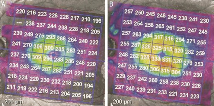
A previous study has demonstrated reversible structural changes in the optic disc, such as the cup-to-disc area ratio, maximum cup depth, and neuroretinal rim volume, following glaucoma surgery[1]. Additionally, prior investigations have also shown decreases in the depth of the lamina cribrosa and thickening of the peripapillary RNFL following successful IOP reduction[2]–[3]. Our study shows a trend towards a significant increase in mean total macular thickness in patients who underwent trabeculectomy or tube shunt surgery (Figure 1).
Figure 1. Macular SD-OCT scans of a patient with substantial macular thickening following surgery.
A: Prior to surgery; B: 14mo following surgery.
It is well established that axial length decreases significantly following glaucoma surgery[2], along with a reduction in the size of the eye. We hypothesize that when IOP is high, the pressure applied on the scleral wall and retinal layers causes diffuse retinal thinning. Once IOP is acutely reduced following surgery, the retina thickens and partially regains its structure. It has also been suggested that peripapillary swelling, due to a sudden postoperative IOP reduction, may cause RNFL thickening[3]. However, this swelling is expected to resolve over time.
Our results agree with recent reports showing macular thickening 1mo following glaucoma surgery[5]–[6]. Nonetheless, in our study, postoperative scans were conducted after an average of 10.8±5.3mo, which is the longest follow-up period to date. Thus, our findings suggest that a change in macular thickness may occur with IOP reduction after surgery, but larger studies are needed to provide stronger evidence.
Acknowledgments
Conflicts of Interest: Pitale PM, None; Chatha U, None; Patel V, None; Gupta L, None; Waisbourd M, None; Pro MJ, None.
REFERENCES
- 1.Lesk MR, Spaeth GL, Azuara-Blanco A, Araujo SV, Katz LJ, Terebuh AK, Wilson RP, Moster MR, Schmidt CM. Reversal of optic disc cupping after glaucoma surgery analyzed with a scanning laser tomograph. Ophthalmology. 1999;106(5):1013–1018. doi: 10.1016/S0161-6420(99)00526-6. [DOI] [PubMed] [Google Scholar]
- 2.Yoshikawa M, Akagi T, Hangai M, Ohashi-Ikeda H, Takayama K, Morooka S, Kimura Y, Nakano N, Yoshimura N. Alterations in the neural and connective tissue components of glaucomatous cupping after glaucoma surgery using swept-source optical coherence tomography. Invest Ophthalmol Vis Sci. 2014;55(1):477–484. doi: 10.1167/iovs.13-11897. [DOI] [PubMed] [Google Scholar]
- 3.Aydin A, Wollstein G, Price LL, Fujimoto JG, Schuman JS. Optical coherence tomography assessment of retinal nerve fiber layer thickness changes after glaucoma surgery. Ophthalmology. 2003;110(8):1506–1511. doi: 10.1016/S0161-6420(03)00493-7. [DOI] [PMC free article] [PubMed] [Google Scholar]
- 4.Huang Y, Gangaputra S, Lee KE, Narkar AR, Klein R, Klein BE, Meuer SM, Danis RP. Signal quality assessment of retinal optical coherence tomography images. Invest Ophthalmol Vis Sci. 2012;53(4):2133–2141. doi: 10.1167/iovs.11-8755. [DOI] [PMC free article] [PubMed] [Google Scholar]
- 5.Sesar A, Cavar I, Sesar AP, Geber MZ, Sesar I, Laus KN, Vatavuk Z, Mandic Z. Macular thickness after glaucoma filtration surgery. Coll Antropol. 2013;37(3):841–845. [PubMed] [Google Scholar]
- 6.Karasheva G, Goebel W, Klink T, Haigis W, Grehn F. Changes in macular thickness and depth of anterior chamber in patients after filtration surgery. Graefes Arch Clin Exp Ophthalmol. 2003;241(3):170–175. doi: 10.1007/s00417-003-0628-6. [DOI] [PubMed] [Google Scholar]


