Abstract
Legumes form symbiotic associations with either nitrogen-fixing bacteria or arbuscular mycorrhizal fungi. Formation of these two symbioses is regulated by a common set of signalling components that act downstream of recognition of rhizobia or mycorrhizae by host plants. Central to these pathways is the calcium and calmodulin-dependent protein kinase (CCaMK)–IPD3 complex which initiates nodule organogenesis following calcium oscillations in the host nucleus. However, downstream signalling events are not fully understood. Here we show that Medicago truncatula DELLA proteins, which are the central regulators of gibberellic acid signalling, positively regulate rhizobial symbiosis. Rhizobia colonization is impaired in della mutants and we provide evidence that DELLAs can promote CCaMK–IPD3 complex formation and increase the phosphorylation state of IPD3. DELLAs can also interact with NSP2–NSP1 and enhance the expression of Nod-factor-inducible genes in protoplasts. We show that DELLA is able to bridge a protein complex containing IPD3 and NSP2. Our results suggest a transcriptional framework for regulation of root nodule symbiosis.
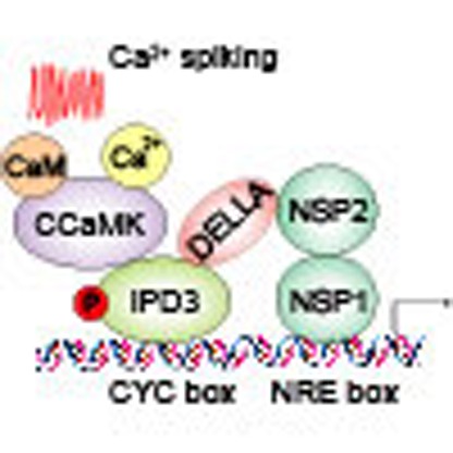 Symbiotic associations between legumes and nitrogen-fixing bacteria are regulated by a CCaMK-IPD3 protein complex which promotes nodule initiation. Here, Jin et al. propose that DELLA proteins positively regulate nodulation by enhancing phosphorylation of IPD3 and acting as a positive regulator of transcription.
The availability of nitrogen is important for plant growth and stress resistance. Legumes are able to form beneficial symbiotic associations with nitrogen-fixing rhizobial bacteria that provide nitrogen to the host plant1,2. Legumes develop a specific organ, called a nodule, which provides the proper microenvironment for ammonia generation through the reduction of molecular dinitrogen by rhizobia and nutrient exchange between both symbionts. Nitrogen fixation is energetically expensive and this symbiotic interaction is precisely regulated by the plant to adapt to environment conditions.
There are two major developmental programs in root nodule symbiosis: nodule primordia originate from root cortical cells which re-activate cell divisions, and simultaneously, root epidermal responses prepare the cell for bacterial infection2. The formation of nitrogen-fixing nodules is controlled by a host genetic programme that synchronizes these two processes3,4. The early development of rhizobial symbiosis is achieved through a chemical communication between plant and rhizobium, present in the rhizosphere. Flavonoids are released by the plant roots as a signal to rhizobia. In turn, rhizobia produce nodulation factors (Nod factors) that are recognized by the host plant to activate a common symbiotic signalling pathway required for mycorrhizal and rhizobial symbioses2,4,5. Over the past 20 years, progress has been made in understanding the genetic programme in Medicago truncatula and Lotus japonicus. Two LysM receptor kinases NFR1/LYK3, NFR5/NFP and a leucine-rich repeat receptor kinase DMI2/SYMRK are necessary for Nod factor perception6,7,8,9,10,11,12. Interestingly, it has been recently shown that NFR1/LYK3 is also involved in mycorrhizal symbiosis and is a new common symbiotic gene13. After perception at the plasma membrane, the remaining components are associated with nucleus, which are involved in both mycorrhizal and rhizobial symbioses. Two cation channels (Castor and Pollux) and three components at the core of the nuclear pore (NENA, NUP85 and NUP133) are located on the nuclear membrane14,15,16,17,18,19. These components are required for Nod-factor-induced calcium oscillations4. Perception of the calcium oscillations requires a nuclear-localized calcium and calmodulin-dependent protein kinase (CCaMK, known as DMI3 in M. truncatula)20,21. The activation of CCaMK is sufficient to induce symbiotic processes, as gain-of-function mutations in CCaMK induce nodulation in the absence of rhizobia22,23,24. CCaMK associates with and phosphorylates CYCLOPS (known as IPD3 in M. truncatula)25,26, which is essential for rhizobial and mycorrhizal colonization. Very recently, phosphorylated CYCLOPS has been shown to bind the promoter of NIN and induce nodulation in the absence of rhizobia27.
Several nuclear-associated transcriptional regulators, including nodule inception (NIN)28,29, an ERF family protein (ERN)30,31, and two GRAS family proteins, Nodulation signaling pathway 1 (NSP1) and NSP2, are required for the expression of Nod-factor-induced genes and initiation of nodulation32,33. NSP1 and NSP2 form a hetero-complex that associates with promoters of Nod-factor-inducible genes, such as ENOD11 and ERN1 (ref. 34). NSP1 and NSP2 were originally thought to function specifically in nodulation. However, it was recently shown that NSP1 and NSP2 are also required for arbuscular mycorrhizal fungi (AMF)-associated lipochitooligosaccharide (LCO) signalling or mycorrhizal infection35,36,37,38. NSP1 contains a DNA-binding domain, whereas there is none in NSP2, which requires an interaction with NSP1 to associate with Nod-factor-inducible promoters34. The NSP1–NSP2 protein complex promotes the expression of NIN and ERN, two transcription factors with nodulation-specific functions. Interestingly, it has recently been shown that NSPs are not required for CYCLOPS-induced NIN expression, but they are required for CYCLOPLS-induced nodule organogenesis27. So it remains unknown how NSP1–NSP2 activity is associated with the action of CCaMK-CYCLOPS or whether the two protein complexes function independently in root nodule symbiosis.
The phytohormone gibberellic acid (GA) has been implicated in many aspects of plant biology, including seed germination, stem elongation, leaf expansion, pollen maturation and induction of flowering39,40. GA is perceived by GID1 (GA insensitive dwarf 1) and promotes the destruction of DELLA (SLR1, slender rice 1, one copy in rice genome) which is a nuclear protein that restrains the cell proliferation and expansion that drive plant growth39,40. DELLA is well known as a transcriptional suppressor that acts by directly binding DNA recognition domain of other transcript factors such as PIF3, 4 and inhibiting their transcriptional activities41,42. DELLA is an integrator of plant responses to hormones and environmental stresses, and DELLA-dependent growth restraints are advantageous in adverse environment stresses43.
DELLA family proteins are required for mycorrhizal symbiosis44,45. The mycorrhizal phenotype of della double mutant in M. truncatula shows DELLAs function at the arbuscule development stage44. Interestingly we recently showed that AMF infection and arbuscule development are both defective in the rice della mutant, slr1, suggesting an early signalling function of DELLAs in symbiosis45. This different mycorrhizal defect of mutants in rice and Medicago could be caused by genetic redundancy of DELLAs in Medicago. The role of GAs in the control of root nodule symbiosis has been investigated for over 60 years46,47. It has been shown that exogenous application of GAs inhibits the formation of infection threads and nodules, but other results show GAs can promote root nodule symbiosis in legumes48,49,50,51.
Here we show that DELLA proteins promote nodule development and infection thread formation during root nodule symbiosis. We provide evidence that DELLAs can promote CCaMK–IPD3/CYCLOPS complex formation and increase the phosphorylation of IPD3/CYCLOPS. We show that DELLAs can form a protein complex with NSP2–NSP1 and are able to bridge a protein complex containing IPD3/CYCLOPS and NSP2. We propose that DELLA proteins represent a missing link in the common symbiotic signalling pathway required for both rhizobial and mycorrhizal symbioses.
Results
GA inhibits root nodule symbiosis
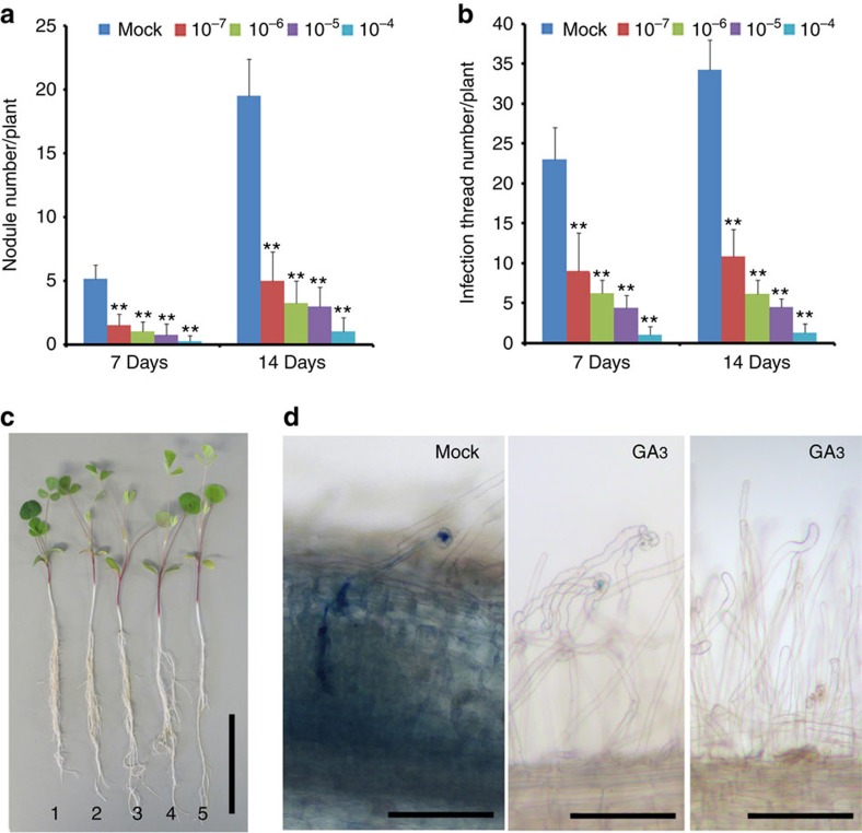
It has previously been shown that gibberellins play an important role in root nodule symbiosis48,49,50. To better understand the nature of this regulation, we initiated studies to assess the effects of GA3 on nodulation efficiency in M. truncatula under our growth conditions. We grew M. truncatula cv. Jemalong line A17 plants in the soil pretreated with 0, 10−7, 10−6, 10−5 and 10−4 M GA3, concentrations of GA3 that are higher than endogenous GAs levels in plants, and quantified nodulation events at 7 and 14 days post inoculation with Sinorhizobium meliloti strain 1021 (Sm1021). As expected the shoots of GA-treated plants displayed increased shoot length and shoot fresh weight (Supplementary Fig. 1a,b). However, root length, lateral root number and root fresh weight of the plants treated with GA3 at concentrations from 10−7 to 10−5 M were not significantly changed compared with control plants (Supplementary Fig. 1c–e). Interestingly, we found that nodulation was severely impaired at 10−6–10−4 M GA3 treatment (Fig. 1a), while the shoot length increased after treated with GA3 (Fig. 1c). The generation of nodules involves a number of processes that may be regulated by GA3, including the re-initiation of cortical cell division to form a nodule primordium and the facilitation of bacterial infection through epidermal and cortical cells. We therefore assessed whether GA3 could regulate bacterial infection of the epidermis by quantifying the number of infection events and nodule development in plants grown at different concentrations of GA3. The infection frequency was also decreased with increasing concentrations of GA3 (Fig. 1b,d), suggesting that GAs regulates both nodule formation and bacterial infection independently of the shoot and root growth phenotype.
Figure 1. GA3 represses root nodulation development and rhizobial infection in M. truncatula.
Wild-type A17 plants were grown with different concentrations of GA3 and upon inoculation with S. meliloti strain 1021 harbouring a LacZ reporter and assayed for nodule development (a), rhizobial infection events (b), and plant development (c). Increasing GA3 concentration promotes plant growth but inhibits nodulation and rhizobial infection. Infection threads were counted following LacZ staining. Twelve plants were analysed for each GA3 concentration. 1:mock, 2:10−7, 3:10−6, 4:10−5, 5:10−4 M GA3. (d) Representative images of infection threads visualized by staining of S. meliloti (strain 1021) expressing the LacZ reporter. The left panel shows infection thread progression in to a nodule primordium. The middle panel shows that a representative image of infection thread progression was inhibited by treatment of 10−4 M GA3 and the right panel shows that a representative image of no infection thread progression in the most plant roots treated with 10−4 M GA3. Scale bars correspond to 2 cm (c) and 100 μm (d). These experiments were repeated three times with similar results. Error bars are standard error. The asterisk indicates a significant decrease to the control with Student's t-test (**P<0.01).
DELLAs are required for root nodule symbiosis
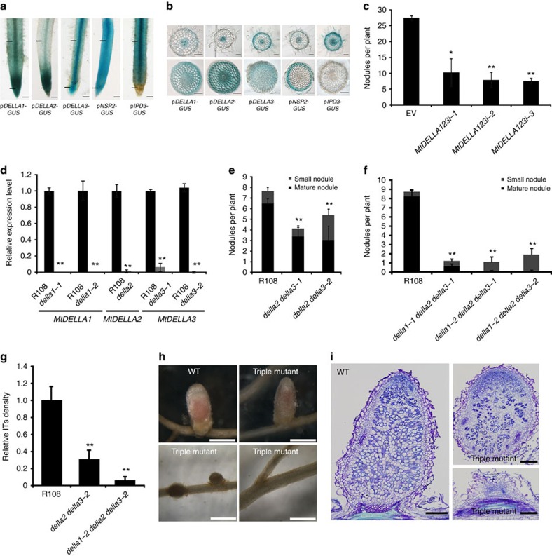
Gibberellin is perceived by GID1 and promotes the interaction of GID1 and DELLA, which in turn leads to the polyubiquitination and subsequent degradation of DELLA by the 26s proteasome in the nucleus39,40. Three DELLA genes and two highly homologous genes were found in the M. truncatula genome database Mt3.5 and Mt4.0 (Supplementary Fig. 2)44. When we transformed functional MtDELLA-GFP (Supplementary Fig. 3) driven by 35S promoter into M. truncatula using hairy root transformation, no green fluorescent protein (GFP) signal was detected in the root, but occasionally very weak GFP signals were detected only in root tip cells, suggesting that the amounts of DELLA proteins are tightly regulated in M. truncatula. Consistent with the degradation of DELLAs by GA treatment in other plant species, GA3 treatment induced the degradation of MtDELLA2 and MtDELLA3 in the nucleus (Supplementary Fig. 3a). Under non-symbiotic conditions, the expression of the three DELLAs was detected in epidermal cells, cortical cells and stele in the rhizobial infection zone (closed to root apical meristem) and elongation zone (Fig. 2a,b). Consistent with our results, expression of DELLA1 and DELLA2 in the epidermal cells (root hair cells) was also found in transcriptional data (http://mtgea.noble.org/v3/blast_result.php?s=226645). Further analysis showed that MtDELLA3 was upregulated 6 h post Nod factor treatment and that MtDELLA1 and MtDELLA2 were induced 7 day post inoculation (d.p.i.) with Sm1021, suggesting a role of DELLAs in root nodule symbiosis (Supplementary Fig. 4a,b). To assess whether DELLAs function in root nodule symbiosis, we generated transformed hairy roots expressing an MtDELLA hairpin allowing RNA-mediated interference of MtDELLAs. We used three different combination of RNA interference (RNAi) targeted three MtDELLAs and observed that expression levels of MtDELLAs were reduced in these RNAi plants (Supplementary Fig. 4e). The total nodule number was significantly decreased in MtDELLAs RNAi plants compared with the empty vector control (Fig. 2c), indicating that DELLA proteins are positive regulators in root nodule symbiosis.
Figure 2. MtDELLAs promote root nodule symbiosis.
(a) Whole-root GUS activity of pDELLA1:GUS, pDELLA2:GUS, pDELLA3:GUS, pNSP2:GUS, pIPD3:GUS are shown. Scale bars correspond to 2 mm. The black bars indicate that the region is selected for the transversal section in b. (b) Thin transversal sections (70 μm) of pDELLA1:GUS, pDELLA2:GUS, pDELLA3:GUS, pNSP2:GUS, pIPD3:GUS roots are shown. Scale bars correspond to 100 μm. (c) Numbers of nodules on MtDELLAs RNAi plant roots 4 weeks post inoculation with S. meliloti. n⩾8, where n denotes the number of plants. (d) qRT-PCR analysis of MtDELLAs transcript levels. The expression levels of MtDELLAs were detected in wild-type (R108) and della1, della2 and della3 mutants. Expression levels were normalized against the reference gene Elongation factor 1-alpha (EF1-α). The RNA was extracted from six individual plants of R108 or mutants. (e,f) Scores of nodule number in della2/della3 double mutants (e) and della1/della2/della3 triple mutants (f). (g) Quantification of infection threads in della double and triple mutants. The infection thread (IT) was visualized by staining of S. meliloti expressing the LacZ reporter and was counted at 7 d.p.i. n⩾13, where n denotes the number of plants. Relative ITs density indicates the number of infection threads per root length per plant, and they were normalized against the wild-type (R108). (h) Nodule development is impaired in della1/della2/della3 triple mutant. There is no nodule formed in 12 plants out of 21 della1-1/della2/della3-1 triple mutants. Occasionally pink nodules were formed in della triple mutants. Scale bars correspond to 500 μm. (i) Nodule sections of the pink nodule and the white nodule in the della triple mutant, nodules were stained with toluidine blue. Scale bars correspond to 200 μm. This is a representative experiment repeated three times in c and d, and twice in e, f and g. Error bars represent s.d. The asterisk indicates a significant decrease relative to wild type or vector control with Student's t-test (*P<0.05; **P<0.01).
To further confirm the RNAi results, M. truncatula lines containing Tnt1 insertions in MtDELLA1, MtDELLA2 and MtDELLA3 were obtained from a mutant population generated at the Samuel Roberts Noble Foundation (Supplementary Fig. 4) (http://medicago-mutant.noble.org/mutant/database.php) and the transcription levels of MtDELLA1, MtDELLA2 and MtDELLA3 were greatly reduced in the della mutants (Fig. 2d). Given genetic redundancy of DELLAs in Medicago, della2/della3 double mutants were generated and showed significantly decreased nodule number at 21 d.p.i. compared with its wild type (R108) (Fig. 2e). However, the root length of double mutant was not different from wild type (Supplementary Fig. 5). While few matured nodules were formed in the della2/della3 double mutants (Fig. 2e). We further generated the della triple mutants and observed very few mature pink nodules in della1/della2/della3 plants (Fig. 2f–i). Intriguingly, no nodules were formed in 12 out of 21 della1-1/della2/della3-1 triple mutants. Further analysis revealed the number of infection threads was greatly reduced in della triple mutants and the pink nodule formed on della triple mutants appeared to be aberrant (Fig. 2f–i). These results indicated that MtDELLAs are essential for root nodule symbiosis.
Gene induction by Nod factors is dependent on DELLAs
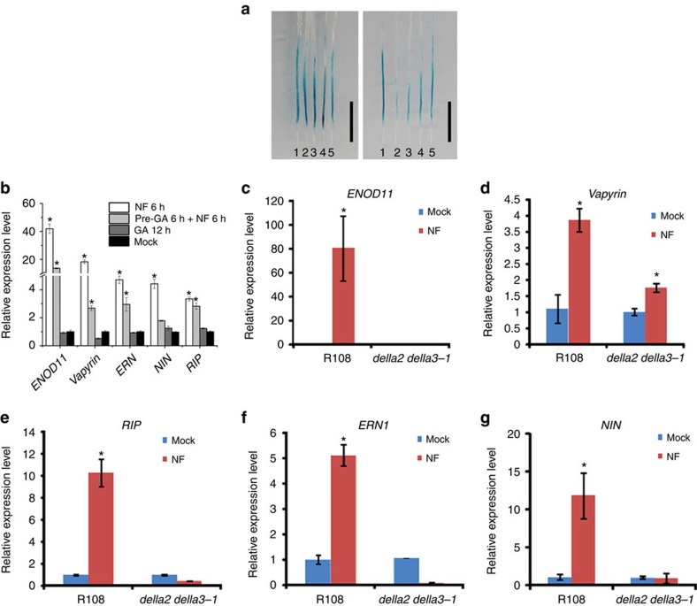
ENOD11 represents one of the earliest marker genes for response to Nod factor and rhizobia. The induction of ENOD11 requires the Nod factor signal transduction pathway and as such provides markers for the regulation of this signalling pathway52. To assess the effects of GA3 on Nod-factor-induced gene expression, we used M. truncatula plants stably transformed with β-glucuronidase (GUS) driven by the ENOD11 promoter. Seven-day-old plants were transferred into liquid BNM medium containing 10−7, 10−6, 10−5 and 10−4 M GA3. Following 6 h of GA3 treatment, 1 nM Nod factor was added to the medium, and after 6 h of Nod factor treatment, the roots were used for GUS staining. Plants without Nod factor treatment only showed GUS activity in root caps, while Nod factor treatment activates ENOD11 in a specific region of the root (Fig. 3a). Pre-treatment with 10−4 M GA3 led to reduced induction of ENOD11 (Fig. 3a,b), but co-treatment with GA3 did not reduce induction of ENOD11 (Fig. 3a,b), suggesting that DELLAs may act at a very early stage in the signal transduction pathway. Consistent with the GA treatment data, we found that the induction of Nod-factor-induced genes was impaired in della2/della3 double mutants (Fig. 3c–g), suggesting that DELLAs may directly regulate the expression of early nodulin genes.
Figure 3. Nod-factor-induced gene expression is promoted by MtDELLAs.
(a) The effect of GA3 on Nod-factor-induced gene expression was assayed using transgenic plants expressing pENOD11:GUS. Note: 6 h pre-treatment of GA3 suppresses expression of ENOD11 (right panel), but GA3 co-treatment with Nod factor does not suppress expression of ENOD11 (left panel). 1:mock, 2:10−4, 3:10−5, 4:10−6, 5:10−7 M GA3. Scale bars correspond to 1 cm. (b) Real-time PCR results revealed that GA3 suppresses expression of Nod-factor-induced genes. Treatment with 1 nM Nod factor for 6 h induced expression of ENOD11, VAPYRIN, NIN, ERN and RIP10 and the induction was reduced upon pre-treatment with 10−5 M GA3 for 6 h. (c–g) Real-time PCR results revealed that MtDELLAs required for induction of nodulin genes. Seven-day-old della2/della3 double mutants and R108 were treated with 1 nM Nod factor or mock (BNM medium) for 6 h. In wild type the nodulin genes were induced, however these genes could not be induced in della2/della3 double mutants after Nod factor treatment. These experiments were repeated three times with similar results, one representative experiment was shown. Error bars are standard error (n=3). The asterisk indicates a significant induction relative to control with Student's t-test (*P<0.01).
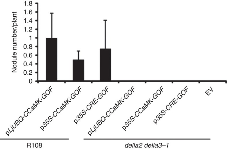
A gain-of-function (GOF) mutation in a CCaMK or a gain-of-function of a cytokinin receptor (CRE1/SNF2) induces spontaneous cortical cells division and leading to spontaneous nodule formation in the absence of rhizobia2. The gain-of-function of CCaMK (1–311aa) or GOF-CRE1 was introduced into hairy roots to determine the effect of GA3-coupled DELLA degradation on the cortical cell division. The transgenic roots treated with GA3 showed a significant reduction in the number of spontaneous nodules in GOF-CRE1 lines (Supplementary Fig. 6a), suggesting that DELLAs are required for spontaneous nodule formation induced by cytokinin and DELLAs may function at the nodule development stage. Spontaneous nodule formation was also inhibited by GA3 treatment in overexpression of GOF-CCaMK plants (Supplementary Fig. 6b). Intriguingly, the spontaneous nodule formation induced by CRE1-GOF or CCaMK1-311-GOF was fully blocked in della2/della3-1 double mutants (Fig. 4), suggesting that DELLA proteins act downstream of CCaMK.
Figure 4. DELLAs promote spontaneous nodule formation induced by CRE1 and CCaMK.
Spontaneous nodule formation induced by gain-of-function CRE1 and CCaMK1-311 was blocked in della double mutants. The nodule number was counted 5 weeks after hairy root transformed plants being transferred to sterile vermiculite with perlite (n⩾6, where n denotes the number of plants).
DELLAs are able to interact with IPD3
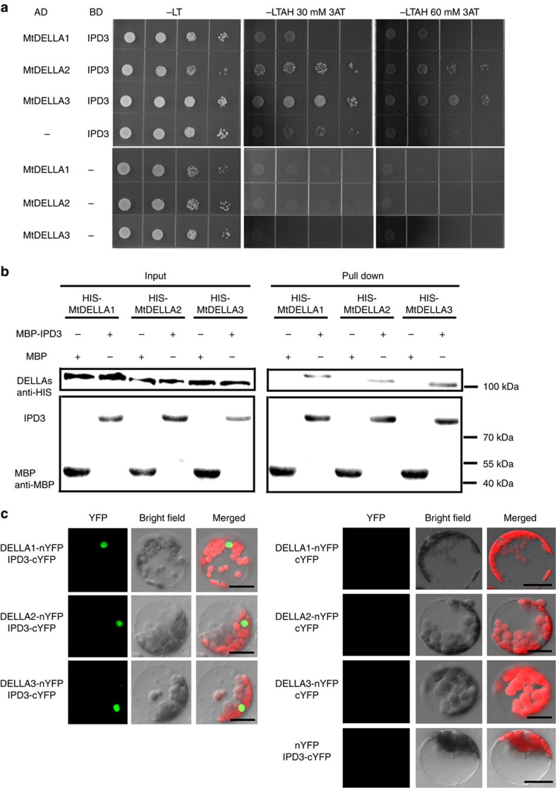
Since DELLA proteins are involved in both root nodule and mycorrhizal symbioses, we propose that DELLAs may interact with the key components of the common signalling pathway. We observed that DELLA proteins MtDELLA1, MtDELLA2 and MtDELLA3 interacted with IPD3, but not CCaMK and NSP1 in a yeast two-hybrid (Y2H) assay, although CCaMK and NSP1 proteins were expressed in yeast (Fig. 5a; Supplementary Fig. 7c,d). MtDELLAs and IPD3 are also able to interact in in vitro pull-down assays (Fig. 5b) and bimolecular fluorescence complementation (BiFC) assays in Arabidopsis protoplasts (Fig. 5c). A strong fluorescence signal was observed in the nucleus of Arabidopsis mesophyll protoplasts co-transformed with IPD3-nYFP and MtDELLAs-cYFP. Protoplasts transformed with MtDELLA-nYFP and the control vector or with IPD3-cYFP and the control vector were used as controls, which showed no positive signal (Fig. 5c). Taken together, these results show that MtDELLAs are able to interact with IPD3.
Figure 5. Interactions between MtDELLAs and MtCYCLOPS/IPD3.
(a) Yeast two-hybrid assays between IPD3 and MtDELLA1, MtDELLA2 or MtDELLA3. Yeast cells carrying different combinatory constructs are listed on the left. Serial dilutions (10 times) of yeast cells expressing the indicated proteins from the pGADT7 (AD) and pGBKT7 (BD) vectors were plated onto SD/-Leu-Trp (-LT) medium or SD/-Leu-Trp-Ade-His (-LTAH) medium with 30 or 60 mM 3-amino-1,2,4-triazole (3AT). (b) Pull-down assays between IPD3 and MtDELLA1, MtDELLA2 or MtDELLA3. MBP-IPD3 fusion protein but not MBP alone bound with HIS-tagged MtDELLA1, MtDELLA2 or MtDELLA3. (c) Detection of protein–protein interactions in Arabidopsis protoplast by BiFC. YFP fluorescence of leaves co-transformed with MtDELLAs-nYFP (N-terminal half of YFP) and IPD3-cYFP (C-terminal half of YFP). No protein interactions were detected in the other three combinations: MtDELLAs-nYFP--cYFP, IPD3-cYFP--nYFP and cYFP--nYFP. Scale bars, 20 μm.
DELLAs can form a protein complex with IPD3 and CCaMK
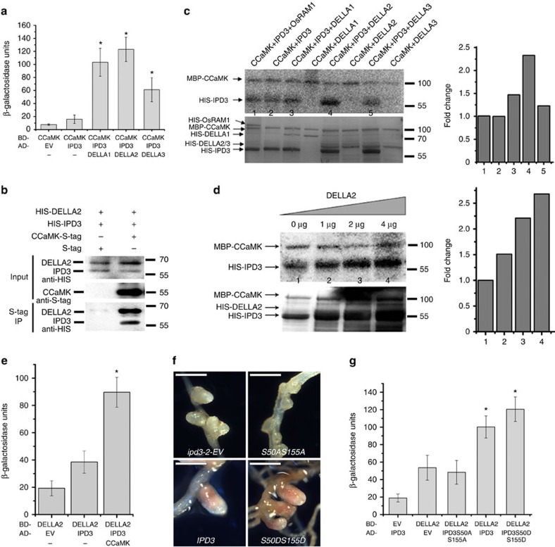
It has been shown that CCaMK interacts with and phosphorylates IPD3/CYCLOPS26. To assess the relationship between CCaMK, IPD3 and MtDELLAs, we performed a yeast three-hybrid (Y3H) assay with the fusion proteins CCaMK-GAL4-DNA-binding domain (GAL4-DBD) and/or IPD3-GAL4 activation domain (GAL4-AD). Additive activation of the reporter was observed when MtDELLA proteins were present (Fig. 6a), suggesting that MtDELLA may form a complex with CCaMK and IPD3. We did not observe the interaction of CCaMK and MtDELLAs in the yeast two-hybrid assay (Supplementary Fig. 7). To further assess whether MtDELLA, CCaMK and IPD3 can form a protein complex, we co-expressed HIS-DELLA, CCaMK-S-tag and HIS-IPD3 in Escherichia coli and found that CCaMK could co-immunoprecipitate both IPD3 and DELLA2 (Fig. 6b), suggesting that MtDELLA, CCaMK and IPD3 are able to form a complex.
Figure 6. CCaMK–IPD3–DELLA protein complex formation.
(a) MtDELLAs increased the interaction between CCaMK and IPD3 in yeast three-hybrid assays. Data are presented as β-galactosidase activity. (b) CCaMK co-immunoprecipitates with IPD3 and DELLA2 in E. coli. CCaMK, IPD3 and MtDELLA2 are co-expressed in E. coli. (c) The effect of MtDELLAs on phosphorylation of IPD3 by CCaMK in vitro phosphorylation assays. The left histogram shows quantitative intensities of IPD3 phosphorylation calculated by ImageJ. The relative intensities were normalized against the control (bar1). (d) MtDELLA2 increased the phosphorylation of IPD3 by CCaMK. An increasing amount of MtDELLA2 was added to the reaction mix. The left histogram shows quantitative intensities of IPD3 phosphorylation calculated by ImageJ. The relative intensities were normalized against the bar1. Autoradiographs show the corresponding phosphorylated IPD3. Autoradiographs of kinase assays (32P) (upper images) and Coomassie staining of the gels (lower image) in c and d. This is a representative experiment that was repeated twice in c and d. (e) CCaMK increased interaction of MtDELLA and IPD3 in yeast three-hybrid assay. Data are presented as β-galactosidase activity. (f) M. truncatula ipd3-2 roots were transformed with IPD3-S50A-S155A, IPD3-S50D-S155D and IPD3. The pink nodules were formed on ipd3-2 mutants contained IPD3 and IPD3-S50D-S155D upon 35 day post inoculation with Sm1021. Scale bars correspond to 500 μm. (g) Yeast three-hybrid assay showed sites S50 and S155 of IPD3 are critical for interaction of IPD3 and MtDELLA. Alanine replacement of S50 and S155 of IPD3 abolished the interaction with MtDELLA. Data are presented as β-galactosidase activity. Results represent the means of three experiments in a, e and g. Error bars represent standard error. The asterisk indicates a significant increase relative to the control with Student's t-test in a, e and g (*P<0.01).
DELLA enhances IPD3 phosphorylation
To analyse whether CCaMK could phosphorylate MtDELLAs, in vitro kinase assays were performed (Fig. 6c,d). We found that CCaMK could not directly phosphorylate MtDELLAs, but addition of MtDELLAs was able to enhance the intensity of phosphorylation of IPD3 by CCaMK (Fig. 6c,d). These results suggest that MtDELLAs may participate in the nodulation signalling pathway by interacting with IPD3 and promoting its phosphorylation. Interestingly we found that CCaMK could further increase the interaction of MtDELLA and IPD3 in a Y3H assay (Fig. 6e), suggesting that calcium could be an important signal for the formation of CCaMK–IPD3–DELLA complex during root nodule symbiosis. The two phosphorylated serine residues within the N-terminal of CYCLOPS/IPD3 are critical for symbiosis in Lotus japonicas27. In M. truncatula, we also found that the two residues IPD3 S50-S155 are essential for symbiosis (Fig. 6f). The phosphoablative mutant version of IPD3 S50A-S155A could not complement the ipd3-2 mutant while the phosphomimetic version of S50D-S155D could fully complement ipd3-2 (Fig. 6f). Interestingly we found that phospoablative mutant version of S50A-S155A also abolished the interaction with MtDELLAs (Fig. 6g; Supplementary Fig. 7). While the precise reason for the phenotype of the phosphoablative mutant remains to be determined, the inability of phospoablative mutant version to interact with MtDELLA, suggests that this interaction may be relevant for symbiosis. Based on these results we propose that phosphorylation of IPD3 by CCaMK promotes assembly of a CCaMK–IPD3–DELLA complex during root nodule symbiosis.
DELLAs–NSP2–NSP1 activates Nod-factor-induced genes
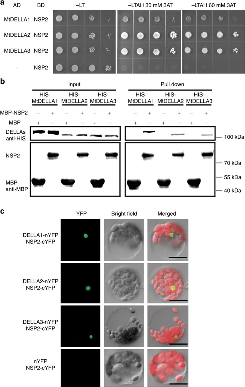
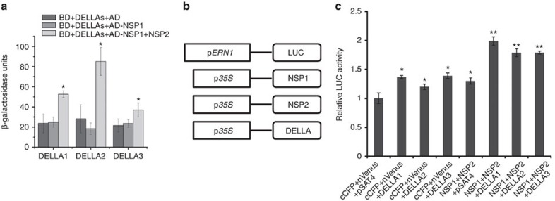
It was recently shown that NSP2 is also required for AMF-associated LCO signalling36,53. We therefore assessed whether MtDELLAs can interact with NSP2, which is a GRAS family member that directly binds to a specific promoter region of Nod-factor-inducible genes through interaction with NSP1. We found that NSP2 could interact with MtDELLA proteins DELLA1, DELLA2 and DELLA3 in Y2H (Fig. 7a), pull-down and BiFC assays (Fig. 7b,c). Intriguingly, we further observed a DELLAs–NSP2–NSP1 protein complex using the Y3H assay (Fig. 8a). It has been shown NSP1 and NSP2 could form complex and activate the expression of ERN134. As DELLA proteins function as positive regulators of root nodule symbiosis, we hypothesized that MtDELLAs could function as transcriptional activators to regulate gene expression. Transient activation analysis in Arabidopsis protoplasts showed that transfection of the protoplasts with NSP1 and NSP2 could induce a low expression level of pERN1-LUC (luciferase reporter gene driven by the ERN1 promoter) compared with the empty vector control (Fig. 8b,c), which is consistent with a previous report54. Co-transfection of NSP1/NSP2 with one of the MtDELLAs led to a much higher additive induction of pERN1-LUC (Fig. 8b,c). These results suggest that MtDELLAs can function as transcription activators and regulate the expression of Nod-factor-induced genes with NSP1/NSP2 in protoplasts.
Figure 7. Interactions between MtDELLAs and NSP2.
(a) Yeast two-hybrid assays between NSP2 and MtDELLA1, MtDELLA2 or MtDELLA3. Yeast cells carrying different combinatory constructs are listed on the left. Serial dilutions (10 times) of yeast cells expressing the indicated proteins from the pDEST-GADT7(AD) and pDEST-GBKT7(BD) vectors were plated onto SD/-Leu-Trp(-LT) medium or SD/-Leu-Trp-Ade-His (-LTAH) medium with 30 or 60 mM 3-amino-1,2,4-triazole (3AT). (b) Pull-down assays between IPD3 and MtDELLA1, MtDELLA2 or MtDELLA3. MBP-IPD3 fusion protein but not MBP alone bound HIS-tagged MtDELLA1, MtDELLA2 or MtDELLA3. (c) Detection of protein–protein interactions in Arabidopsis protoplast by BiFC. YFP fluorescence of leaves co-transformed with MtDELLAs-nYFP and NSP2-cYFP. There is no protein interaction in the other three combinations: MtDELLAs-nYFP--cYFP, NSP2-cYFP--nYFP and cYFP--nYFP (Fig. 5c). Scale bars, 20 μm.
Figure 8. The effect of DELLAs on NSP2–NSP1 induced expression of ERN1.
(a) Yeast three-hybrid assay assessing combinations of BD-MtDELLAs, AD-NSP1 and NSP2, showing MtDELLAs form a protein complex with NSP1 and NSP2. Data are presented as β-galactosidase activity. The asterisk indicates a significant increase relative to the control with Student's t-test (*P<0.05). (b,c) A transient reporter assay was used to test the trans-activation effects of NSP1, NSP2 and MtDELLAs on the expression of ERN1 in Arabidopsis protoplasts. (b) Schematic represent of the effectors and the reporter. (c) The activity caused by vector control, NSP1–NSP2 and MtDELLAs. The asterisk (*) indicates a significant increase relative to empty vector control (P<0.05), the asterisk (**) indicates a significant increase relative to empty vector control and NSP1–NSP2 (P<0.01). Results represent the means of three experiments. Error bars represent standard error.
DELLA can bridge a protein complex containing IPD3 and NSP2
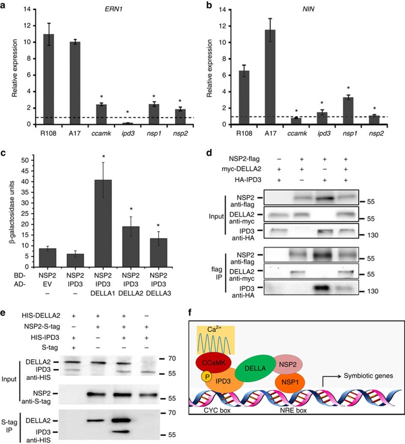
CCaMK–IPD3 and NSP2–NSP1 can both independently regulate the expression of Nod-factor-induced genes in Nicotiana benthamiana, suggesting that the two protein complexes may function independently27,34,55. However, the fact that we found that expression of both NIN and ERN1 was lower in ccamk, ipd3, nsp1 and nsp2 mutants (Fig. 9a,b) suggests that the CCaMK–IPD3 and NSP2–NSP1 complexes may both regulate the expression of common Nod-factor-induced genes. We assessed whether MtDELLAs may be able to link IPD3 and NSP2 using Y3H in combination with the fusion proteins NSP2-GAL4-DNA-binding domain (GAL4-DBD) and/or IPD3-GAL4 activation domain (GAL4-AD). Activation of the reporter was observed only when MtDELLA protein was present, suggesting that MtDELLA bridges an interaction between IPD3 and NSP2 (Fig. 9c). A protein complex containing MtDELLA, IPD3 and NSP2 was also suggested by co-immunoprecipitation in N. benthamiana leaves (Fig. 9d). Interestingly, we found that IPD3 can co-immunoprecipitate with NSP2 in the absence of MtDELLA in N. benthamiana, possibly because the endogenous NbDELLA or another protein may also be able to form a protein complex with NSP2 and IPD3. To further test whether MtDELLA is able to bridge a protein complex containing IPD3 and NSP2, we co-expressed NSP2-S-tag and HIS-IPD3 in E. coli and found that NSP2 could not co-immunoprecipitate with IPD3, however NSP2 can co-immunoprecipitate with IPD3 in the presence of MtDELLA (Fig. 9e). Taken together, our results suggest that MtDELLA may be able to bridge a protein complex containing IPD3 and NSP2 (Fig. 9f).
Figure 9. MtDELLAs can link IPD3 and NSP2.
(a,b) The dmi3-1, ipd3-2, nsp1-2 and nsp2-1 mutants show defect in NIN and ERN1 expression. The RNA was extracted from 6 individual plants of wild type or mutants. Error bars represent standard error (n=3, where n denotes the number of technical repetition). The asterisk indicates a significant decrease relative to the control with Student's t-test (* P<0.05) (c) Yeast three-hybrid assay assessing combinations of BD-NSP2, AD-IPD3 and MtDELLAs, indicating MtDELLA proteins bridge a protein complex containing IPD3 and NSP2. Data are presented as β-galactosidase activity. The asterisk indicates a significant increase relative to the control with Student's t-test (*P<0.05) (d) NSP2 co-immunoprecipitates with IPD3 and DELLA2 in N. benthamiana. NSP2, IPD3 and MtDELLA2 are co-expressed in N. benthamiana. (e) NSP2 co-immunoprecipitates with IPD3 only in the presence of MtDELLA. NSP2, IPD3 and MtDELLA are co-expressed in E. coli. (f) Proposed model showing MtDELLAs bridge a bigger protein complex containing CCaMK–IPD3 and NSP2–NSP1.
Discussion
Previous studies have shown that GA biosynthetic genes were upregulated during arbuscule mycorrhizal symbiosis and both positive and negative roles of GAs in mycorrhizal symbiosis have been suggested56,57,58,59,60. The role of GAs in legume root nodule symbiosis has been assayed for over 60 years46,47. The upregulation of GA biosynthetic genes at different stages of nodule development suggests an important role for GA in early infection and the mature stages of nodulation, such as nodule primordium development and the correct establishment of nodulation zones49,50,51. The Pisum sativum GA biosynthetic mutant, na-1, develops significantly fewer and underdeveloped nodules than its wild-type parent and GA application and grafting suggested that this nodulation phenotype in the na-1 mutant is due to reduced GA level in the roots51, suggesting that GAs play a positive role in cortical cell divisions or nodule maturation. Here we found that DELLA is a positive regulator at the infection and the root nodule development stages. Taken together, these results suggest that strict regulation of DELLA proteins levels is required for normal development of mycorrhizal and root nodule symbioses, but the underlying mechanism needs to be further understood.
Legume plants share a common symbiotic signalling pathway to perceive LCO signalling molecules from rhizobial bacteria and mycorrhizal fungi, and activate the symbiotic signalling pathway4,61. It has been shown previously that DELLAs are essential for mycorrhizal symbiosis in rice and M. truncatula44,45. Here we show that MtDELLAs are also required for root nodule symbiosis and therefore represent a new common component of the signalling pathways required for root nodule and mycorrhizal symbioses. There are five DELLA proteins in Arabidopsis and only one DELLA protein in rice, suggesting that the DELLA family of proteins may have undergone several duplications during the evolution of Arabidopsis. Based on the available genome information for M. truncatula, we found three proteins containing DELLA domains and two GRAS proteins which fall into the cluster with DELLA proteins. We found that GA3 treatment severely inhibited rhizobial infection and root nodule development programmes. Consistent with this, we found that the nodule number was further reduced in della triple mutants. A total of 14% nodules still formed in triple mutants compared with wild type possibly because unknown MtDELLAs or other highly homologous GRAS proteins may also function during root nodule development and compensate for the mutation of the MtDELLAs we describe here.
Here we showed that MtDELLAs can increase the interaction between CCaMK and IPD3/CYCLOPS and lead to a higher level phosphorylation of IPD3 by CCaMK, indicating that MtDELLAs may be involved in assembly of a functional CCaMK-IPD3-DELLA protein complex. It has been shown that phosphorylated versions of CYCLOPS/IPD3 can bind the promoter of NIN and activate NIN expression27. Interestingly we found that a phospoablative mutant version of IPD3 (S50A-S155A) abolished the interaction with MtDELLAs and was unable to rescue an ipd3 mutant. A phosphomimetic version (S50D-S155D) increased the interaction with DELLAs. These results are consistent with a model where the interaction of MtDELLAs and IPD3 is relevant for symbiosis. Furthermore we found CCaMK could further increase the interaction of MtDELLA and IPD3, suggesting that assembly of the CCaMK–IPD3–DELLA complex could be activated by CCaMK during root nodule symbiosis.
In vertebrates, CaMKIV contributes to phosphorylation of CREB on S133. Once phosphorylated on S133, CREB recruits its co-activator CBP, then CaMKIV phosphorylates CBP and thereby stimulates CREB-dependent transcription62. Our data suggests that the CCaMK/IPD3/DELLA complex could act via a similar mechanism in root nodule symbiosis as CaMKIV/CREB/CBP. Our data suggests that CCaMK can phosphorylate IPD3/CYCLOPS and that the phosphorylated version of IPD3 can interact with MtDELLAs. We found that DELLA can also increase the interaction between CCaMK and IPD3/CYCLOPS and stimulate higher levels of IPD3/CYCLOPS phosphorylation. However, we did not observe CCaMK phosphorylation of MtDELLAs in vitro, which is different from the calcium decoding system in vertebrates.
DELLAs are key negative regulators of gibberellin signalling40. It has been shown that DELLA interacts with other transcription factors, such as PIF3 and PIF4 to block their DNA-binding abilities of targeting gene promoters and inhibit their transcriptional activities41,42. Here we show that MtDELLAs are required for the expression of Nod-factor-induced genes, indicating MtDELLAs function as positive regulators in this context. Consistent with their role in root nodule symbiosis, we found that MtDELLAs can interact with another GRAS transcription factor, NSP2. Furthermore, MtDELLA, NSP2 and NSP1 were able to form a protein complex (Fig. 9f). Transient assays in Arabidopsis protoplasts showed that a luciferase reporter controlled by the ERN1 promoter was additively transactivated by MtDELLAs, suggesting that MtDELLAs may function as transcriptional activators during root nodule symbiosis.
The binding sites for the CYCLOPS/IPD3 proteins (CYC-box) and NSP1 (NRE-box) in the NIN promoter are separated by gaps of 8 and 54 bps (Supplementary Note 1). This close proximity in DNA-binding sites suggest an interaction between the NSP2–NSP1 and CCaMK–IPD3 protein complexes may be possible. Consistent with this idea, we found that MtDELLAs are able to bridge a protein complex containing IPD3 and NSP2. The NRE-box and CYC-box may not always be associated in promoters of Nod-factor-induced genes, suggesting that NSP2–NSP1 and CCaMK–IPD3 many also function independently. We propose that NSP2–NSP1, CCaMK–IPD3 and other transcription factors may act in combination to regulate the expression of early nodulin genes with appropriate spatial and temporal patterns, and based on phenotype, that the MtDELLAs act during the early stages of signal transduction.
The legume–rhizobia symbiosis is the most important symbiotic association in terms of biological nitrogen fixation63,64, however nitrogen fixation is energetically expensive to the plant, so a number of additional external and internal factors negatively regulate nodulation in legumes64. Here, we show that the transcriptional repressor MtDELLA proteins, which are central regulators of GA signalling promote nodulation in M. truncatula and may act as crosstalk nodes to integrate hormone signals with rhizobial infection and nodule development.
Methods
Rhizobia inoculation and GA3 treatment
M. truncatula seeds were treated with 98% sulfuric acid and plated onto 1% agar medium at 4 °C. After about 3 days, they were moved to 22 °C overnight for germination. Then the seedlings were moved to a mixed soil containing 1:1 ratio of sand and perlite. After a minimum of 3 days, plants were inoculated with S. meliloti. Seeds of R108 or A17 were used as the wild type. Plants were grown in a greenhouse at 22 °C with 16/8 h of light/dark cycle at 22 °C. S. meliloti strain Sm1021 was incubated in liquid Luria broth overnight with 400 μg ml−1 streptomycin selection. Bacteria were pelleted at 2,000g for 15 min and resuspended in H2O to OD600=0.03.
A concentration of 10 mM GA3 stock solution (GA3; G-7645-1G; Sigma) was dissolved using ethanol and diluted with ultra-pure H2O to a final working solution of 10−4 or 10−5 M GA3. Ethanol (0.1%) was used as mock solution. Three days after germination, 1 ml GA3 working solution or mock solution was applied daily to each plant of WT or mutants.
MtDELLAs RNAi M. truncatula plants were produced by Agrobacterium rhizogenes (Arqual) mediated hairy root transformation65. The candidate sequences that can be targeted by RNAi were selected according to clontech RNAi Target Sequence Selector (http://bioinfo.clontech.com/rnaidesigner/sirnaSequenceDesign.do;jsessionid=c0a8fe1e501a50bb6ab39948a9982afb46cf4fb4dd) and were amplified by PCR (primers seen in Supplementary Table 1) from wild-type gDNA. Fragments were cloned to the vector pK7GWIWGII-R, which carry a dsRed fluorescent tag for selection of transgenic hairy roots. The plants were transferred to sand with perlite about 1 month after transformation, and nodules were scored 3 weeks post inoculation with Sm1021.
Genotyping of della mutants
We obtained M. truncatula R108 Tnt1 transposon insertion lines of Mtdella1-1 (NF4215), Mtdella1-2(NF5155), Mtdella2 (NF14614), Mtdella3-1 (NF11992) and Mtdella3-2 (NF15025) from the Noble Foundation Tnt1 database (http://medicago-mutant.noble.org/mutant/database.php). The location of Tnt1 insertions in MtDELLAs were shown in Supplementary Fig. 4. These mutants were grown and genotyped by PCR using Tnt1-R1 and DELLAs gene-specific primers (Supplementary Table 1). NF14614 was crossed with NF11992 or NF15025 to generate double mutants. Then della2/della3 double mutants were crossed with NF5155 or NF4215 and della1/della2/della3 triple mutants were segregated from population of della triple heterozygous mutants.
Vector information
For yeast two-hybrid constructs, the appropriate genes were cloned into the Gateway donor vector pEntry-topo-SD (appropriate primers see Supplementary Table 1), sequenced and then recombined into the pGBKT7GW and pGADT7GW vectors by LR reactions (Invitrogen). For yeast three-hybrid constructs, pBridge was employed and all the genes were introduced into MCSI and MCSII with ligation high (TOYOBO) except DELLA3, which was recombined to the MCSII by ClonExpressTM II One Step Cloning Kit (Vazyme). For the BiFC vectors, the genes of interest were cloned into the pSAT4-nYFP-N1 and pSAT4-cYFP-N1 with appropriate restriction enzymes cleavage sites (Supplementary Table 1). For protein expression in E. coli, pET28a, pET32a, and pMal-C2X were used (for primers see Supplementary Table 1). For transient reporter assays, the 2.4 kb ERN1 promoter region was amplified by PCR from M. truncatula R108 genomic DNA and cloned by replacing the 2 × 35S promoter in the pSAT1-cCFP-C (pE3242), a protoplast expression vector. The firefly luciferase gene was amplified by PCR from pH35S-LUC-GW plasmid (kindly provide by Hongtao Liu) and fused with MtERN1 promoter in pSAT1-cCFP-C (pE3242). NSP1 and NSP2 were cloned in to pSAT1-cCFP-C (pE3242) and pSAT1-nVenus-C (pE3228). MtDELLAs fused with an asRed (a fluorescent protein from Anemonia sulcata) tag were cloned by replacing the EYFP in the pSAT4-cEYFP-C1-B vector. NSP2 cDNA was amplified and cloned into pUB-GFP-C-3 × FLAG at the XbaI/KpnI sites. 3 × HA-IPD3 was amplified and inserted into the Gateway donor vector pEntry-topo-SD and then recombined into the pK7GW2-R vectors by LR reactions. Myc-tagged DELLA2 was amplified and inserted into pUB-GFP-c'-3 × FLAG vector, in which 3 × FLAG tag was not expressed, since DELLA2-myc-R primer contains a stop code. For GUS staining assay, promoters were cloned to the vector pEntry-topo-SD. Then they were recombined into pBGWFS7. For complementary experiment, DELLA1/2/3 fused with EGFP (enhanced GFP) at N-terminal was amplified from DELLA1/2/3-pK7WGF2.0, and cloned to the vector pEntry-topo-SD and then were recombined into pK7WG2-R by LR reactions. All primers, restriction enzyme cleavage sites, and short descriptions are listed in Supplementary Table 1 and all the plasmids were sequenced.
Nod factor treatments
Seven-day-old della2/della3 double mutants and R108 were incubated in liquid BNM or BNM containing 10−5M GA3. After 6 h, 10−9M Nod factor or H2O were added into the medium. After a further 6 h the roots of each group (each group contains 6–10 plants) were harvested and frozen in liquid nitrogen for RNA extraction. AVG was not used in all the media.
RNA extraction and real-time PCR
Total RNA was isolated with TransZol UP reagent (TransGen) and treated with Ambion DNA Removal Kit. First strand cDNA was synthesized with oligo (dT)18 from 1 μg RNA with M-MLV reverse transcriptase (Takara). The qRT-PCR experiments were performed on a BioRad real-time PCR detection system (iQ5) using SYBR Green reagent (Toyobo). The PCR cycle number was determined for each primer pair to the point at which amplification was in the linear phase.
Yeast two hybrid and yeast three hybrid
The yeast strain AH109 was transformed with the destination vector pGBKT7GW and pGADT7GW containing IPD3, NSP2, MtDELLA1, MtDELLA2, MtDELLA3, NSP1, CCaMK or their mutants according to the LiAc transformation method66. The expressed proteins were then examined for interaction by dropping 7 μl of yeast suspension on minimal synthetic dropout (SD) agar medium containing the dropout supplement (DO) His-, Leu-, Trp-, Ade- and different concentrations (as stated) of 3-amino-1,2,4-triazole (Clontech). Yeast growth was monitored for up to 7 d. The liquid β-galactosidase assay was performed according to the yeast handbook PT3024-1 (Clontech). The transformation method of Y3H was the same as Y2H, 0.005 mM Met was added into the selection medium.
BiFC assays
Plasmids were extracted according to the manual of NucleoBond Xtra Midi Plus (Macherey-Nagel). Isolating Arabidopsis mesophyll protoplasts and transient gene expression were performed as previously described67. YFP fluorescence was recorded by a confocal laser scanning microscope (FluoView FV1000; Olympus).
Pull-down assays
The coding sequences of MtDELLA1, MtDELLA2 or MtDELLA3 were cloned into pCold-TF (Takara) for production of HIS-tagged fusion proteins, HIS-MtDELLA1, HIS-MtDELLA2 or HIS-MtDELLA3 in E. coli, respectively. The coding sequences of NSP2 or IPD3 were cloned into pMAL-C2X (NEB) for production of MBP-tagged fusion proteins, MBP-NSP2 or MBP-IPD3 in E. coli, respectively. The HIS-tagged fusion proteins were purified using Ni-NTA Agarose (QIAGEN). The MBP-tagged fusion proteins were purified uing amylose resin (NEB). To test the interaction between NSP2 and MtDELLA1, MtDELLA2 or MtDELLA3, the MBP-NSP2 resin was used to capture the purified HIS-MtDELLA1, His-MtDELLA2 or His-MtDELLA3 protein. To test the interaction between IPD3 and MtDELLA1, MtDELLA2 or MtDELLA3, the MBP-IPD3 resin was used to capture the purified HIS-MtDELLA1, HIS-MtDELLA2 or HIS-MtDELLA3 protein. The monoclonal anti-MBP (Ambmart) or anti-HIS (CW Biotech) antibody was used for western blotting analysis (The information of antibodies in Supplementary Table 2 and Uncropped versions of blots in Supplementary Fig. 8).
DELLA2 and DELLA3 localization and promoter::GUS analysis
Genomic MtDELLA2, MtDELLA3 were transferred from the pENTRY vector, described above, into the pK7WGF2 vector by LR reaction. The resulting clone was transformed into A. rhizogenes (Arqua1) and for hairy root transformation in M. truncatula. Three weeks after hairy root transformation sub-cellular localization was monitored using a confocal Zeiss Axio Imager M1 upright microscope.
Roots of promoter-GUS transgenic plants were used for GUS staining assay. The roots were put into the staining buffer and vacuumed for 10 min then incubated for 0.5 to 3 h at 37 °C. After staining, roots were included in 3% low gelling agarose and sliced into 70 μm sections using a VT 1000S vibratome (Leica). Slices were observed and taken pictures in bright field light microscopy (Nikon SMZ1500).
In vitro phosphorylation
IPD3 was expressed from pET28a in E. coli strain Rosetta (TransGen Biotech). Expression products were affinity purified via nickel-agarose (Qiagen) under the denaturing conditions by using 8 M urea. Denatured proteins were refolded by stepwise dialysis26. Purification of MBP-CCaMK, HIS-IPD3, HIS-MtDELLA1, HIS-MtDELLA2 and HIS-MtDELLA3 was performed according to the protocol of E8200 (New England Biolabs) and of BioSprint96 (Qiagen), respectively. IPD3 was phosphorylated in vitro by CCaMK as described26. Each reaction was carried out by using 1 μg MBP-CCaMK protein, 0.2 mM CaCl2, 0.5 μM Bovine CaM, 2 μg full length and 6 × HIS-IPD3 as substrate. Phosphorylation reactions were performed at 30 °C for 30 min in the presence of DELLA1, DELLA2 and DELLA3. Relative intensities were then calculated for each band by ImageJ.
Co-immunoprecipitation assay
Co-immunoprecipitation studies of CCaMK/IPD3/DELLA2, IPD3/DELLA2/NSP2 and DELLA2/NSP2/NSP1 were performed. Proteins were expressed in E. coli strain Rosetta (TransGen Biotech) using pETDuet-1 and RSFDuet-1 vectors. Immunoprecipitation of proteins used anti-S-tag antibody (MBL). Protein A agarose (Merck-Millipore) was used to precipitate the immunoprotein complexes with 50 ml RIPA buffer (150 mM NaCl, 50 mM Tris-HCl, pH 7.5, 1 mM DTT, 1 mM PMSF, 0.1%Triton X-100, 0.1% pellet of cocktail (Roche)) in 50 ml IP buffer. After immunoprecipitation, beads were washed five times with IP buffer. Proteins were then released and collected by boiling in 1 × SDS loading buffer for 5 min. Immunoprecipitation products were detected by immunoblotting with anti-HIS antibody.
For co-immunoprecipitaion in N. benthamiana, Flag-tagged NSP2, Myc-tagged DELLA and HA-tagged IPD3 were expressed from N. Benthamiana leaves through coinfiltration of A. tumefaciens strains GV3101 harbouring proper constructs. Three days after infiltration, N. benthamiana leaves were powdered in liquid nitrogen. For each sample, 0.3 g powder was lysed in 600 μl immunoprecipitaion buffer containing 50 mM Tris-HCl (pH 7.5), 150 mM NaCl, 1 mM EDTA, 0.2% Triton X-100, and 1 × EDTA-free protease inhibitor cocktail (Roche). Samples were incubated on ice for 30 min, followed by centrifuge twice at 15,000g for 20 min at 4 °C. Partial supernatant (50 μl) was retained for input samples to ensure proteins were normally expressed in all samples. The rest of the supernatant was incubated with a 20 μl slurry of anti-flag M2 agarose beads (sigma) for 2.5 h at 4 °C with a roller shaker. The beads were collected by centrifuge gently and washed once with the immunoprecipitaion buffer. Proteins were eluted by boiling the beads in 60 μl 1 × SDS loading buffer containing 5% β-mercaptoethanol and separated on SDS–PAGE gels. Co-immunoprecipitated proteins were detected by western blot analysis using anti-Flag (sigma), anti-Myc (millipore) or anti-HA (sigma) antibody (the information of antibodies in Supplementary Table 2 and uncropped versions of blots in Supplementary Fig. 8).
Protoplast transient assay
The protoplast transient assay was performed as previously described67. The 2,410 bp ERN1 promoter was amplified by PCR from A17 genomic DNA and fused with the firefly luciferase gene was used as a transcriptional activity reporter (15 μg of plasmid per transfection). pRNL (a Renilla luciferase reporter plasmid) was used as an internal control to normalize transfection efficiency in protoplast assays (2 μg per transfection). Other effectors of transcription factor constructs were used at 12 μg per transfection. Protoplasts were incubated in WI solution for about 13 h. Protoplasts were harvested by centrifugation at 50g for 12 min, resuspended with 200–700 μl reporter lysis buffer (RLB), and vortexed for several seconds. Then they were frozen in liquid nitrogen, thawed at 37°C, centrifuged at 13,500g for 1 min and the supernatant (lysis solution) was used for further analysis. Luciferase Assay Reagent (100 μl; Promega) was added to 20 μl of the lysis solution, and luciferase activity was measured with GloMax 20/20 Luminometera.
Data availability
The authors declare that the data supporting the findings of this study are available within the article and its Supplementary Information files or are available from the corresponding author upon request.
Additional information
How to cite this article: Jin, Y. et al. DELLA proteins are common components of symbiotic rhizobial and mycorrhizal signalling pathways. Nat. Commun. 7:12433 doi: 10.1038/ncomms12433 (2016).
Supplementary Material
Supplementary Figures 1 - 8, Supplementary Tables 1 and 2 and Supplementary Note 1
Acknowledgments
We thank Dr. Ben Miller at University of East Anglia for critical reading of the manuscript and helpful suggestions. We thank Dr. Fernanda de Carvalho-Niebel for providing the ERN1 promoter and Dr. Xiaodong Xu for providing the Renilla luciferase reporter plasmid. This work was supported by 973 National Key Basic Research Programme in China (No. 2015CB158300), the Strategic Priority Research Program of the Chinese Academy of Sciences (XDA08010401), National Science Foundation (31522007), the ‘1000-talents Plan' for young researchers from China, NKLPMG to EW, Ministry of Agriculture of China for Transgenic Research (2014ZX08009-003-003) to J.Y. and Project funded by China Postdoctoral Science Foundation (2014M551464) to Y.J. The M. truncatula Tnt1 insertion mutant lines were obtained from The Samuel Roberts Noble Foundation, Inc. and were created through research funded, in part, by a grant from the National Science Foundation, NSF-0703285.
Footnotes
Author contributions Conceived and designed the experiments: E.W., N.Y., Y.J. and H.L. Performed the experiments: Y.J., H.L., D.L., N.Y. and C.W. Contributed new analytic/computational tools: X.W., J.Y. and H.D. Analysed data and wrote the manuscript: E.W., Y.J. and H.L.
References
- Oldroyd G. E., Murray J. D., Poole P. S. & Downie J. A. The rules of engagement in the legume-rhizobial symbiosis. Annu. Rev. Genet. 45, 119–144 (2011). [DOI] [PubMed] [Google Scholar]
- Oldroyd G. E. & Downie J. A. Coordinating nodule morphogenesis with rhizobial infection in legumes. Annu. Rev. Plant Biol. 59, 519–546 (2008). [DOI] [PubMed] [Google Scholar]
- Popp C. & Ott T. Regulation of signal transduction and bacterial infection during root nodule symbiosis. Curr. Opin. Plant Biol. 14, 458–467 (2011). [DOI] [PubMed] [Google Scholar]
- Oldroyd G. E. D. Speak, friend, and enter: signalling systems that promote beneficial symbiotic associations in plants. Nat. Rev. Microbiol. 11, 252–263 (2013). [DOI] [PubMed] [Google Scholar]
- Geurts R., Fedorova E. & Bisseling T. Nod factor signaling genes and their function in the early stages of Rhizobium infection. Curr. Opin. Plant Biol. 8, 346–352 (2005). [DOI] [PubMed] [Google Scholar]
- Endre G. et al. A receptor kinase gene regulating symbiotic nodule development. Nature 417, 962–966 (2002). [DOI] [PubMed] [Google Scholar]
- Stracke S. et al. A plant receptor-like kinase required for both bacterial and fungal symbiosis. Nature 417, 959–962 (2002). [DOI] [PubMed] [Google Scholar]
- Amor B. B. et al. The NFP locus of Medicago truncatula controls an early step of Nod factor signal transduction upstream of a rapid calcium flux and root hair deformation. Plant J. 34, 495–506 (2003). [DOI] [PubMed] [Google Scholar]
- Limpens E. et al. LysM domain receptor kinases regulating rhizobial Nod factor-induced infection. Science 302, 630–633 (2003). [DOI] [PubMed] [Google Scholar]
- Madsen E. B. et al. A receptor kinase gene of the LysM type is involved in legume perception of rhizobial signals. Nature 425, 637–640 (2003). [DOI] [PubMed] [Google Scholar]
- Radutoiu S. et al. Plant recognition of symbiotic bacteria requires two LysM receptor-like kinases. Nature 425, 585–592 (2003). [DOI] [PubMed] [Google Scholar]
- Antolin-Llovera M., Ried M. K., Binder A. & Parniske M. Receptor kinase signaling pathways in plant-microbe interactions. Annu. Rev. Phytopathol. 50, 451–473 (2012). [DOI] [PubMed] [Google Scholar]
- Zhang X. et al. The receptor kinase CERK1 has dual functions in symbiosis and immunity signalling. Plant J. 81, 258–267 (2015). [DOI] [PubMed] [Google Scholar]
- Ane J. M. et al. Medicago truncatula DMI1 required for bacterial and fungal symbioses in legumes. Science 303, 1364–1367 (2004). [DOI] [PubMed] [Google Scholar]
- Imaizumi-Anraku H. et al. Plastid proteins crucial for symbiotic fungal and bacterial entry into plant roots. Nature 433, 527–531 (2005). [DOI] [PubMed] [Google Scholar]
- Saito K. et al. NUCLEOPORIN85 is required for calcium spiking, fungal and bacterial symbioses, and seed production in Lotus japonicus. Plant Cell 19, 610–624 (2007). [DOI] [PMC free article] [PubMed] [Google Scholar]
- Charpentier M. et al. Lotus japonicus CASTOR and POLLUX are ion channels essential for perinuclear calcium spiking in legume root endosymbiosis. Plant Cell 20, 3467–3479 (2008). [DOI] [PMC free article] [PubMed] [Google Scholar]
- Groth M. et al. NENA, a Lotus japonicus homolog of Sec13, is required for rhizodermal infection by arbuscular mycorrhiza fungi and rhizobia but dispensable for cortical endosymbiotic development. Plant Cell 22, 2509–2526 (2010). [DOI] [PMC free article] [PubMed] [Google Scholar]
- Capoen W. et al. Nuclear membranes control symbiotic calcium signaling of legumes. Proc. Natl Acad. Sci. USA 108, 14348–14353 (2011). [DOI] [PMC free article] [PubMed] [Google Scholar]
- Levy J. et al. A putative Ca2+ and calmodulin-dependent protein kinase required for bacterial and fungal symbioses. Science 303, 1361–1364 (2004). [DOI] [PubMed] [Google Scholar]
- Mitra R. M. et al. A Ca2+/calmodulin-dependent protein kinase required for symbiotic nodule development: Gene identification by transcript-based cloning. Proc. Natl Acad. Sci. USA 101, 4701–4705 (2004). [DOI] [PMC free article] [PubMed] [Google Scholar]
- Gleason C. et al. Nodulation independent of rhizobia induced by a calcium-activated kinase lacking autoinhibition. Nature 441, 1149–1152 (2006). [DOI] [PubMed] [Google Scholar]
- Tirichine L. et al. Deregulation of a Ca2+/calmodulin-dependent kinase leads to spontaneous nodule development. Nature 441, 1153–1156 (2006). [DOI] [PubMed] [Google Scholar]
- Takeda N., Maekawa T. & Hayashi M. Nuclear-localized and deregulated calcium- and calmodulin-dependent protein kinase activates rhizobial and mycorrhizal responses in Lotus japonicus. Plant Cell 24, 810–822 (2012). [DOI] [PMC free article] [PubMed] [Google Scholar]
- Messinese E. et al. A novel nuclear protein interacts with the symbiotic DMI3 calcium- and calmodulin-dependent protein kinase of Medicago truncatula. Mol. Plant Microbe Interact. 20, 912–921 (2007). [DOI] [PubMed] [Google Scholar]
- Yano K. et al. CYCLOPS, a mediator of symbiotic intracellular accommodation. Proc. Natl Acad. Sci. USA 105, 20540–20545 (2008). [DOI] [PMC free article] [PubMed] [Google Scholar]
- Singh S., Katzer K., Lambert J., Cerri M. & Parniske M. CYCLOPS, a DNA-binding transcriptional activator, orchestrates symbiotic root nodule development. Cell Host Microbe 15, 139–152 (2014). [DOI] [PubMed] [Google Scholar]
- Schauser L., Roussis A., Stiller J. & Stougaard J. A plant regulator controlling development of symbiotic root nodules. Nature 402, 191–195 (1999). [DOI] [PubMed] [Google Scholar]
- Marsh J. F. et al. Medicago truncatula NIN is essential for rhizobial-independent nodule organogenesis induced by autoactive calcium/calmodulin-dependent protein kinase. Plant Physiol. 144, 324–335 (2007). [DOI] [PMC free article] [PubMed] [Google Scholar]
- Andriankaja A. et al. AP2-ERF transcription factors mediate Nod factor dependent Mt ENOD11 activation in root hairs via a novel cis-regulatory motif. Plant Cell 19, 2866–2885 (2007). [DOI] [PMC free article] [PubMed] [Google Scholar]
- Middleton P. H. et al. An ERF transcription factor in Medicago truncatula that is essential for Nod factor signal transduction. Plant Cell 19, 1221–1234 (2007). [DOI] [PMC free article] [PubMed] [Google Scholar]
- Kalo P. et al. Nodulation signaling in legumes requires NSP2, a member of the GRAS family of transcriptional regulators. Science 308, 1786–1789 (2005). [DOI] [PubMed] [Google Scholar]
- Smit P. et al. NSP1 of the GRAS protein family is essential for rhizobial Nod factor-induced transcription. Science 308, 1789–1791 (2005). [DOI] [PubMed] [Google Scholar]
- Hirsch S. et al. GRAS proteins form a DNA binding complex to induce gene expression during nodulation signaling in Medicago truncatula. Plant Cell 21, 545–557 (2009). [DOI] [PMC free article] [PubMed] [Google Scholar]
- Liu W. et al. Strigolactone biosynthesis in Medicago truncatula and rice requires the symbiotic GRAS-type transcription factors NSP1 and NSP2. Plant Cell 23, 3853–3865 (2011). [DOI] [PMC free article] [PubMed] [Google Scholar]
- Maillet F. et al. Fungal lipochitooligosaccharide symbiotic signals in arbuscular mycorrhiza. Nature 469, 58–63 (2011). [DOI] [PubMed] [Google Scholar]
- Delaux P. M., Becard G. & Combier J. P. NSP1 is a component of the Myc signaling pathway. New Phytol. 199, 59–65 (2013). [DOI] [PubMed] [Google Scholar]
- Nagae M., Takeda N. & Kawaguchi M. Common symbiosis genes CERBERUS and NSP1 provide additional insight into the establishment of arbuscular mycorrhizal and root nodule symbioses in Lotus japonicus. Plant Signal Behav. 9, e28544 (2014). [DOI] [PMC free article] [PubMed] [Google Scholar]
- Ueguchi-Tanaka M., Nakajima M., Motoyuki A. & Matsuoka M. Gibberellin receptor and its role in gibberellin signaling in plants. Annu. Rev. Plant Biol. 58, 183–198 (2007). [DOI] [PubMed] [Google Scholar]
- Sun T. P. The molecular mechanism and evolution of the GA-GID1-DELLA signaling module in plants. Curr. Biol. 21, R338–R345 (2011). [DOI] [PubMed] [Google Scholar]
- de Lucas M. et al. A molecular framework for light and gibberellin control of cell elongation. Nature 451, 480–484 (2008). [DOI] [PubMed] [Google Scholar]
- Feng S. et al. Coordinated regulation of Arabidopsis thaliana development by light and gibberellins. Nature 451, 475–479 (2008). [DOI] [PMC free article] [PubMed] [Google Scholar]
- Achard P. et al. Integration of plant responses to environmentally activated phytohormonal signals. Science 311, 91–94 (2006). [DOI] [PubMed] [Google Scholar]
- Floss D. S., Levy J. G., Levesque-Tremblay V., Pumplin N. & Harrison M. J. DELLA proteins regulate arbuscule formation in arbuscular mycorrhizal symbiosis. Proc. Natl Acad. Sci. USA 110, E5025–E5034 (2013). [DOI] [PMC free article] [PubMed] [Google Scholar]
- Yu N. et al. A DELLA protein complex controls the arbuscular mycorrhizal symbiosis in plants. Cell Res. 24, 130–133 (2014). [DOI] [PMC free article] [PubMed] [Google Scholar]
- Dullaart J. & Duba L. I. Presence of gibberellin-like substances and their possible role in auxin bioproduction in root nodules and roots of Lupinus-Luteus L. Acta Bot. Neerl. 19, 877–883 (1970). [Google Scholar]
- Hayashi S., Gresshoff P. M. & Ferguson B. J. Mechanistic action of gibberellins in legume nodulation. J. Integr. Plant Biol. 56, 971–978 (2014). [DOI] [PubMed] [Google Scholar]
- Ferguson B. J., Ross J. J. & Reid J. B. Nodulation phenotypes of gibberellin and brassinosteroid mutants of pea. Plant Physiol. 138, 2396–2405 (2005). [DOI] [PMC free article] [PubMed] [Google Scholar]
- Lievens S. et al. Gibberellins are involved in nodulation of Sesbania rostrata. Plant Physiol. 139, 1366–1379 (2005). [DOI] [PMC free article] [PubMed] [Google Scholar]
- Maekawa T. et al. Gibberellin controls the nodulation signaling pathway in Lotus japonicus. Plant J. 58, 183–194 (2009). [DOI] [PubMed] [Google Scholar]
- Ferguson B. J., Foo E., Ross J. J. & Reid J. B. Relationship between gibberellin, ethylene and nodulation in Pisum sativum. New Phytol. 189, 829–842 (2011). [DOI] [PubMed] [Google Scholar]
- Journet E. P. et al. Medicago truncatula ENOD11: a novel RPRP-encoding early nodulin gene expressed during mycorrhization in arbuscule-containing cells. Mol. Plant Microbe Interact. 14, 737–748 (2001). [DOI] [PubMed] [Google Scholar]
- Gobbato E. et al. A GRAS-type transcription factor with a specificfunction in mycorrhizal signaling. Curr. Biol. 22, 2236–2241 (2012). [DOI] [PubMed] [Google Scholar]
- Cerri M. R. et al. Medicago truncatula ERN transcription factors: regulatory interplay with NSP1/NSP2 GRAS factors and expression dynamics throughout rhizobial infection. Plant Physiol. 160, 2155–2172 (2012). [DOI] [PMC free article] [PubMed] [Google Scholar]
- Limpens E. & Bisseling T. CYCLOPS: a new vision on rhizobium-induced nodule organogenesis. Cell Host Microbe 15, 127–129 (2014). [DOI] [PubMed] [Google Scholar]
- Shaul-Keinan O. et al. Hormone concentrations in tobacco roots change during arbuscular mycorrhizal colonization with Glomus intraradices. New Phytol. 154, 501–507 (2002). [DOI] [PubMed] [Google Scholar]
- Garrido J. M. G., Morcillo R. J. L., Rodriguez J. A. M. & Bole J. A. O. Variations in the mycorrhization characteristics in roots of wild-type and ABA-deficient tomato are accompanied by specific transcriptomic alterations. Mol. Plant Microbe Interact. 23, 651–664 (2010). [DOI] [PubMed] [Google Scholar]
- Ortu G. et al. Plant genes related to gibberellin biosynthesis and signaling are differentially regulated during the early stages of AM fungal interactions. Mol. Plant 5, 951–954 (2012). [DOI] [PubMed] [Google Scholar]
- Takeda N. et al. Gibberellins interfere with symbiosis signaling and gene expression and alter colonization by arbuscular mycorrhizal fungi in Lotus japonicus. Plant Physiol. 167, 545–U442 (2015). [DOI] [PMC free article] [PubMed] [Google Scholar]
- Martin-Rodriguez J. A. et al. Role of gibberellins during arbuscular mycorrhizal formation in tomato: new insights revealed by endogenous quantification and genetic analysis of their metabolism in mycorrhizal roots. Physiol. Plant. 154, 66–81 (2015). [DOI] [PubMed] [Google Scholar]
- Parniske M. Arbuscular mycorrhiza: the mother of plant root endosymbioses. Nat. Rev. Microbiol. 6, 763–775 (2008). [DOI] [PubMed] [Google Scholar]
- Wayman G. A., Lee Y. S., Tokumitsu H., Silva A. J. & Soderling T. R. Calmodulin-kinases: modulators of neuronal development and plasticity. Neuron 59, 914–931 (2008). [DOI] [PMC free article] [PubMed] [Google Scholar]
- Peoples M. B. et al. The contributions of nitrogen-fixing crop legumes to the productivity of agricultural systems. Symbiosis 48, 1–17 (2009). [Google Scholar]
- Ferguson B. J. et al. Molecular analysis of legume nodule development and autoregulation. J. Integr. Plant Biol. 52, 61–76 (2010). [DOI] [PubMed] [Google Scholar]
- Boisson-Dernier A. et al. Agrobacterium rhizogenes-transformed roots of Medicago truncatula for the study of nitrogen-fixing and endomycorrhizal symbiotic associations. Mol. Plant Microbe Interact. 14, 695–700 (2001). [DOI] [PubMed] [Google Scholar]
- Gietz R. D. & Woods R. A. Transformation of yeast by lithium acetate/single-stranded carrier DNA/polyethylene glycol method. Methods Enzymol. 350, 87–96 (2002). [DOI] [PubMed] [Google Scholar]
- Yoo S. D., Cho Y. H. & Sheen J. Arabidopsis mesophyll protoplasts: a versatile cell system for transient gene expression analysis. Nat. Protoc. 2, 1565–1572 (2007). [DOI] [PubMed] [Google Scholar]
Associated Data
This section collects any data citations, data availability statements, or supplementary materials included in this article.
Supplementary Materials
Supplementary Figures 1 - 8, Supplementary Tables 1 and 2 and Supplementary Note 1
Data Availability Statement
The authors declare that the data supporting the findings of this study are available within the article and its Supplementary Information files or are available from the corresponding author upon request.


