Abstract
Purpose
To investigate the role of the zinc finger e-box binding homeobox 1 (ZEB1) transcription factor in posterior polymorphous corneal dystrophy 3 by demonstrating its ability to regulate type IV collagen gene transcription via binding to putative E2 box motifs.
Methods
Putative E2 box motifs were identified by in silico analysis within the promoter region of collagen, type IV, alpha3 (COL4A3) and collagen, type IV, alpha4 (COL4A4). To test the ability of ZEB1 to bind to each identified E2 box, electrophoretic mobility shift assays were performed by incubating ZEB1-enriched nuclear extracts with DIG-labeled probes containing one of each of the identified E2 box motifs. Dual-luciferase reporter assays were performed to test the effects of ZEB1 on the luciferase activity of COL4A3 and cadherin 1 (CDH1) promoter constructs, and to determine the effect of a ZEB1 truncating mutation on CDH1 promoter activity.
Results
ZEB1 exhibited binding to six of the nine COL4A3 E2 box probes, whereas no binding was observed for either of the two COL4A4 E2 box probes. ZEB1 overexpression resulted in reduced activity of the COL4A3 promoter construct containing all identified E2 box motifs, whereas a truncating ZEB1 mutation led to the loss of ZEB1-dependent repression of the CDH1 promoter.
Conclusions
COL4A3 gene expression is negatively regulated by ZEB1 binding to E2 box motifs in the COL4A3 promoter region. Therefore, the altered expression of type IV collagens, particularly COL4A3, in the corneal endothelium in individuals with PPCD3 is likely due to reduced transcriptional repression in the setting of a single functional ZEB1 allele.
Keywords: ZEB1, COL4A3, COL4A4, posterior polymorphous corneal dystrophy 3, corneal endothelium
Posterior polymorphous corneal dystrophy (PPCD) is a rare, bilateral, autosomal dominant disorder resulting in phenotypic features that range from asymptomatic corneal endothelial changes to visual impairment secondary to corneal edema and glaucoma.1,2 Posterior polymorphous corneal dystrophy has been mapped to two different chromosomal loci: chromosome 20 (known as the PPCD1 locus) and chromosome 10 (known as the PPCD3 locus).3 Although PPCD1 (OMIM 609140) has recently been associated with promoter region mutations in the ovo-like 2 (OVOL2) gene, PPCD3 (OMIM 609140) was associated more than a decade ago with mutations in the zinc finger E-box binding homeobox 1 (ZEB1) gene.4–11 Although approximately 35 distinct frameshift and nonsense ZEB1 mutations have been identified in families with PPCD, the mechanisms underpinning development of the disease have yet to be fully elucidated. To date, all reported PPCD3-associated ZEB1 mutations are predicted to result in the truncation or loss of the encoded protein from the mutant allele.12 Thus, ZEB1 haploinsufficiency is hypothesized to be the cause of PPCD3, with subsequent altered corneal endothelial expression of genes regulated by ZEB1.6,7,12–15
ZEB1 encodes a two-handed zinc finger homeodomain transcription factor that is known to repress gene expression via binding to promoter region E2 box motifs, and has been implicated in regulating the epithelial-to-mesenchymal transition (EMT) pathway.16–19 Several groups have reported alterations in the corneal expression of the type IV collagens in individuals with PPCD and in a ZEB1-deficient mouse model of PPCD3, with elevated COL4A3 protein expression in corneal fibroblasts and endothelial cells.6,20,21 Taken together, these results suggest that type IV collagens, and in particular COL4A3, are differentially expressed in PPCD3 due to reduced wild-type ZEB1 levels.
We have previously demonstrated ZEB1 binding to two E2 box motifs in the COL4A3 promoter region.6,22 However, with the identification of additional putative E2 box motifs by in silico analysis within the promoter regions of COL4A3 and collagen, type IV, alpha 4 (COL4A4), predicted to be coregulated with COL4A3 due to their proximity to each other in the genome, we investigated the ability of ZEB1 to independently bind to each E2 box within the COL4A3 and COL4A4 promoter regions.23 In addition, we confirmed the results of these DNA binding assays by determining the ability of ZEB1 to regulate COL4A3 promoter activity.
Methods
In Silico Identification of E2 Boxes
Putative E2 box motifs were identified within the COL4A3 and COL4A4 promoters (5000-bp region upstream of the transcriptional start sites) with the two following filtering criteria: having a JASPAR (http://jaspar.genereg.net/) threshold cutoff score ≥90% within the JASPAR MA0103.2 transcription factor model; and/or containing previously published consensus sequences (GT[CACCTG]T, TG[CACCTG]T, or [C/T]ACCT[G/T]T).24,25
HEK293T Cell Culture
HEK293T cells (HCL4517; GE Healthcare, Pittsburgh, PA, USA) were grown in Dulbecco's modified Eagle's medium (Life Technologies, Grand Island, NY, USA) supplemented with 10% (vol/vol) fetal bovine serum (Atlanta Biologicals, Flowery Branch, GA, USA), 100 U/mL penicillin (Life Technologies), and 100 μg/mL streptomycin (Life Technologies). The cells were maintained in a humidified chamber containing 5% CO2.
Generation of DIG-Labeled E2 Box Probes
Custom DNA probes (Table), each containing one of the E2 box motifs identified in the COL4A3 and COL4A4 promoter regions, were labeled with Digoxigenin (DIG) using the DIG Gel Shift Kit, second Generation (Roche, Mannheim, Germany) as per the manufacturer's recommendations.
Table.
List of COL4A3 and COL4A4 E2 Box Probes

ZEB1 Overexpression and Preparation of Nuclear Extracts
The overexpression construct, pCMV6-XL5-ZEB1 (OriGene, Rockville, MD, USA), which expresses full-length ZEB1, was transfected into HEK293T cells using a lipid-based transfection method (Lipofectamine LTX; Life Technologies) per the manufacturer's recommendation. Seventy-two hours after transfection, HEK293T cell nuclear extracts were isolated using a procedure based on Current Protocols in Molecular Biology, Supplement 75, Unit 12.1.26
In Vitro DNA Binding Assay
Electrophoretic mobility shift assay (EMSA) experiments were performed with the DIG Gel Shift Kit, second Generation (Roche) according to manufacturer's protocols. Briefly, in a 10 μL total volume, 1 μg ZEB1-enriched HEK293T cell nuclear extract was incubated with 31 mM of custom DIG-labeled probes corresponding to each of the putative COL4A3 and COL4A4 E2 box motifs. After gel electrophoresis and transfer onto a nylon membrane, protein-bound DIG-labeled probes were immunologically detected using the anti-DIG–Alkaline Phosphatase conjugate (Roche) and CSPD chemiluminescent substrate (Roche). Competitive inhibition was performed with a 100-fold molar excess of each unlabeled probe. To validate ZEB1-specific binding to each DIG-labeled E2 box probe, 2 μg anti-ZEB1 antibody (D80D3; Cell Signaling Technology, Danvers, MA, USA) and 2 μg normal rabbit IgG (sc-2027X; Santa Cruz Biotechnology, Dallas, TX, USA) were added to abrogate competitive inhibition. The anti-ZEB1 antibody and each of the unlabeled probes were incubated with the ZEB1-enriched HEK293T cell nuclear extract for 15 minutes at room temperature before the addition of each corresponding DIG-labeled probe. Densitometric analysis was performed using ImageJ (http://imagej.nih.gov/ij/; provided in the public domain by the National Institutes of Health, Bethesda, MD, USA).27
Plasmid Construction
Two luciferase reporter constructs, containing different lengths of the COL4A3 promoter, were generated. The first reporter construct, COL4A3 P−307, contained the first 307 bp upstream of the transcriptional start site (TSS) and included the COL4A3 core promoter region but did not contain any identified E2 box motifs. The second reporter construct, COL4A3 P−5000, contained the 5000 bp upstream of the TSS and included the COL4A3 core promoter region and all of the identified COL4A3 E2 box motifs. As ZEB1 repression of cadherin 1 (CDH1) via ZEB1 binding to the two E2 box motifs in the CDH1 promoter region is well characterized, a third reporter construct was generated, CDH1 P−601, that contains the first 601 base pairs upstream of the TSS (including the two E2 boxes) as a positive control for ZEB1 activity. The promoter regions included in each of the three reporter constructs (COL4A3 P−307, COL4A3 P−5000 and CDH1 P−601) were amplified from human genomic DNA using custom primers designed with KpnI or HindIII restriction sites (Supplementary Table S1). Promoter amplicons were codigested with KpnI and HindIII restriction enzymes (New England Biolabs, Ipswich, MA, USA) and cloned into a luciferase vector (pGL4.11[luc2P]; Promega, Madison, WI, USA). A ZEB1 mutant construct (pReceiver MO2-ZEB1R325*) encoding the p.(Arg325*) mutant protein previously associated with PPCD3 was generated using mutation-specific primers (Supplementary Table S1) and the Phusion Site-Directed Mutagenesis kit (Thermo Fisher Scientific, Waltham, MA, USA).7 The ZEB1 mutation was introduced into a commercially available pEZ-M02 expression vector (pReceiver M02-ZEB1WT, EX-F0876-M02; GeneCopoeia, Rockville, MD, USA) containing ZEB1 variant 2 cDNA (NCBI accession number: NM_030751.5). All plasmid constructs were purified using PerfectPrep Spin Mini Kit (5 PRIME; Fisher Scientific, Pittsburgh, PA, USA) and HiPure Plasmid Filter Maxiprep kit (Life Technologies). Each of the constructs was sequenced to confirm the fidelity of the amplified insert with the wild-type sequence.
Plasmid Transfection and Dual Luciferase Reporter Assay
HEK293T cells were seeded at approximately 30% confluency into 24-well plates. The following day, the cells were transfected using Lipofectamine LTX (Life Technologies) according to the manufacturer's recommendations. To test for ZEB1-dependent regulation of COL4A3, HEK293T cells were cotransfected with 250 ng of each of the three luciferase promoter constructs (CDH1 P−601, COL4A3 P−307, COL4A3 P−5000) and either a ZEB1 overexpression construct (pReceiver M02-ZEB1, 250 ng) or an empty vector (pCMV6-Entry, PS100001; OriGene, 250 ng). To test the impact of a PPCD3-associated ZEB1 truncating mutation on ZEB1 activity, 250 ng of a luciferase construct containing CDH1 P−601 was cotransfected into HEK293T cells with 250 ng of either a ZEB1 (pReceiver M02-ZEB1) or ZEB1R325* (pReceiver MO2-ZEB1R325*) construct. To account for differences in transfection efficiency between wells, 250 ng of pGL4.75[hRluc_CMV] (Promega) was added to each transfection for a total of 750 ng of plasmid DNA per transfection. Cells were lysed at 48 hours after transfection with 100 μL Passive Lysis Buffer (Promega). Protein quantification of cell lysates was performed using the Pierce BCA Protein Assay Kit (Life Technologies) and FilterMax F5 microplate reader (Molecular Devices, Sunnyvale, CA, USA). Promoter activity was measured using the Dual-luciferase Reporter Assay System (Promega) according to the manufacturer's instructions. Each trial was normalized to Renilla firefly luminescence to account for variability in transfection efficiency.
ZEB1 Overexpression Validation by Immunoblotting
Ten micrograms HEK293T whole-cell lysate from the dual-luciferase activity assays was subjected to SDS-PAGE and subsequently transferred onto a 0.45-μm PVDF membrane. ZEB1 protein was detected using an anti-ZEB1 antibody (D80D3; Cell Signaling Technology) at a 1:500 dilution in TBST containing 0.1% skim milk. For the detection of both full-length and truncated ZEB1 protein, anti-ZEB1 antibody (sc-10572; Santa Cruz Biotechnology) at a 1:100 dilution in TBST containing 0.1% skim milk was used. Immunoblotting for α-tubulin was performed as a loading control using an anti-α-tubulin antibody (DM1A, Cell Signaling Technology) at a 1:4000 dilution. Anti-rabbit IgG (AP132P; EMD Millipore, Temecula, CA, USA), anti-goat IgG (AB324P, EMD Millipore), and anti-mouse IgG (170-5047, Bio-Rad, Hercules, CA, USA) secondary antibodies conjugated to horseradish peroxidase (HRP) were used at dilution factors of 1:20000, 1:30000, and 1:40000, respectively. Chemiluminescence was initiated by incubation with the Luminata Forte HRP substrate (EMD Millipore) and visualized on Hyperfilm (GE Healthcare).
Statistical Analyses
The mean and standard error (SEM) were graphed for each group of luminescence values determined by a Dual-luciferase Reporter Assay System (Promega). A 2-way ANOVA followed by Bonferroni multiple comparisons test were performed to determine statistically significant (P < 0.05) differences in the means of luminescence for the experiments using the three distinct promoter reporter constructs (CDH1 P−601, COL4A3 P−307, and COL4A3 P−5000) with or without ZEB1 overexpression. A t-test was performed to identify statistically significant differences in the means of luminescence generated from the CDH1 P−601 reporter construct combined with ZEB1 or ZEB1R325*. Graphs were generated and statistical tests were performed with GraphPad Prism 5.0f for Mac (GraphPad, Inc., La Jolla, CA, USA).
Results
Putative E2 Box Sequence Motifs Identified in COL4A3 and COL4A4 Promoter Regions
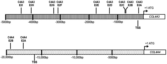
An in silico analysis and manual search using previously identified E2 box sequence motifs revealed nine E2 box motifs within the promoter region (5-kb region upstream of the transcriptional start site) of COL4A3 and two E2 box motifs within the promoter region of COL4A4 (Table; Fig. 1A).18,24,25,28
Figure 1.
Schematic depicting the locations of the putative nine E2 box motifs (C4A3, E2A-E2I) and two E2 box motifs (C4A4, E2A and E2B) identified within the 5-kb region upstream of the transcriptional start sites (TSS) for COL4A3 and COL4A4, respectively.
ZEB1 Binds to Multiple COL4A3 E2 Box Motifs In Vitro
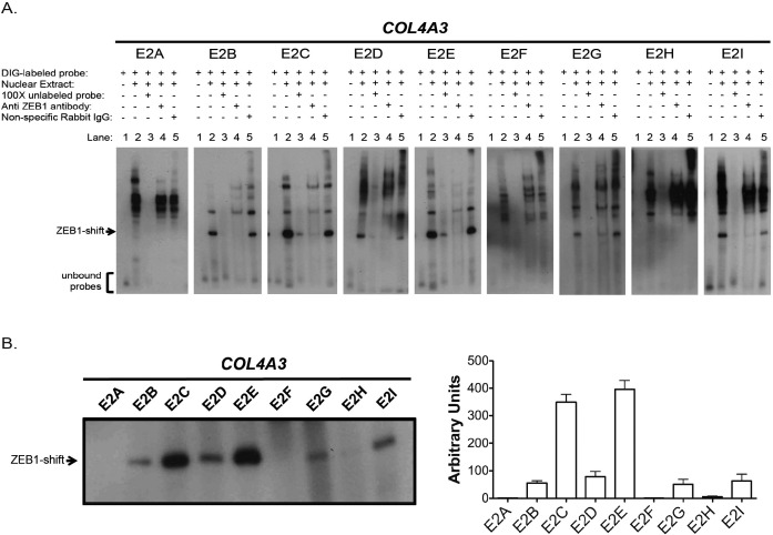
Electrophoretic mobility shift assays were performed to determine to which of the nine putative E2 box motifs identified in the COL4A3 promoter ZEB1 binds (Fig. 2). In the absence of ZEB1-enriched HEK293 nuclear extract, each of the nine COL4A3 promoter probes demonstrated a single band representing the unbound probe (Fig. 2A, Lane 1). When the nuclear extract was added, multiple shifted bands, representing different protein-probe complexes, were observed for each of the nine COL4A3 DIG-labeled probes. However, a distinct lower band (Fig. 2A, Lane 2, denoted by a black arrow) was observed with six probes (C4E3-E2B, C4E3-E2C, C4E3-E2D, C4E3-E2E, C4E3-E2G, and C4E3-E2I), which was presumed to represent binding of each probe to isolated ZEB1 protein. A side-by-side comparison showed that C4A3-E2C and C4A3-E2E had the highest relative levels of the lower shifted bands, whereas C4A3-E2B, C4A3-E2D, C4A3-E2G, and C4A3-E2I exhibited moderate relative levels (Fig. 2B). This suggested that, of the nine identified COL4A3 E2 box motifs, ZEB1 bound the strongest to the E2 box motifs represented by the C4A3-E2C and C4A3-E2E probes. Competition assays using a 100-fold molar excess of each respective unlabeled probe effectively diminished the intensity of each lower shifted band and confirmed the specificity of each COL4A3 DIG-labeled probe (Fig. 2A, Lane 3).
Figure 2.
ZEB1 protein from HEK293T nuclear extracts bound to six of the nine identified COL4A3 E2 box motifs. (A) Electrophoretic mobility shift assay performed using HEK293T nuclear extracts as a source of ZEB1 protein and DIG-labeled oligonucleotide probes containing each of the nine identified COL4A3 E2 box motifs. Lane 1: only a single band represented by unbound probes was visible and no complex was formed with the addition of DIG-labeled probes only. Lane 2: protein(s) in the HEK293T nuclear extracts interacted with DIG-labeled E2 box probes causing multiple shifted bands. Lane 3: the addition of unlabeled E2 box probes in 100-fold excess compared with the DIG-labeled E2 box probes resulted in the disappearance of the multiple bands observed in lane 2. Lane 4: the addition of ZEB1 antibody (D80D3; Cell Signaling Technology) to the HEK293T nuclear lysates resulted in the disappearance of the lower shifted band observed with six COL4A3 E2 box probes (ZEB1-shift). Lane 5: the addition of nonspecific rabbit IgG to the nuclear lysates did not cause the disappearance of the lower shifted band. (B) Electrophoretic mobility shift assay blot and corresponding graph show the relative intensities of the ZEB1-shifted bands from each of the nine DIG-labeled COL4A3 E2 box probes.
To identify any ZEB1-specific gel shifted bands within the multiple bands that appeared with the addition of nuclear extract, an anti-ZEB1 antibody was added to the reactions (Fig. 2A, Lane 4). The lower shifted bands (denoted by a black arrow) seen for six of the nine COL4A3 DIG-labeled probes were each abolished when incubated with a ZEB1 antibody (Fig. 2A, Lane 4), whereas incubation with a nonspecific rabbit IgG had a minimal, if any, impact on the shifted bands (Fig. 2A, Lane 5).
ZEB1 Does Not Directly Bind to Any Identified COL4A4 E2 Box Motif In Vitro
ZEB1-enriched HEK293T nuclear extracts and custom DIG-labeled probes were used in EMSA experiments to determine if ZEB1 binds to one or both of the identified COL4A4 E2 box motifs. In the absence of ZEB1-enriched HEK293T nuclear extract, neither of the COL4A4 probes demonstrated a band (Supplementary Fig. S1, Lane 1). In the presence of the nuclear extract, multiple bands were observed with both COL4A4 DIG-labeled probes. However, a band presumed to represent binding of each probe to isolated ZEB1 protein, as observed with the COL4A3 DIG-labeled probes, was not observed (Supplementary Fig. S1, Lane 2). Incubation with a 100-fold molar excess of each of the unlabeled probes abolished each band observed in lane 2, confirming the specificity of each COL4A4 DIG-labeled probe (Supplementary Fig. S1, Lane 3). For both COL4A4 probes, the addition of an anti-ZEB1 or nonspecific IgG showed a minimal impact on the banding pattern, indicating that the protein-probe complexes do not contain ZEB1 bound to either of the two identified COL4A4 E2 box motifs (Supplementary Fig. S1, Lanes 4–5).
ZEB1 Overexpression Suppresses COL4A3 Promoter Activity
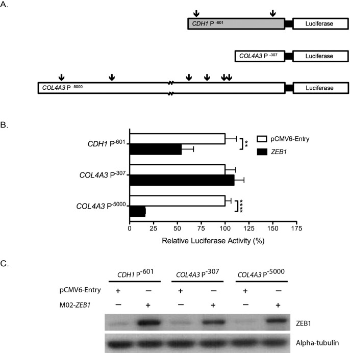
Dual-luciferase reporter assays were performed to determine the effects of ZEB1 overexpression on COL4A3 promoter activity (Fig. 3). The CDH1 gene encodes a cell adhesion protein, and the ability of ZEB1 to repress CDH1 transcription via its binding to two E2 box motifs in the CDH1 promoter region is a well-characterized phenomenon.29,30 Thus, the CDH1 reporter construct, CDH1 P−601, which contains the first 601 base pairs upstream of the TSS, including the two E2 boxes (denoted by black arrows in Fig. 3A), was used as a positive control for ZEB1 activity. Interaction of ZEB1 with the proximal CDH1 E2 box motif (CDH1 E2prox) was demonstrated by EMSA (Supplementary Fig. S2). Overexpression of ZEB1 with the pReceiver MO2-ZEB1 construct resulted in a statistically significant decrease in promoter activity for both CDH1 P−601 and COL4A3 P−5000 when compared with cotransfection with an empty vector, pCMV6-Entry (Fig. 3B). However, no decrease in promoter activity was identified when HEK293T cells were cotransfected with ZEB1 and the COL4A3 P−307 reporter construct. Overexpression of ZEB1 in the HEK293T cells was verified by Western blotting (Fig. 3C).
Figure 3.
Effect of ZEB1 overexpression on COL4A3 promoter activity. (A) Schematic diagram of luciferase reporter constructs (not drawn to scale). Black arrows represent E2 box motifs that have been shown to bind ZEB1 in vitro. (B) Relative luciferase activity of CDH1 and COL4A3 promoter constructs was measured when cotransfected into HEK293T cells with an empty vector, pCMV6-Entry, or a ZEB1 cDNA containing construct, pReceiver MO2-ZEB1. Reporter constructs CDH1 P−601 and COL4A3 P−5000, which contain identified E2 box motifs, exhibited significant decreases in luciferase activity when cotransfected into HEK293T cells with ZEB1 compared to being cotransfected with pCMV6-Entry. The reporter construct COL4A3 P−307 did not demonstrate a decrease in luciferase activity when cotransfected with ZEB1. **P < 0.05, ****P < 0.0001, n = 6, error bars denote SEM). (C) Anti-ZEB1 (D80D3; Cell Signaling Technology) immunoblotting verified ZEB1 overexpression in each of the HEK293T cell lysates used in the luciferase activity assays cotransfected with pReceiver MO2-ZEB1.
A ZEB1 Truncating Mutation Abrogates ZEB1-Dependent Repression of CDH1
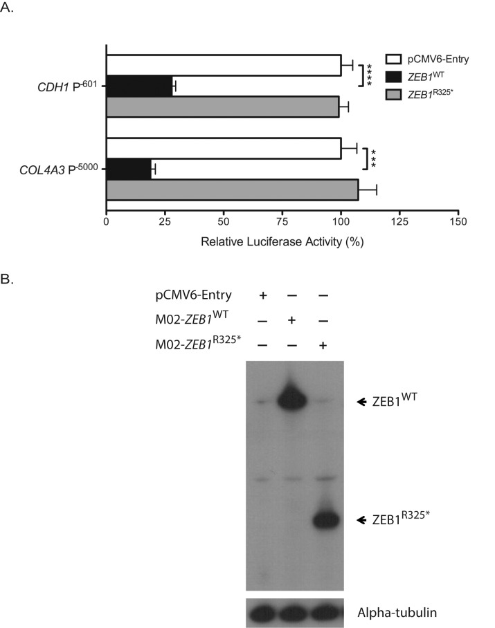
In a dual-luciferase reporter assay, CDH1 P−601 and COL4A3 P−5000 luciferase reporter constructs each were cotransfected into HEK293T cells with an overexpression construct containing either ZEB1WT or ZEB1R325* cDNA. Although the promoter activities of both the CDH1 P−601 and COL4A3 P−5000 reporter constructs were significantly reduced when cotransfected with ZEB1WT, the promoter activities of CDH1 and COL4A3 did not decrease when cotransfected with ZEB1R325* (Fig. 4A). The expressions of ZEB1WT and ZEB1R325* protein were confirmed to be similar by Western blotting (Fig. 4B).
Figure 4.
Impact of ZEB1 truncating mutations on CDH1 and COL4A3 promoter activities. (A) The relative luciferase activities of CDH1 and COL4A3 promoter constructs measured when cotransfected into HEK293T cells with either an empty vector (pCMV6-Entry), a ZEB1WT construct or a ZEB1R325* mutant construct. Relative to cotransfections with the empty vector, a significant reduction in both the CDH1 and COL4A3 promoter activities were observed when cotransfected with the ZEB1WT construct, while no significant change in promoter activities was noted when cotransfectioned with the ZEB1R325* mutant construct. ***P < 0.001, ****P < 0.0001, n = 6, error bars denote SEM. (B) ZEB1WT (MO2-ZEB1WT) and ZEB1R325* (MO2- ZEB1R325*) expression in the HEK293T cells was confirmed by Western blotting using a ZEB1 antibody (sc-10572; Santa Cruz Biotechnology).
Discussion
Although PPCD1 and PPCD3 are associated with mutations in two different genes (OVOL2 and ZEB1) on two different chromosomes, the indistinguishable clinical features of the individuals in families mapped to the PPCD1 locus and those with ZEB1 truncating mutations leads to the logical conclusion that alterations in a common pathway involving OVOL2 and ZEB1 are responsible for producing the common phenotype. We previously identified OVOL2 as a potential causal gene for PPCD1 (Le DJ, et al. IOVS 2015;56:ARVO E-Abstract 2521), and a separate group independently confirmed the presence of pathogenic OVOL2 promoter variants in several families previously mapped to the PPCD1 locus.11 A proposed role for OVOL2 in the pathogenesis of PPCD comes from its maintenance of the transcriptional program of cells derived from surface ectoderm, such as human corneal epithelial cells, via activation of epithelial genes and repression of mesenchymal genes.31 In cells derived from neuroectoderm, such as the corneal endothelium, an absence of OVOL2 expression is accompanied by high expression levels of some mesenchymal genes, including ZEB1, whereas epithelial gene expression is repressed.6,31 As the OVOL2 promoter region mutation (c.-307T > C) that we and other investigators have identified in three families with PPCD1 results in the formation of a binding site motif for the transcription factor FOXO3, a promoter of gene transcription, we hypothesize that this mutation results in increased expression of OVOL2, a known repressor of ZEB1 and ZEB2.32–36 Thus, in PPCD1, a presumed derepression of OVOL2 transcription by a OVOL2 promoter mutation in corneal endothelial-precursor cells, perhaps at the level of the neuroectoderm, may result in the activation of epithelial genes and development of epithelial cell characteristics.31 Accordingly, OVOL2 overexpression in murine neural progenitor cells leads to the development of epithelial cell–like cell-cell junctions and the expression of epithelial-specific genes, features characteristic of PPCD, and decreased expression of mesenchymal genes, such as Zeb1 and Zeb2.31 We predict that the resultant decrease in expression of ZEB1 has the same effect as the truncating mutations identified in individuals with PPCD3, namely ZEB1 insufficiency.
As a major regulator of EMT, ZEB1 is capable of acting as either a transcriptional activator or repressor and aberrant expression of ZEB1 is associated with tumorigenesis and disease.19,37–40 During EMT, the expression of CDH1, a classic epithelial marker that is involved in cell-cell adhesion and in the maintenance of apicobasal polarity, is repressed by ZEB1 via its binding to two E2 box motifs in the CDH1 promoter region.29,30 Although altered ZEB1 levels have been shown to affect CDH1 expression, we have demonstrated that the CDH1 transcript is not present in ex vivo human corneal endothelial cells (and presumably also absent in in vivo endothelium).41–43 Therefore, ZEB1 haploinsufficiency likely results in changes in the expression of other genes that are the direct targets of ZEB1 in the corneal endothelium. Although it has not yet been definitively determined which of these differentially expressed genes are involved in the pathogenesis of PPCD, altered expression of type IV collagens, particularly COL4A3, has been demonstrated in the corneal endothelium in individuals with PPCD3 and in ZEB1-null mice.6,20 In addition to the E2-box motifs in the promoter of COL4A3 initially reported by Krafchak and colleagues,6 we have identified seven additional COL4A3 E2 box motifs and demonstrated the ability of ZEB1 to bind to six of the nine E2 boxes. Although we also identified two E2 box motifs within the promoter region of COL4A4, no ZEB1 binding was observed, indicating that ZEB1 may not directly regulate COL4A4 expression. The reason ZEB1 bound to E2-box motifs in COL4A3, but not COL4A4 may be explained by the fact that the nucleotide sequences flanking the core motifs can affect ZEB1 binding.44 Therefore, further investigation of sequences that flank E2 box motifs will likely lead to a better refinement of identifying E2 box motifs by in silico analysis that demonstrate binding in vitro. Using promoter activity assays, we demonstrated that ZEB1 has a significant effect on the activity of the COL4A3 promoter, mediated through ZEB1 binding to E2 box motifs. Although we have demonstrated that ZEB1 is capable of binding to the COL4A3 promoter and repressing its activity in HEK293T cells, many factors influence whether ZEB1 functions as an activator or repressor of gene expression, including the presence of cofactors that may function as either corepressors or coactivators, Wnt signaling activity, cell type, and the stage of development.45 Although HEK293T cells were used for the EMSAs and dual luciferase reporter assays given several experimental considerations including ease of cell transfection, ideally such experiments would be performed in human corneal endothelial cells to confirm the role that ZEB1 plays in the regulation of COL4A3 expression.
To better understand the molecular basis of PPCD3, we previously investigated the impact of PPCD3-associated ZEB1 mutations and demonstrated that ZEB1 truncating mutations may result in impaired nuclear localization and/or a significant decrease in the production of ZEB1 protein, reinforcing the hypothesis that PPCD3 is caused by ZEB1 haploinsufficiency.12 However, neither of these mechanisms explains how a mutant ZEB1 protein such as p.(Arg325*), which primarily localizes to the nucleus (in spite of lacking the nuclear localization signal at position 892-998) and is expressed at levels similar to the wild-type ZEB1, leads to ZEB1 haploinsufficiency. The demonstration that p.(Arg325*) is unable to repress transcription of CDH1, a known ZEB1 target gene, and COL4A3 provides evidence of a third mechanism via which truncating ZEB1 mutations may lead to PPCD3, that is, loss of E2 box binding ability, and subsequent loss of transcriptional repression of genes whose expression is regulated by ZEB1. Therefore, the truncating ZEB1 mutations identified to date in individuals with PPCD3 may lead to one or a combination of events that include altered nuclear localization, reduced ZEB1 protein production and/or abrogated binding to the promoter regions of target genes. Ultimately, each mechanism is likely to impact corneal endothelial gene expression by reducing ZEB1-dependent transcriptional regulation in the context of only one functional ZEB1 allele. Therefore, studies to compare the corneal endothelial cell transcriptome in individuals with PPCD3 to controls are ongoing in an effort to identify other proteins besides COL4A3 whose expression is regulated by ZEB1. We acknowledge that the mechanisms via which ZEB1 truncating mutations lead to PPCD3 are unlikely to involve just one gene downstream of ZEB1 (i.e., COL4A3). However, the demonstration of the ability of ZEB1 to directly regulate COL4A3 expression provides the basis for investigation into the ability of ZEB1 to regulate the expression of other type IV collagens and genes involved in cell adhesion to gain further insight into the role of ZEB1 in corneal endothelial cell function and dysfunction.
Acknowledgments
The authors thank Aleck Cervantes and Katherine Gee for their technical assistance in generating the ZEB1R325* mutant expression construct.
Supported by National Eye Institute Grants 1R01 EY022082 (AJA) and P30 EY000331 (core grant), the Walton Li Chair in Cornea and Uveitis (AJA), the Stotter Revocable Trust, and an unrestricted grant from Research to Prevent Blindness.
Disclosure: D.-W.D. Chung, None; R.F. Frausto, None; S. Chiu, None; B.R. Lin, None; A.J. Aldave, None
References
- 1. Krachmer JH. Posterior polymorphous corneal dystrophy: a disease characterized by epithelial-like endothelial cells which influence management and prognosis. Trans Am Ophthalmol Soc. 1985; 83: 413–475. [PMC free article] [PubMed] [Google Scholar]
- 2. Weiss JS,, Moller HU,, Aldave AJ,, et al. IC3D classification of corneal dystrophies--edition 2. Cornea. 2015; 34: 117–159. [DOI] [PubMed] [Google Scholar]
- 3. Aldave AJ,, Han J,, Frausto RF. Genetics of the corneal endothelial dystrophies: an evidence-based review. Clin Genet. 2013; 84: 109–119. [DOI] [PMC free article] [PubMed] [Google Scholar]
- 4. Heon E,, Mathers WD,, Alward WL,, et al. Linkage of posterior polymorphous corneal dystrophy to 20q11. Hum Mol Genet. 1995; 4: 485–488. [DOI] [PubMed] [Google Scholar]
- 5. Gwilliam R,, Liskova P,, Filipec M,, et al. Posterior polymorphous corneal dystrophy in Czech families maps to chromosome 20 and excludes the VSX1 gene. Invest Ophthalmol Vis Sci. 2005; 46: 4480–4484. [DOI] [PubMed] [Google Scholar]
- 6. Krafchak CM,, Pawar H,, Moroi SE,, et al. Mutations in TCF8 cause posterior polymorphous corneal dystrophy and ectopic expression of COL4A3 by corneal endothelial cells. Am J Hum Genet. 2005; 77: 694–708. [DOI] [PMC free article] [PubMed] [Google Scholar]
- 7. Aldave AJ,, Yellore VS,, Yu F,, et al. Posterior polymorphous corneal dystrophy is associated with TCF8 gene mutations and abdominal hernia. Am J Med Genet A. 2007; 143A: 2549–2556. [DOI] [PubMed] [Google Scholar]
- 8. Yellore VS,, Papp JC,, Sobel E,, et al. Replication and refinement of linkage of posterior polymorphous corneal dystrophy to the posterior polymorphous corneal dystrophy 1 locus on chromosome 20. Genet Med. 2007; 9: 228–234. [DOI] [PubMed] [Google Scholar]
- 9. Hosseini SM,, Herd S,, Vincent AL,, Heon E. Genetic analysis of chromosome 20-related posterior polymorphous corneal dystrophy: genetic heterogeneity and exclusion of three candidate genes. Mol Vis. 2008; 14: 71–80. [PMC free article] [PubMed] [Google Scholar]
- 10. Liskova P,, Gwilliam R,, Filipec M,, et al. High prevalence of posterior polymorphous corneal dystrophy in the Czech Republic; linkage disequilibrium mapping and dating an ancestral mutation. PLoS One. 2012; 7: e45495. [DOI] [PMC free article] [PubMed] [Google Scholar]
- 11. Davidson AE,, Liskova P,, Evans CJ,, et al. Autosomal-dominant corneal endothelial dystrophies CHED1 and PPCD1 are allelic disorders caused by non-coding mutations in the promoter of OVOL2. Am J Hum Genet. 2016; 98: 75–89. [DOI] [PMC free article] [PubMed] [Google Scholar]
- 12. Chung DW,, Frausto RF,, Ann LB,, Jang MS,, Aldave AJ. Functional impact of ZEB1 mutations associated with posterior polymorphous and Fuchs' endothelial corneal dystrophies. Invest Ophthalmol Vis Sci. 2014; 55: 6159–6166. [DOI] [PMC free article] [PubMed] [Google Scholar]
- 13. Bakhtiari P,, Frausto RF,, Roldan AN,, Wang C,, Yu F,, Aldave AJ. Exclusion of pathogenic promoter region variants and identification of novel nonsense mutations in the zinc finger E-box binding homeobox 1 gene in posterior polymorphous corneal dystrophy. Mol Vis. 2013; 19: 575–580. [PMC free article] [PubMed] [Google Scholar]
- 14. Evans CJ,, Liskova P,, Dudakova L,, et al. Identification of six novel mutations in ZEB1 and description of the associated phenotypes in patients with posterior polymorphous corneal dystrophy 3. Ann Hum Genet. 2015; 79: 1–9. [DOI] [PubMed] [Google Scholar]
- 15. Liskova P,, Evans CJ,, Davidson AE,, et al. Heterozygous deletions at the ZEB1 locus verify haploinsufficiency as the mechanism of disease for posterior polymorphous corneal dystrophy type 3. Eur J Hum Genet. 2015; October 28 2016; 24: 985–991. [DOI] [PMC free article] [PubMed] [Google Scholar]
- 16. Behrens J,, Lowrick O,, Klein-Hitpass L,, Birchmeier W. The E-cadherin promoter: functional analysis of a G.C-rich region and an epithelial cell-specific palindromic regulatory element. Proc Natl Acad Sci U S A. 1991; 88: 11495–11499. [DOI] [PMC free article] [PubMed] [Google Scholar]
- 17. Giroldi LA,, Bringuier PP,, de Weijert M,, Jansen C,, van Bokhoven A,, Schalken JA. Role of E boxes in the repression of E-cadherin expression. Biochem Biophys Res Commun. 1997; 241: 453–458. [DOI] [PubMed] [Google Scholar]
- 18. Remacle JE,, Kraft H,, Lerchner W,, et al. New mode of DNA binding of multi-zinc finger transcription factors: deltaEF1 family members bind with two hands to two target sites. EMBO J. 1999; 18: 5073–5084. [DOI] [PMC free article] [PubMed] [Google Scholar]
- 19. Vandewalle C,, Van Roy F,, Berx G. The role of the ZEB family of transcription factors in development and disease. Cell Mol Life Sci. 2009; 66: 773–787. [DOI] [PMC free article] [PubMed] [Google Scholar]
- 20. Liu Y,, Peng X,, Tan J,, Darling DS,, Kaplan HJ,, Dean DC. Zeb1 mutant mice as a model of posterior corneal dystrophy. Invest Ophthalmol Vis Sci. 2008; 49: 1843–1849. [DOI] [PMC free article] [PubMed] [Google Scholar]
- 21. Merjava S,, Liskova P,, Jirsova K. Immunohistochemical characterization of collagen IV in control corneas and in corneas obtained from patients suffering from posterior polymorphous corneal dystrophy [in Slovak]. Cesk Slov Oftalmol. 2008; 64: 115–119. [PubMed] [Google Scholar]
- 22. Yellore VS,, Rayner SA,, Nguyen CK,, et al. Analysis of the role of ZEB1 in the pathogenesis of posterior polymorphous corneal dystrophy. Invest Ophthalmol Vis Sci. 2012; 53: 273–278. [DOI] [PMC free article] [PubMed] [Google Scholar]
- 23. Momota R,, Sugimoto M,, Oohashi T,, Kigasawa K,, Yoshioka H,, Ninomiya Y. Two genes COL4A3 and COL4A4 coding for the human alpha3(IV) and alpha4(IV) collagen chains are arranged head-to-head on chromosome 2q36. FEBS Lett. 1998; 424: 11–16. [DOI] [PubMed] [Google Scholar]
- 24. Mathelier A,, Zhao X,, Zhang AW,, et al. JASPAR 2014: an extensively expanded and updated open-access database of transcription factor binding profiles. Nucleic Acids Res. 2014; 42: D142–D147. [DOI] [PMC free article] [PubMed] [Google Scholar]
- 25. Ikeda K,, Kawakami K. DNA binding through distinct domains of zinc-finger-homeodomain protein AREB6 has different effects on gene transcription. Eur J Biochem. 1995; 233: 73–82. [DOI] [PubMed] [Google Scholar]
- 26. Abmayr SM,, Yao T,, Parmely T,, Workman JL. Preparation of Nuclear and Cytoplasmic Extracts from Mammalian Cells. Current Protocols in Molecular Biology: Hoboken, NJ: John Wiley & Sons, Inc.; 2001: 12.1.1–12.1.10. [DOI] [PubMed] [Google Scholar]
- 27. Schneider CA,, Rasband WS,, Eliceiri KWNIH. Image to ImageJ: 25 years of image analysis. Nat Methods. 2012; 9: 671–675. [DOI] [PMC free article] [PubMed] [Google Scholar]
- 28. Murray D,, Precht P,, Balakir R,, Horton WE,, Jr. The transcription factor deltaEF1 is inversely expressed with type II collagen mRNA and can repress Col2a1 promoter activity in transfected chondrocytes. J Biol Chem. 2000; 275: 3610–3618. [DOI] [PubMed] [Google Scholar]
- 29. Pena C,, Garcia JM,, Garcia V,, et al. The expression levels of the transcriptional regulators p300 and CtBP modulate the correlations between SNAIL, ZEB1, E-cadherin and vitamin D receptor in human colon carcinomas. Int J Cancer. 2006; 119: 2098–2104. [DOI] [PubMed] [Google Scholar]
- 30. Perez-Moreno M,, Jamora C,, Fuchs E. Sticky business: orchestrating cellular signals at adherens junctions. Cell. 2003; 112: 535–548. [DOI] [PubMed] [Google Scholar]
- 31. Kitazawa K,, Hikichi T,, Nakamura T,, et al. OVOL2 maintains the transcriptional program of human corneal epithelium by suppressing epithelial-to-mesenchymal transition. Cell Rep. 2016; 15: 1359–1368. [DOI] [PubMed] [Google Scholar]
- 32. Lee B,, Villarreal-Ponce A,, Fallahi M,, et al. Transcriptional mechanisms link epithelial plasticity to adhesion and differentiation of epidermal progenitor cells. Dev Cell. 2014; 29: 47–58. [DOI] [PMC free article] [PubMed] [Google Scholar]
- 33. Paik JH,, Kollipara R,, Chu G,, et al. FoxOs are lineage-restricted redundant tumor suppressors and regulate endothelial cell homeostasis. Cell. 2007; 128: 309–323. [DOI] [PMC free article] [PubMed] [Google Scholar]
- 34. Roca H,, Hernandez J,, Weidner S,, et al. Transcription factors OVOL1 and OVOL2 induce the mesenchymal to epithelial transition in human cancer. PLoS One. 2013; 8: e76773. [DOI] [PMC free article] [PubMed] [Google Scholar]
- 35. Watanabe K,, Villarreal-Ponce A,, Sun P,, et al. Mammary morphogenesis and regeneration require the inhibition of EMT at terminal end buds by Ovol2 transcriptional repressor. Dev Cell. 2014; 29: 59–74. [DOI] [PMC free article] [PubMed] [Google Scholar]
- 36. Le DJ,, Chung DW,, Frausto RF,, Kim MJ,, Aldave AJ. Identification of potentially pathogenic variants in the posterior polymorphous corneal dystrophy 1 locus. PLoS One. 2016; 11: e0158467. [DOI] [PMC free article] [PubMed] [Google Scholar]
- 37. Watanabe Y,, Kawakami K,, Hirayama Y,, Nagano K. Transcription factors positively and negatively regulating the Na, K-ATPase alpha 1 subunit gene. J Biochem. 1993; 114: 849–855. [DOI] [PubMed] [Google Scholar]
- 38. Liu Y,, El-Naggar S,, Darling DS,, Higashi Y,, Dean DC. Zeb1 links epithelial-mesenchymal transition and cellular senescence. Development. 2008; 135: 579–588. [DOI] [PMC free article] [PubMed] [Google Scholar]
- 39. Cho JH,, Gelinas R,, Wang K,, et al. Systems biology of interstitial lung diseases: integration of mRNA and microRNA expression changes. BMC Med Genomics. 2011; 4: 8. [DOI] [PMC free article] [PubMed] [Google Scholar] [Research Misconduct Found]
- 40. Preca BT,, Bajdak K,, Mock K,, et al. A self-enforcing CD44s/ZEB1 feedback loop maintains EMT and stemness properties in cancer cells. Int J Cancer. 2015; 137: 2566–2577. [DOI] [PubMed] [Google Scholar]
- 41. Hashiguchi M,, Ueno S,, Sakoda M,, et al. Clinical implication of ZEB-1 and E-cadherin expression in hepatocellular carcinoma (HCC). BMC Cancer. 2013; 13: 572. [DOI] [PMC free article] [PubMed] [Google Scholar]
- 42. Zhang J,, Lu C,, Zhang J,, Kang J,, Cao C,, Li M. Involvement of ZEB1 and E-cadherin in the invasion of lung squamous cell carcinoma. Mol Biol Rep. 2013; 40: 949–956. [DOI] [PubMed] [Google Scholar]
- 43. Frausto RF,, Le DJ,, Aldave AJ. Transcriptomic analysis of cultured corneal endothelial cells as a validation for their use in cell-replacement therapy. Cell Transplant. 2016; 25: 1159–1176. [DOI] [PMC free article] [PubMed] [Google Scholar]
- 44. Gordan R,, Shen N,, Dror I,, et al. Genomic regions flanking E-box binding sites influence DNA binding specificity of bHLH transcription factors through DNA shape. Cell Rep. 2013; 3: 1093–1104. [DOI] [PMC free article] [PubMed] [Google Scholar]
- 45. Sanchez-Tillo E,, de Barrios O,, Valls E,, Darling DS,, Castells A,, Postigo A. ZEB1 and TCF4 reciprocally modulate their transcriptional activities to regulate Wnt target gene expression. Oncogene. 2015; 34: 5760–5770. [DOI] [PubMed] [Google Scholar]


