

Abstract
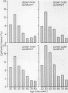
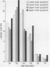
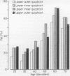
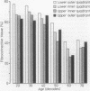
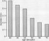
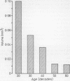
Ageing changes in the normal human female breast were studied to determine their significance for the evolution of mammary cancer. Employing the morphometric techniques of point counting and planimetry, objective quantitative measurements were made of the structure of the normal female breast in 58 subjects from the prepubertal to late postreproductive period. The relative amounts of epithelial and connective tissue varied with age, and the epithelial elements (combined lobular and extralobular) were unevenly distributed within the gland, with lower containing more than upper quadrants. The upper outer quadrant, however, usually contained the largest proportion of lobular units, which may relate to the higher incidence of lobular carcinoma found in this quadrant. Involution was shown to be a premenopausal rather than postmenopausal phenomenon. Mammary dysplastic changes were uncommon in all age groups.
Full text
PDF
Images in this article
Selected References
These references are in PubMed. This may not be the complete list of references from this article.
- FRANTZ V. K., PICKREN J. W., MELCHER G. W., AUCHINCLOSS H., Jr Indicence of chronic cystic disease in so-called "normal breasts; a study based on 225 postmortem examinations. Cancer. 1951 Jul;4(4):762–783. doi: 10.1002/1097-0142(195107)4:4<762::aid-cncr2820040414>3.0.co;2-v. [DOI] [PubMed] [Google Scholar]
- Ozzello L. Epithelial-stromal junction of normal and dysplastic mammary glands. Cancer. 1970 Mar;25(3):586–600. doi: 10.1002/1097-0142(197003)25:3<586::aid-cncr2820250314>3.0.co;2-1. [DOI] [PubMed] [Google Scholar]
- Ozzello L. Proceedings: Electron microscopic study of functional and dysfunctional human mammary glands. J Invest Dermatol. 1974 Jul;63(1):19–26. doi: 10.1111/1523-1747.ep12677303. [DOI] [PubMed] [Google Scholar]
- Ozzello L. Ultrastructure of the human mammary gland. Pathol Annu. 1971;6:1–59. [PubMed] [Google Scholar]
- Stirling J. W., Chandler J. A. The fine structure of ducts and subareolar ducts in the resting gland of the female breast. Virchows Arch A Pathol Anat Histol. 1977 Mar 11;373(2):119–132. doi: 10.1007/BF00432157. [DOI] [PubMed] [Google Scholar]
- Stirling J. W., Chandler J. A. The fine structure of the normal, resting terminal ductal-lobular unit of the female breast. Virchows Arch A Pathol Anat Histol. 1976 Dec 27;372(3):205–226. doi: 10.1007/BF00433280. [DOI] [PubMed] [Google Scholar]
- Wellings S. R., Jensen H. M., Marcum R. G. An atlas of subgross pathology of the human breast with special reference to possible precancerous lesions. J Natl Cancer Inst. 1975 Aug;55(2):231–273. [PubMed] [Google Scholar]
- Wellings S. R., Jensen H. M. On the origin and progression of ductal carcinoma in the human breast. J Natl Cancer Inst. 1973 May;50(5):1111–1118. doi: 10.1093/jnci/50.5.1111. [DOI] [PubMed] [Google Scholar]