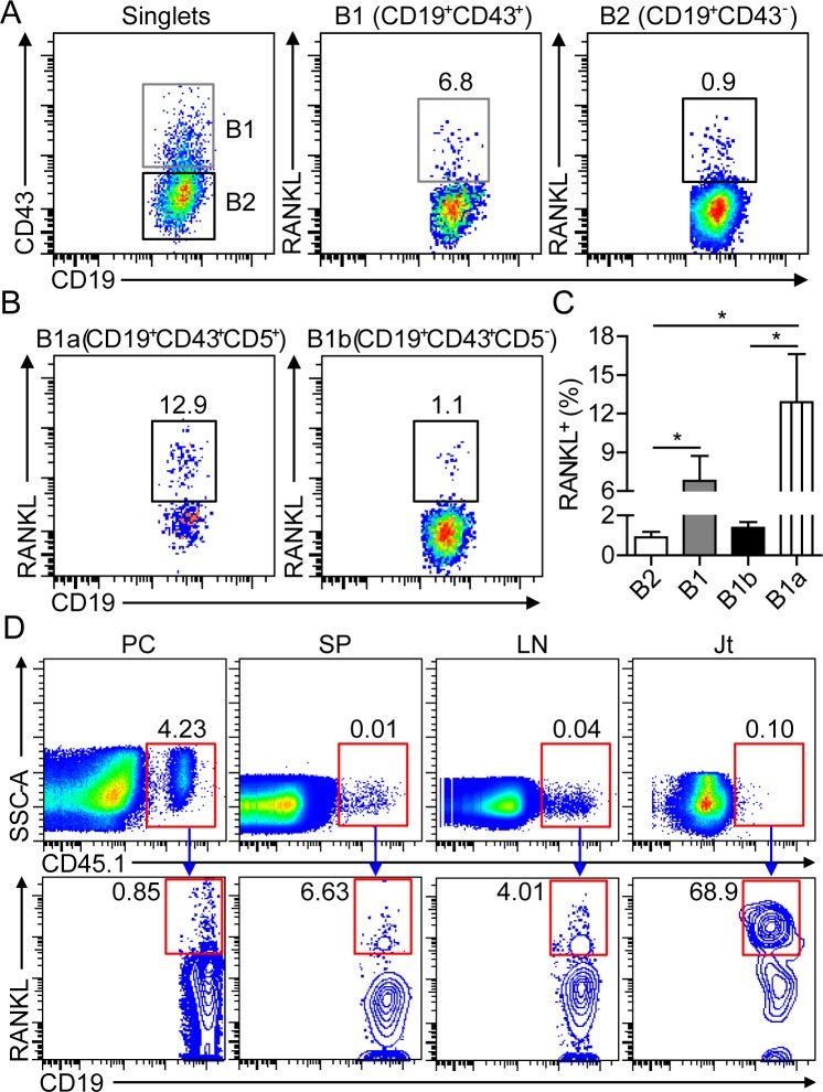
Figure 4. B1a cells express RANKL in the inflamed joint of CIA mice.
A., B. Flow cytometric analysis of RANKL expression on CD19+CD43− B2 and CD19+CD43+ B1 cells in A, CD19+CD43+CD5+ B1a and CD19+CD43+CD5− B1b cells in B in the inflamed joint of CIA mice on day 30 post 1st CII-immunization. C. The frequencies of RANKL+ B2, B1, B1a and B1b cells were enumerated from three independent experiments (n = 5-7 per group in each experiment). D. Peritoneal B1a cells from CD45.1+ BoyJ mice were intraperitoneally transferred into CD45.2+ C57 mice and followed by CII immunization for CIA induction. On day 17 after cell transfer, RANKL expression on donor CD45.1+ B1a cells in the PC, SP, LN and joint tissue (Jt) were detected by flow cytometry. The flow profiles are representative data from three independent experiments with similar results.

