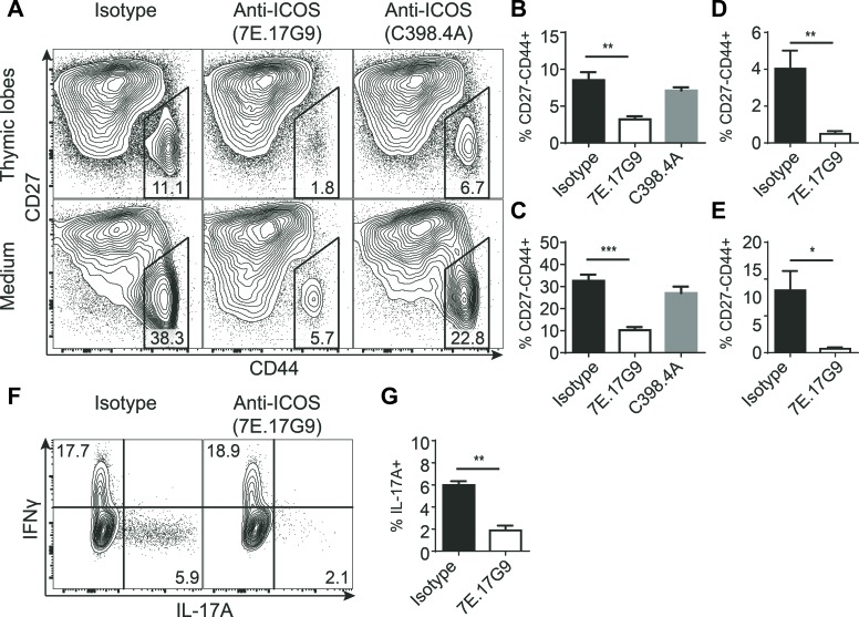
Figure 3. Treatment with ICOS specific antibodies severely reduces the development of IL-17-producing γδ T cells.
FTOC treated with anti-ICOS antibodies and cultured for 7 or 11 days. A. Representative flow cytometric plots showing CD27 and CD44 expression within γδ T cells harvested from the lobes (top) or culture medium (bottom) of e15 + 7 days FTOC. B.-E. Quantification of the CD44+CD27− population percentages within γδ T cells harvested from thymic lobes (B, D) or culture medium (C, E) from e15 + 7 days FTOC (B, C) or e14 + 11 days FTOC (D, E) (n = 4). F. Representative flow cytometric plots showing intracellular IL-17A and IFNγ expression in γδ T cells from e14 + 11 days FTOC thymic lobes stimulated for 4 hours with PMA and ionomycin. G. Quantification of the IL-17A+ fraction of e14 + 11 days FTOC γδ T cells stimulated for 4 hours with PMA and ionomycin (n = 3). Each sample was pooled from a well containing 4-5 individual lobes. Bars denote mean percentage ± SEM of the gated populations.

