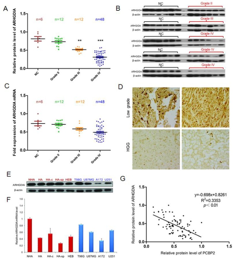
Figure 1. ARHGDIA protein but not mRNA is frequently downregulated in glioma tissues compared to control brain tissues.
A. Relative ARHGDIA protein levels in 6 control brain tissues,12×II grade, 12×III grade, and 48×IV glioma tissues. B. Representative western blot showing ARHGDIA protein levels in 6 control brain tissues, 6×II grade, 6×III grade, and 48×IV glioma tissues. β-actin was used as a loading control. C. Real-time PCR analysis of relative ARHGDIA mRNA expression in 6 control brain tissues, 12×II grade, 12×III grade, and 48×IV glioma tissues. GAPDH was used as a control. n.s., nonsignificant. D. Representative immunohistochemical staining of ARHGDIA in 16 low-grade glioma tissues and 19 high-grade glioma tissues using anti-human ARHGDIA antibodies. Original magnification,×400. E. Western blotting showed ARHGDIA protein levels in 4 human astrocyte cell lines (HA, NHA, HA-c, HA-sp), 1 human embryonic brain cell line (HEB), and 4 glioma cell lines (T98G, U87 MG, A172, U251). F. Real-time PCR analysis of ARHGDIA mRNA in 4 human astrocyte lines, 1 human embryonic brain cell line and the 4 indicated glioma cell lines. G. Relative ARHGDIA and PCBP2 protein levels in 6 control brain tissues,12×II grade, 12×III grade, and 48×IV glioma tissues.

