

Abstract
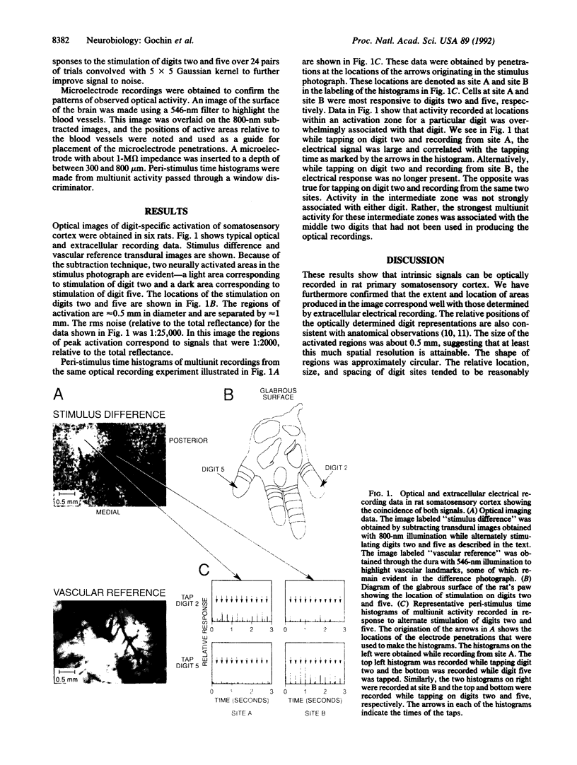
The responses of somatosensory cortex (S-I) to tactile stimulation of the forepaw were assessed by intrinsic signal optical imaging. The tips of digits two or five were alternately touched with mechanical tappers while video photographs were taken of S-I illuminated by an 800-nm light source. The resulting images showed two highlighted areas about 300 microns in diameter and 500 microns apart. Generation of these images required less than 1 hr. Electrode penetrations placed in the areas highlighted during stimulation provided multiunit recordings with receptive fields appropriate for the stimulated digit and not the other digit. Penetrations between the high-lighted areas yielded receptive fields on intervening digits. These results demonstrate that intrinsic signal optical images are obtainable in S-I and confirm the functional somatotopy previously reported using electrical recording. Furthermore, the short time required to produce the images and the obtainable spatial resolution suggest that optical recording could be employed for the study of cortical reorganization in this brain region.
Full text
PDF
Images in this article
Selected References
These references are in PubMed. This may not be the complete list of references from this article.
- Blasdel G. G., Salama G. Voltage-sensitive dyes reveal a modular organization in monkey striate cortex. Nature. 1986 Jun 5;321(6070):579–585. doi: 10.1038/321579a0. [DOI] [PubMed] [Google Scholar]
- Chapin J. K., Lin C. S. Mapping the body representation in the SI cortex of anesthetized and awake rats. J Comp Neurol. 1984 Oct 20;229(2):199–213. doi: 10.1002/cne.902290206. [DOI] [PubMed] [Google Scholar]
- Chapin J. K., Sadeq M., Guise J. L. Corticocortical connections within the primary somatosensory cortex of the rat. J Comp Neurol. 1987 Sep 15;263(3):326–346. doi: 10.1002/cne.902630303. [DOI] [PubMed] [Google Scholar]
- Dawson D. R., Killackey H. P. The organization and mutability of the forepaw and hindpaw representations in the somatosensory cortex of the neonatal rat. J Comp Neurol. 1987 Feb 8;256(2):246–256. doi: 10.1002/cne.902560205. [DOI] [PubMed] [Google Scholar]
- Frostig R. D., Lieke E. E., Ts'o D. Y., Grinvald A. Cortical functional architecture and local coupling between neuronal activity and the microcirculation revealed by in vivo high-resolution optical imaging of intrinsic signals. Proc Natl Acad Sci U S A. 1990 Aug;87(16):6082–6086. doi: 10.1073/pnas.87.16.6082. [DOI] [PMC free article] [PubMed] [Google Scholar]
- Grinvald A., Frostig R. D., Lieke E., Hildesheim R. Optical imaging of neuronal activity. Physiol Rev. 1988 Oct;68(4):1285–1366. doi: 10.1152/physrev.1988.68.4.1285. [DOI] [PubMed] [Google Scholar]
- Orbach H. S., Cohen L. B., Grinvald A. Optical mapping of electrical activity in rat somatosensory and visual cortex. J Neurosci. 1985 Jul;5(7):1886–1895. doi: 10.1523/JNEUROSCI.05-07-01886.1985. [DOI] [PMC free article] [PubMed] [Google Scholar]
- Ratzlaff E. H., Grinvald A. A tandem-lens epifluorescence macroscope: hundred-fold brightness advantage for wide-field imaging. J Neurosci Methods. 1991 Feb;36(2-3):127–137. doi: 10.1016/0165-0270(91)90038-2. [DOI] [PubMed] [Google Scholar]
- Ts'o D. Y., Frostig R. D., Lieke E. E., Grinvald A. Functional organization of primate visual cortex revealed by high resolution optical imaging. Science. 1990 Jul 27;249(4967):417–420. doi: 10.1126/science.2165630. [DOI] [PubMed] [Google Scholar]
- Welker C. Microelectrode delineation of fine grain somatotopic organization of (SmI) cerebral neocortex in albino rat. Brain Res. 1971 Mar 5;26(2):259–275. [PubMed] [Google Scholar]