

Abstract
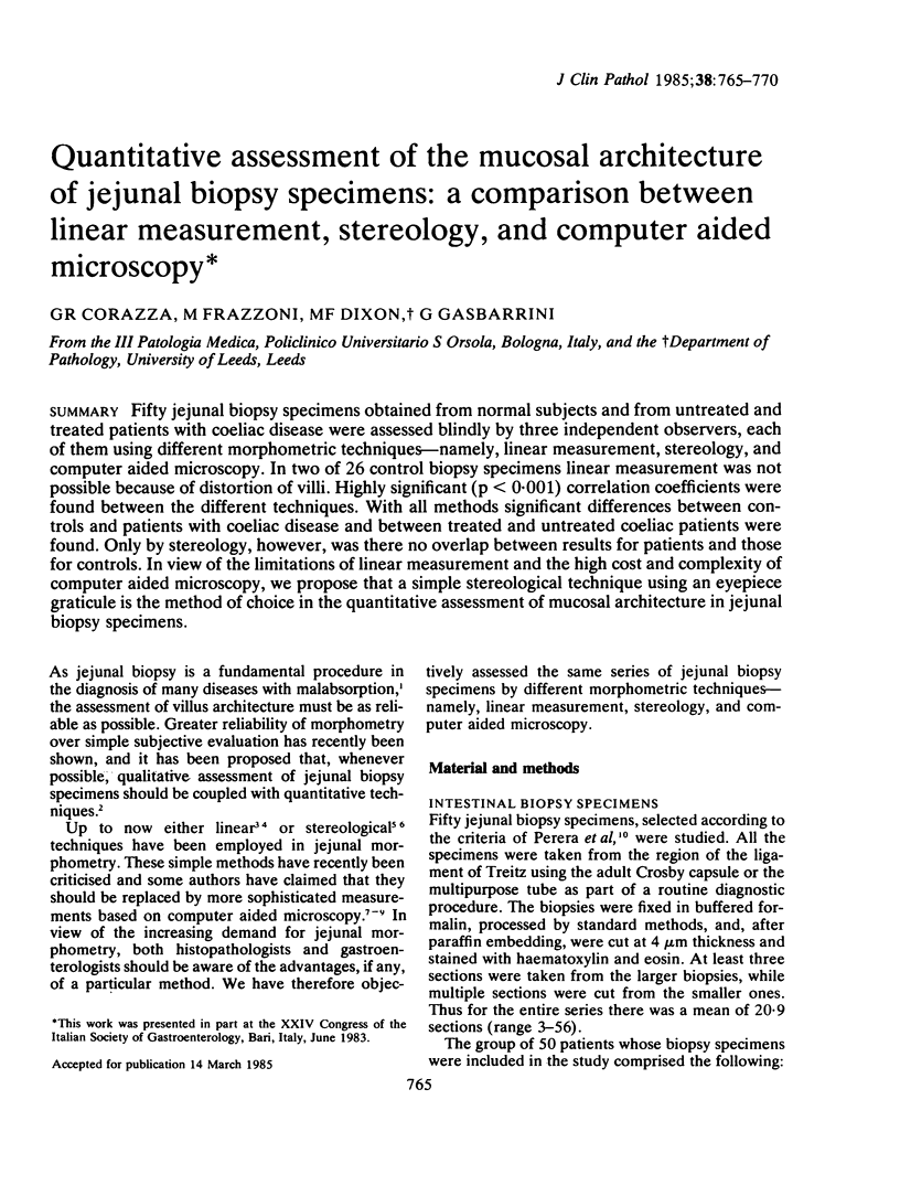
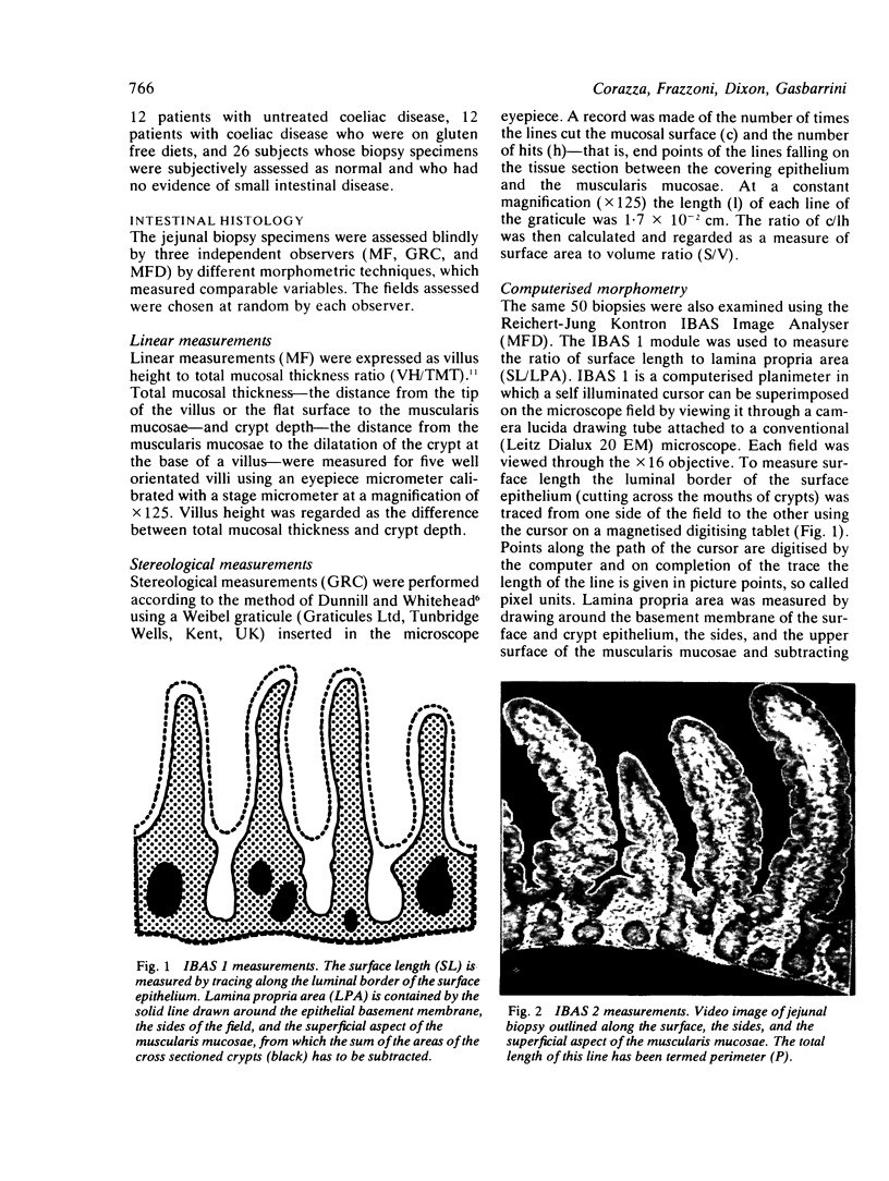
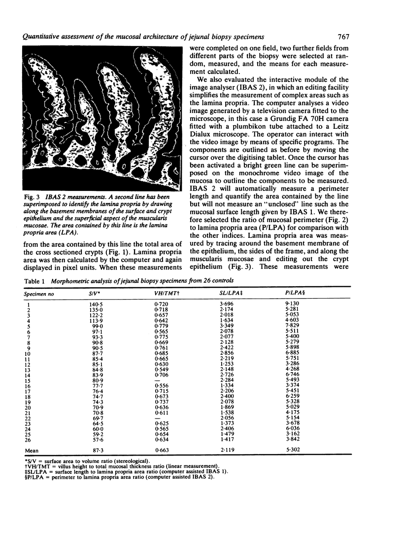
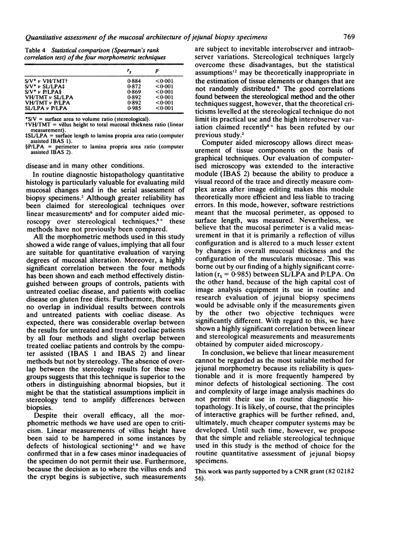
Fifty jejunal biopsy specimens obtained from normal subjects and from untreated and treated patients with coeliac disease were assessed blindly by three independent observers, each of them using different morphometric techniques-namely, linear measurement, stereology, and computer aided microscopy. In two of 26 control biopsy specimens linear measurement was not possible because of distortion of villi. Highly significant (p less than 0.001) correlation coefficients were found between the different techniques. With all methods significant differences between controls and patients with coeliac disease and between treated and untreated coeliac patients were found. Only by stereology, however, was there no overlap between results for patients and those for controls. In view of the limitations of linear measurement and the high cost and complexity of computer aided microscopy, we propose that a simple stereological technique using an eyepiece graticule is the method of choice in the quantitative assessment of mucosal architecture in jejunal biopsy specimens.
Full text
PDF
Images in this article
Selected References
These references are in PubMed. This may not be the complete list of references from this article.
- Brandborg L. L. Histologic diagnosis of diseases of malabsorption. Am J Med. 1979 Dec;67(6):999–1006. doi: 10.1016/0002-9343(79)90641-7. [DOI] [PubMed] [Google Scholar]
- Chalkley H. W., Cornfield J., Park H. A Method for Estimating Volume-Surface Ratios. Science. 1949 Sep 23;110(2856):295–297. doi: 10.1126/science.110.2856.295. [DOI] [PubMed] [Google Scholar]
- Corazza G. R., Bonvicini F., Frazzoni M., Gatto M., Gasbarrini G. Observer variation in assessment of jejunal biopsy specimens. A comparison between subjective criteria and morphometric measurement. Gastroenterology. 1982 Dec;83(6):1217–1222. [PubMed] [Google Scholar]
- Dunnill M. S., Whitehead R. A method for the quantitation of small intestinal biopsy specimens. J Clin Pathol. 1972 Mar;25(3):243–246. doi: 10.1136/jcp.25.3.243. [DOI] [PMC free article] [PubMed] [Google Scholar]
- Meinhard E. A., Wadbrook D. G., Risdon R. A. Computer card morphometry of jejunal biopsies in childhood coeliac disease. J Clin Pathol. 1975 Feb;28(2):85–93. doi: 10.1136/jcp.28.2.85. [DOI] [PMC free article] [PubMed] [Google Scholar]
- Perera D. R., Weinstein W. M., Rubin C. E. Symposium on pathology of the gastrointestinal tract-Part II. Small intestinal biopsy. Hum Pathol. 1975 Mar;6(2):157–217. doi: 10.1016/s0046-8177(75)80176-6. [DOI] [PubMed] [Google Scholar]
- RUBIN C. E., BRANDBORG L. L., PHELPS P. C., TAYLOR H. C., Jr Studies of celiac disease. I. The apparent identical and specific nature of the duodenal and proximal jejunal lesion in celiac disease and idiopathic sprue. Gastroenterology. 1960 Jan;38:28–49. [PubMed] [Google Scholar]
- SHINER M., DONIACH I. Histopathologic studies in steatorrhea. Gastroenterology. 1960 Mar;38:419–440. [PubMed] [Google Scholar]
- Scott B. B., Losowsky M. S. Patchiness and duodenal-jejunal variation of the mucosal abnormality in coeliac disease and dermatitis herpetiformis. Gut. 1976 Dec;17(12):984–992. doi: 10.1136/gut.17.12.984. [DOI] [PMC free article] [PubMed] [Google Scholar]
- Slavin G., Sowter C., Robertson K., McDermott S., Paton K. Measurement in jejunal biopsies by computer-aided microscopy. J Clin Pathol. 1980 Mar;33(3):254–261. doi: 10.1136/jcp.33.3.254. [DOI] [PMC free article] [PubMed] [Google Scholar]
- Stewart J. S., Pollock D. J., Hoffbrand A. V., Mollin D. L., Booth C. C. A study of proximal and distal intestinal structure and absorptive function in idiopathic steatorrhoea. Q J Med. 1967 Jul;36(143):425–444. [PubMed] [Google Scholar]
- Tulloh E. A., Baylis J. M., Challacombe D. N. Automated analysis of morphological change in the duodenal mucosa of children with coeliac disease. Arch Dis Child. 1981 Nov;56(11):860–863. doi: 10.1136/adc.56.11.860. [DOI] [PMC free article] [PubMed] [Google Scholar]