Abstract
Introduction: Gastroparesis, or delayed gastric emptying, can be diagnosed with gastric emptying scintigraphy. Manometric studies of patients with gastroparesis show increased pyloric tone (pylorospasm). Among the recent endoscopic therapies for pylorospasm is peroral endoscopic pylorotomy (POP). In this study, we explored the effect of POP on gastric emptying in healthy pigs.
Material and methods: Four mini-pigs underwent POP following general anaesthesia. The mucosal entrance was situated 5 cm above the pylorus. POP was performed through a submucosal tunnel dissection. The duration of gastric emptying was assessed by scintigraphy before and after the procedure. The pigs were then euthanised for necropsy and pathologic assessment of the pylorus.
Results: The mean duration of the procedure was 55 (± 4 SD) min. All surgeries were performed in their entirety with 100 % feasibility. There were no cases of bleeding. The one case of perforation had no clinical significance. The duration of gastric emptying was 2.22-fold shorter after POP compared with before POP (T½ post-POP = 84.5 [± 35.7 SD] min vs. T½ pre-POP = 188.4 [± 87.3 SD] min; P = 0.029). In agreement with the endoscopic observations, sectioning of the pyloric muscle in each pig was histologically complete.
Conclusion: The efficacy of the procedure provides indirect proof of the involvement of the pyloric ring in delayed gastric emptying and suggests new therapies for patients with gastroparesis. Our protocol combining gastric emptying scintigraphy and POP validated the use of anaesthetised mini-pigs as a learning and training model for POP or other endoscopic/surgical procedures related to gastric emptying.
Introduction
Gastroparesis is characterised by an association with upper gastrointestinal symptoms and objective delayed gastric emptying in the absence of mechanical obstruction 1. It occurs in 1.8 % of the population 2, with a higher prevalence in women. Symptoms include early satiety, postprandial fullness, nausea, vomiting, and upper abdominal pain. These symptoms are essentially induced by eating and often lead to an altered quality of life 3 or nutritional deficiencies. The gold standard for the diagnosis of gastroparesis is gastric emptying scintigraphy, performed after the consumption of a standardised radiolabelled meal 4.
Gastroparesis is most often idiopathic, but it can also develop in the setting of diabetes or postoperatively due to vagal damage during surgery 5. Most treatments of gastroparesis, such as prokinetics, target the symptoms and thus are frequently ineffective. Surgical therapies include gastric electric stimulation, but evidence for the efficacy of this approach is lacking. Recent manometric studies have shown that pyloric pressure, including phasic and tonic contractions, is elevated in patients with gastroparesis 6 7. This phenomenon, termed pylorospasm, has resulted in new therapeutic approaches in patients who develop gastroparesis after surgical pyloroplasty 8. Inspired by the success of peroral endoscopic myotomy (POEM) 9 in patients with achalasia, the use of peroral pyloromyotomy (POP) in a patient with refractory gastroparesis was first described in 2013 10. However, POP is difficult to perform, especially by physicians without experience in POEM. Here, we describe a porcine model suitable for training physicians in POP. We also explore the effect of POP on gastric emptying in healthy pigs.
Material and methods
Animals
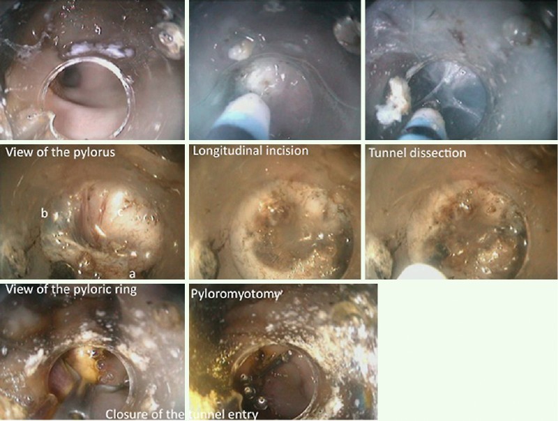
Four Landrace mini-pigs (three castrated males and one female) with an average weight of 25.5 kg (22 – 29 kg) were used in this study. The study was approved by the Institutional Animal Care and Use Committee. All pigs underwent a first gastric emptying scintigraphy under light sedation. Two days later, POP was performed on each pig (Fig. 1). For organisational reasons, a second gastric emptying was carried out 5 days after the endoscopic procedure to evaluate its impact on gastric emptying. Finally, the pigs were euthanised after 10 days of follow-up to allow pathological assessment of the pylorus. Duration of the follow-up was determined according to economic purposes.
Fig. 1.

Peroral pyloromyotomy. a, pyloric ring; b, submucosa of the bulb; c, mucosa of the bulb.
Gastric emptying scintigraphy
The pigs were fasted for 24 hours and then lightly sedated using intramuscular ketamine (6 g). A laryngoscope was used to establish a nasogastric feeding tube to administer a meal consisting of one scrambled egg mixed with 68 MBq Tc99-m, 125 g of pellet food and 80 mL of water. Before the scintigraphy was started, the pigs were lightly anaesthetised with intravenous propofol at a low dose (5 mg/kg/h) to avoid an impact of the drug on gastric emptying. The pigs were then placed in a natural position in front of the camera (Axis Model, Philips Medical). Data were acquired for 90 min. The half emptying time (T½) is expressed in minutes.
Endoscopic procedure
Two days after scintigraphy, POP was performed as described previously 11. All procedures were carried out with the animals under general anaesthesia using intravenous propofol (200 mg/h) after premedication with ketamine (6 g) and endotracheal intubation. A gastroscope (Karl Stortz 13821PKS, Karl Stortz Endoscopy, Tuttlingen, Germany) with a distal cap attached (conical cap DH-28GR, Fujifilm, Japan) was used to define a mucosotomy site 4 cm above the pylorus, on the anterior wall of the great curvature. After the submucosal injection of a saline solution containing indigo carmine, a longitudinal mucosal incision was made using a T-type HybridKnife (ERBE Medical, Tübingen, Germany), assisted by air insufflation and using the Endocut-mode (cut duration: 2; effect: 2; cut interval: 2) via an electrosurgical generator (VIO 300D; ERBE Medical, Germany). Submucosal tunnel dissection using the SWIFT COAG setting (50 W, effect: 4) revealed the pylorus. After identification of the pyloric muscle, its circumferential muscular fibres were progressively cut using the HybridKnife or a HookKnife (Olympus, Tokyo, Japan), until cutting was considered to be complete. Myotomy was considered endoscopically complete when the full white pyloric ring was cut with visualisation of the muscle circular layer of the duodenum.
Finally, the tunnel entry was closed using haemoclips (Boston Resolution, Boston Scientific, Natick, Massachusetts, United States). In the only case of perforation, antibiotics were injected intraperitoneally during surgery and then again 4 hours later.
Statistical analysis
A descriptive statistical analysis of the data using means ± SD was employed. Comparison of linked samples was performed using the Student test for paired data. Fisher’s exact test was used to compare qualitative data. A P value less than 0.05 was considered to indicate statistical significance.
Results (Table 1)
Table 1. Results for peroral endoscopic pylorotomy and gastric emptying.
| Pig 1 | Pig 2 | Pig 3 | Pig 4 | Mean | |
| Procedure duration, min | 55 | 50 | 55 | 60 | 55 (± 4.08 SD) |
| Complications, n | |||||
| Perforation | 1 | 0 | 0 | 0 | 25 % |
| Bleeding | 0 | 0 | 0 | 0 | 0 % |
| Anticipated sacrifice | 0 | 0 | 0 | 0 | 0 % |
| T½ gastric emptying, min | |||||
| Before POP | 301.89 | 89 | 180 | 183 | 188.47 (± 87.3 SD) |
| After POP | 125 | 38 | 85 | 90 | 84.5 (± 35.74 SD) P = 0.03 (CI 95 %: 20.15 – 187.8) |
| Technical difficulty | Loop + + + | Loop + | Loop + + | Loop + + | |
| Feasibility, n | 1 | 1 | 1 | 1 | 100 % |
| Pathological analysis | |||||
| % section of pyloric muscle, % | > 90 | > 90 | > 90 | > 90 | > 90 |
POP, peroral endoscopic pylorotomy.
Feasibility
Endoscopic pyloromyotomy was technically possible in all four pigs (feasibility: 100 %). The pylorus muscle and its thick white fibres were readily identified in each pig (Table 1).
The average duration of the pyloromyotomy was 55 (± 4 SD) minutes. No bleeding occurred during or after the procedure. A perforation occurred in the first pig during the myotomy phase, but the endoscopic procedure was completed successfully. The perforation was treated with intraperitoneal injection of delayed action amoxicillin (25 mg/kg) administered immediately, as defined in the study protocol. There were no fatalities during the procedure or during the 10 days of follow-up.
The mean weight of the animals was 25.5 kg before and 24 kg after the POP. During follow-up, no sign of suffering or a change in behaviour was noted.
Gastric emptying
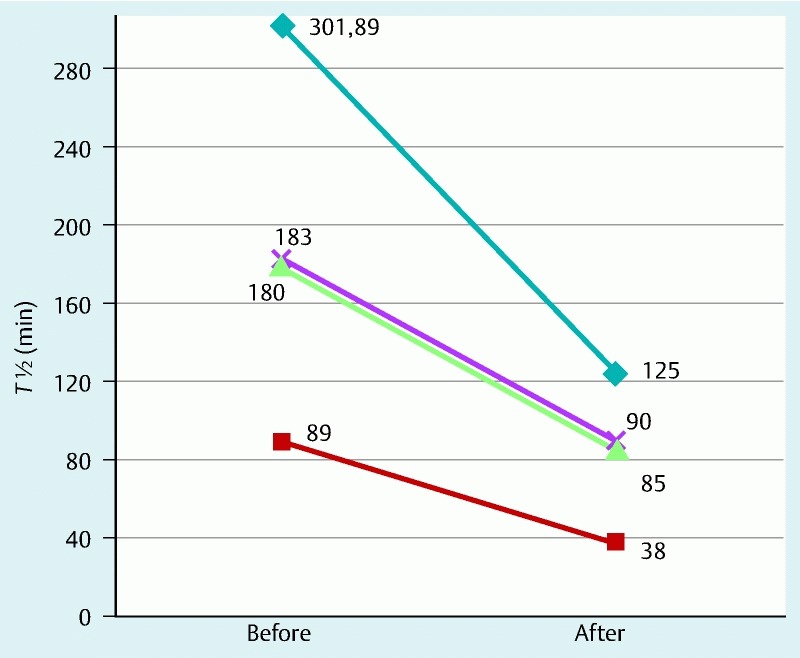
The mean duration of gastric emptying (T½) in the four pigs before POP was 188.47 minutes (± 87.03 SD). After POP, the T½ was 2.2-fold shorter, with an average duration of 84.5 minutes (± 35.74 SD). This difference was statistically significant (P = 0.029). The duration of gastric emptying in each of the four pigs before and after POP is shown in Fig. 2.
Fig. 2.

Individual results for gastric emptying scintigraphy showing T½ values before and after peroral endoscopic pylorotomy (POP).
Histology
Necropsy confirmed the only perforation that developed during endoscopy as a pneumoperitoneum without peritonitis.
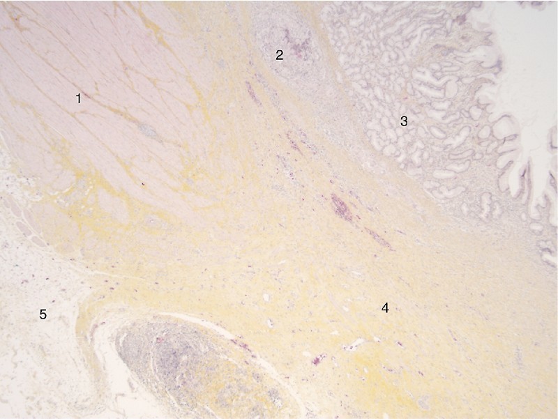
In agreement with the endoscopic observations, sectioning of the pyloric muscle in each pig was histologically complete (> 90 % of the length and thickness of the muscle). An example of pathologic assessment of the pylorus is shown in Fig. 3.
Fig. 3.

Pathologic assessment of a complete section of the pylorus (horizontal axis and haematoxylin and eosin (H&E) staining): 1, normal gastric muscularis propria; 2, granuloma replacing the submucosa (tunnel dissection); 3, normal gastric mucosa; 4, fibrosis replacing the pyloric muscle (area of the pyloromyotomy); 5, serosa and subserosa.
Discussion
Peroral endoscopic pyloromyotomy is a new functional technique that evolved from endoscopic submucosal dissection (ESD) and POEM. In Europe, the anaesthetised pig is considered to be a valid training model and recommended for ESD learning. Our study demonstrates that the same model can be adapted for POP learning and development. The endoscopists who participated in this study had no previous experience with either POP or POEM, but each had already conducted 60 ESDs in animal models and 30 ESDs in human patients. Thus, even among these inexperienced operators, the feasibility of POP was 100 %. The duration of the procedure was typically 55 minutes. Because the anatomy and histology of pigs and humans are similar, this porcine model not only allows training in this technique but also familiarises physicians with the muscular fibrous section of the pylorus. The success of this learning approach was demonstrated by the ability of operators with experience only in ESD to identify the muscular fibres easily during each procedure. This was confirmed by histologic analysis, which verified the completeness of the myotomy in all four pigs.
These encouraging results on animals recommend this model for surgeons who plan to operate on humans. In fact, endoscopy is more difficult in pigs than in humans because of differences in their gastric anatomy. The porcine stomach is U-shaped, resulting in higher pressure on the gastroscope, and it has larger gastric loops. Seen in retrovision, the pylorus is positioned next to the cardia, which renders POP more difficult. The J-shaped stomach of humans should facilitate the procedure.
The safety of POP was indicated by the occurrence of a perforation in only one pig, which was uncomplicated. The perforation occurred during endoscopy and was inconsequential because of the establishment of a submucosal dissection tunnel, which allowed the perforation site to be isolated from the gastric secretions. However, this complication was due to the many gastric loops in the U-shaped porcine stomach. In humans, the systematic use of CO2 insufflation and the J-shape of the stomach would minimise the risk of perforation during the procedure.
There is currently no animal model validated for the evaluation of endoscopic or surgical procedures aimed to treat gastroparesis. The simplicity of our study protocol, combined with the availability of a gastric emptying study, highlight the feasibility of using mini-pigs as an animal model. Our study therefore provides a proof of concept of the beneficial effect of POP on gastric emptying, even in healthy pigs. Moreover, it is the first study to use scintigraphy for thorough evaluation of the procedure.
In 2011, in the first animal study on POP, Kawai et al. 12 used manometry to assess its effects on pyloric pressure, but they were unable to evaluate the impact on gastric emptying. We determined that the T½ of gastric emptying in healthy pigs was accelerated more than twofold after POP, confirming the efficiency of the procedure.
The variation of gastric emptying duration between each pig before and after POP could be compared with healthy humans. Indeed a wide inter-individual variation has been described in pivotal studies. But, as it is the first study using gastric emptying scintigraphy in pigs, there is no reference concerning these data 13.
To better characterise and understand the mechanism underlying the efficacy of POP, we are currently working with a porcine model of gastroparesis after surgical vagotomy. By examining gastric emptying under these conditions, we will be able to assess the efficiency not only of POP but also of other endoscopic/surgical procedures.
Pylorospasm is a common finding in patients with gastroparesis 7; thus, POP may be the treatment of choice for refractory gastroparesis.
POP could also represent an alternative procedure in hypertrophic pyloric stenosis if suppliers are able to miniaturize their devices for use in infants (operative channel of paediatric gastroscopes: 2 mm).
Conclusion
A twofold decrease in T½ of gastric emptying was achieved after POP, as verified by gastric emptying scintigraphy. The efficacy of the procedure provides indirect proof of the involvement of the pyloric ring in delayed gastric emptying and suggests new therapies for patients with gastroparesis.
Our protocol combining gastric emptying scintigraphy and POP validated the use of anaesthetised mini-pigs as a learning and training model for POP or other endoscopic/surgical procedures related to gastric emptying. Pathologic confirmation of pyloric sectioning, the low complication rate, and the absence of mortality demonstrate the feasibility and safety of POP, even when performed by physicians without experience in POEM.
A prospective study on gastroparetic pigs after surgical vagotomy is ongoing in parallel with prospective work on patients with refractory gastroparesis and a significantly impaired quality of life.
Footnotes
Competing interests: None
References
- 1.Camilleri M, Parkman H P, Shafi M A. et al. American College of Gastroenterology. Clinical guideline: management of gastroparesis. Am J Gastroenterol. 2013;108:18–37; quiz 38. doi: 10.1038/ajg.2012.373. [DOI] [PMC free article] [PubMed] [Google Scholar]
- 2.Jung H-K, Choung R S, Locke G R. et al. The incidence, prevalence, and outcomes of patients with gastroparesis in Olmsted County, Minnesota, from 1996 to 2006. Gastroenterology. 2009;136:1225–1233. doi: 10.1053/j.gastro.2008.12.047. [DOI] [PMC free article] [PubMed] [Google Scholar]
- 3.de la Loge C, Trudeau E, Marquis P. et al. Cross-cultural development and validation of a patient self-administered questionnaire to assess quality of life in upper gastrointestinal disorders: the PAGI-QOL. Qual Life Res. 2004;13:1751–1762. doi: 10.1007/s11136-004-8751-3. [DOI] [PubMed] [Google Scholar]
- 4.Szarka L A, Camilleri M. Gastric emptying. Clin Gastroenterol Hepatol. 2009;7:823–827. doi: 10.1016/j.cgh.2009.04.011. [DOI] [PubMed] [Google Scholar]
- 5.Hasler W L. Gastroparesis. Curr Opin Gastroenterol. 2012;28:621–628. doi: 10.1097/MOG.0b013e328358d619. [DOI] [PubMed] [Google Scholar]
- 6.Nguyen L A, Snape W J. Clinical presentation and pathophysiology of gastroparesis. Gastroenterol Clin North Am. 2015;44:21–30. doi: 10.1016/j.gtc.2014.11.003. [DOI] [PubMed] [Google Scholar]
- 7.Gourcerol G, Ducrotté P. Editorial: impaired fasting pyloric compliance in gastroparesis and the benefits of therapeutic pyloric dilatation – authors’ reply. Aliment Pharmacol Ther. 2015;41:908. doi: 10.1111/apt.13157. [DOI] [PubMed] [Google Scholar]
- 8.Sarosiek I, Davis B, Eichler E. et al. Surgical approaches to treatment of gastroparesis: gastric electrical stimulation, pyloroplasty, total gastrectomy and enteral feeding tubes. Gastroenterol Clin North Am. 2015;44:151–167. doi: 10.1016/j.gtc.2014.11.012. [DOI] [PubMed] [Google Scholar]
- 9.Youn Y H, Minami H, Chiu P WY. et al. Peroral endoscopic myotomy for treating achalasia and esophageal motility disorders. J Neurogastroenterol Motil. 2016;22:14–24. doi: 10.5056/jnm15191. [DOI] [PMC free article] [PubMed] [Google Scholar]
- 10.Khashab M A, Stein E, Clarke J O. et al. Gastric peroral endoscopic myotomy for refractory gastroparesis: first human endoscopic pyloromyotomy (with video) Gastrointest Endosc. 2013;78:764–768. doi: 10.1016/j.gie.2013.07.019. [DOI] [PubMed] [Google Scholar]
- 11.Shlomovitz E, Pescarus R, Cassera M A. et al. Early human experience with per-oral endoscopic pyloromyotomy (POP) Surg Endosc. 2015;29:543–551. doi: 10.1007/s00464-014-3720-6. [DOI] [PubMed] [Google Scholar]
- 12.Kawai M, Peretta S, Burckhardt O. et al. Endoscopic pyloromyotomy: a new concept of minimally invasive surgery for pyloric stenosis. Endoscopy. 2012;44:169–173. doi: 10.1055/s-0031-1291475. [DOI] [PubMed] [Google Scholar]
- 13.Tougas G, Eaker E Y, Abell T L. et al. Assessment of gastric emptying using a low fat meal: establishment of international control values. Am J Gastroenterol. 2000;95:1456–1462. doi: 10.1111/j.1572-0241.2000.02076.x. [DOI] [PubMed] [Google Scholar]


