. 2016 Aug 23;5:e13424. doi: 10.7554/eLife.13424

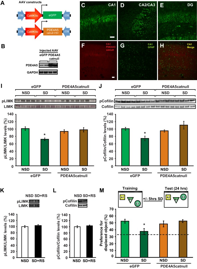
Figure 5. Expression of catalytically inactive PDE4A5 in hippocampal neurons prevents memory deficits and alterations in the cAMP-PKA-LIMK-cofilin signaling pathway associated with sleep deprivation.

(A) Mice were injected with pAAV9-CaMKIIα0.4-eGFP or pAAV9-CaMKIIα0.4-PDE4A5catnull-VSV into the hippocampus to drive neuronal expression of eGFP or catalytically inactive full-length PDE4A5 (PDE4A5catnull). (B) Robust PDE4A5catnull expression was observed at the expected molecular weight, 108 kDa, in hippocampal lysates. (CE) PDE4A5catnullexpression was observed in all 3 subregions of the hippocampus. (FH) PDE4A5catnullwas not expressed in astrocytes reflected by a lack of co-labeling between PDE4A5catnull and GFAP expression. (I) Sleep deprivation causes a reduction in LIMK serine 596 phosphorylation in the hippocampus that is prevented by PDE4A5catnull expression (n = 7–8; two-way ANOVA, effect of virus F1,27 = 3.299, p=0.08; effect of sleep deprivation F1,27 = 6.124, p=0.02; interaction effect F1,27 = 11.336, p=0.002; eGFP SD group versus other groups p<0.05). (J) Sleep deprivation causes a reduction in cofilin phosphorylation in the hippocampus that is prevented by PDE4A5catnull expression (n = 9–10; two-way ANOVA, effect of virus F1,35 = 4.122, p=0.05; effect of sleep deprivation F1,35 = 2.885, p=0.1; interaction effect F1,35 = 9.416, p=0.004; eGFP SD group versus other groups p<0.05). (K, L) Three hours of recovery sleep after five hours of sleep deprivation restores hippocampal LIMK phosphorylation at serine 596 and cofilin phosphorylation at serine 3 to those observed in non-sleep deprived controls (p>0.45 for both comparisons). (M) Mice expressing eGFP or PDE4A5catnull were trained in the hippocampus-dependent object-place recognition task and immediately sleep deprived for 5 hr after training (SD) or left undisturbed (NSD). Hippocampal PDE4A5catnull expression prevents memory deficits caused by sleep deprivation (n = 8–10; two-way ANOVA, effect of virus F1,33 = 2.626, p=0.115; effect of sleep deprivation F1,33 = 2.311, p=0.138; interaction effect F1,33 = 7.485, p=0.01; posthoc Dunnet test eGFP SD group versus other groups p<0.05). In all blots, each lane represents one individual animal. NSD: non-sleep deprived, SD: sleep deprived, SD+RS: sleep deprived plus recovery sleep. Scale bar, 100 µm. Values represent the mean ± SEM. *p<0.05 by posthoc Dunnet’s posthoc test. See also Figure 5—figure supplement 1.

DOI: http://dx.doi.org/10.7554/eLife.13424.018

Figure 5—source data 1. Recovery sleep following sleep deprivation restores LIMK and cofilin phosphorylation levels in the hippocampus, and expression of an inactive version of PDE4A5 in hippocampal neurons prevents memory deficits associated with sleep deprivation.
(K) The data source file contains the relative optical density values (in arbitrary units) of the pLIMK and LIMK western blots for each individual animal of both the non-sleep deprived control group (NSD) and the group that underwent 5 hr of sleep deprivation followed by 3 hr of recovery sleep (SD + RS). (L) The data source file contains the relative optical density values (in arbitrary units) of the pcofilin and cofilin western blots for each individual animal of both the non-sleep deprived control group (NSD) and the group that underwent 5 hr of sleep deprivation followed by 3 hr of recovery sleep (SD + RS). (M) The data source file contains the object exploration times for the displaced (DO) and non-displaced objects (NDO1, NDO2) for each individual animal of each group.
DOI: 10.7554/eLife.13424.019

Figure 5.

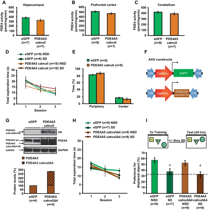
Figure 5—figure supplement 1. Expression of catalytically null PDE4A5 in the hippocampus: Catalytically inactive PDE4A5 without the unique N-terminal localization domain fails to prevent memory deficits associated with sleep loss.

Figure 5—figure supplement 1.

(A) PDE4A5catnull expression in hippocampal neurons did not significantly affect PDE4 activity in the hippocampus (n = 7, Student’s t test p=0.097). (B) PDE4 activity in the prefrontal cortex was not altered by expression of the catalytically inactive PDE4A5catnull in the hippocampus (n = 7–8, Student’s t test p=0.162). (C) PDE4 activity in the cerebellum was not changed by expression of the catalytically inactive PDE4A5catnull in the hippocampus (n = 7–8, Student’s t test p=0.293). (D) Expression of the catalytically inactive PDE4A5catnull in hippocampal neurons did not alter the total time spent exploring objects during training in the object-place recognition task (n = 8–10, two-way ANOVA, effect of virus F1,33 = 0.043, p=0.873). All groups show a decrease in the total object exploration time during consecutive training sessions (two-way ANOVA effect of session F2,66 = 32.777, p=0.0001). Mice expressing PDE4A5catnull had a slightly but non-significantly lower object exploration time during the first training session, and a slightly but non-significantly higher object exploration time during the last training session (interaction effect F2,66 = 4.875, p=0.011, one way ANOVAs per session, p>0.05). (E) Mice expressing PDE4A5catnull spend a similar time in the periphery of the open field as mice expressing eGFP in hippocampal neurons (n = 8, Student’s t test, p=0.292). (F) Mice were injected with pAAV9-CaMKIIα0.4-eGFP or pAAV9-CaMKIIα0.4-PDE4A5catnullΔ4-VSV into the hippocampus to drive neuronal expression of eGFP or catalytically inactive full-length PDE4A5 which lacked the N-terminal domain unique for PDE4A5 (PDE4A5catnullΔ4). A VSV-tag was included to discriminate between endogenous PDE4A5 and the truncated PDE4A5catnullΔ4. (G) PDE4A5catnullΔ4 protein levels in the hippocampus 4 weeks after viral injection. A sample blot probed with an isoform-nonspecific PDE4A antibody revealed the presence of both wild-type PDE4A5 protein and truncated PDE4A5catnullΔ4 protein. Probing the blot with an antibody for the HA-tag confirmed that the truncated protein is indeed the N-terminal lacking catalytically inactive PDE4A5catnullΔ4. Each band represents an individual animal (H) Expression of the catalytically inactive PDE4A5catnullΔ4 lacking the N-terminal domain in hippocampal neurons did not affect total object exploration time during training in the object-place recognition task (n = 7–9, two-way ANOVA effect of virus F1,29 = 0.470, p=0.498). All groups show decreased total object exploration times during consecutive training sessions (two-way ANOVA effect of session F2,58 = 13.597, p=0.0001; interaction effect F2,58 = 0.555, p=0.557). (I) Mice expressing eGFP or the N-terminal domain lacking inactive form of PDE4A5catnullΔ4 were trained in the hippocampus-dependent object-place recognition task. Sleep deprivation causes memory deficits in both eGFP and PDE4A5catnullΔ4 mice (n = 7–9; two-way ANOVA effect of sleep deprivation F1,29 = 18.131, p=0.0001; effect of virus F1,29 = 1.064, p=0.311; interaction effect F1,29 = 0.001, p=0.986; eGFP NSD versus EGFP SD, posthoc Tukey’ t test p=0.0054; PDE4A5catnullΔ4 NSD versus PDE4A5catnullΔ4 SD, posthoc Tukey’ t test p=0.0037). Dotted line indicates chance level performance. NSD: non-sleep deprived, SD: sleep deprived. Values represent the mean ± SEM. #p<0.01 by Tukeys t test.
Figure 5—figure supplement 1—source data 1. Exploratory activity in mice expressing catalytically inactive PDE4A5 or PDE4A5Δ4 in hippocampal excitatory neurons.
(D) The data source file contains the total object exploration times during the three training sessions for each individual animal of all four groups. (E) The data source file contains the time spent in the periphery and center of the open field for each individual animal of both groups. (H) The data source file contains the total object exploration times during the three training sessions for each individual animal of all four groups. (I) The data source file contains the object exploration times for the displaced (DO) and non-displaced objects (NDO1, NDO2) for each individual animal of each group.
DOI: 10.7554/eLife.13424.021