

Abstract
Intralobar pulmonary sequestration is a rare malformation that predisposes to recurrent respiratory infections. It is difficult to diagnose unless a more extensive directed investigation (to the vasculature and pulmonary parenchyma) is take on. Failure to diagnose and treat this condition can lead to recurrent pneumonia and fatal hemoptysis. Most cases are diagnosed before the age of 20 years. In this report, we present an extremely rare case of elderly woman with initial diagnosis of intralobar sequestration, and to our knowledge, this case represents the oldest diagnosed patient in the literature.
Keywords: Intralobar, Lung, Sequestration
Introduction
Pulmonary sequestration is a relatively rare entity comprising 0.15%-6.4% of all congenital pulmonary malformations and defined as an area of dysplastic and nonfunctioning pulmonary tissue that is not in normal continuity with the tracheobronchial tree and that derives its blood supply from systemic vessels [1], [2]. It has been classically described in 2 forms—intralobar sequestration (ILS) located within the visceral pleura and surrounded by normal lung, and extralobar sequestration which have a separate pleural covering. Both types are supplied with blood from the aorta or its branches. The venous return of the ILS is usually via the pulmonary veins, whereas extralobar sequestrations generally have systemic venous drainage.
ILS accounts for 75% of pulmonary sequestrations [3]. Half of the patients with ILS are diagnosed before the age of 20 years with symptoms of recurrent pulmonary infection or cardiac disease, whereas a small portion of patients are asymptomatic and diagnosed incidentally [4]. It is rarely diagnosed after 40 years of age and a delay of up to 7 years between the onset of symptoms and the diagnosis was reported [5]. Only few published cases in the literature report the initial diagnosis of this condition in patients over 50 years [6].
At pathologic examination, ILS is characterized by inflammation and fibrosis. At radiologic examination, ILS typically appears as a consolidation or mass, with or without cavitation, within a lower lobe. In many cases, cystic change may be present within the affected lobe. Identification of a systemic arterial supply supports the diagnosis. Patients are treated with surgical excision, and prognosis is favorable.
Traditionally, the diagnosis of pulmonary sequestration requires arteriography to identify abnormal systemic vessels feeding the abnormal portion of the lung. More recently, other procedures have been advocated as a less invasive means of identifying the anomalous artery. Contrast-enhanced (CE) multidetector-row computed tomography (MDCT) and CE magnetic resonance (MR) images with multiplanar and three-dimensional (3D) reconstructions improve the definition of the arterial and venous vasculature of the malformation, without the need for further invasive diagnostic procedures.
In this report, we describe the use of CE-MDCT to image the aberrant systemic artery in a rare case of ILS in an elderly woman.
Case report
A 80-year-old nonsmoker woman with a history of pulmonary infection was referred to our department for imaging. The medical history included many hospitalizations for pneumonia. Now, she presented with persistent dry cough and fever for previous 2 weeks despite antibiotic treatment. Laboratory results revealed mild leukocytosis. Chest radiograph was done, but it was unremarkable apart from mild infiltration at the left lung base (Fig. 1). CE-MDCT was performed (Fig. 2) and revealed an irregular margins nonhomogenous pulmonary mass, 2 cm in diameter, with tiny central cystic appearing air space involving the medial basal segment of the left lower lobe and adjacent to this, particularly in the basal segments, there was a fluid-density areas with multiple dispersed tiny consolidating areas, suggesting ongoing pneumonic process. A 3D-multiplanar reconstructions (MPR) obtained from CE-MDCT images depicted a systemic arterial supply to the lesion. The aberrant artery measuring 1 cm in diameter and originated from the lower thoracic descending aorta. A venous vessel draining from the lesion into the left lower pulmonary vein was visualized. Hence, a diagnosis of intralobar pulmonary sequestration was made. Surgery was not considered at the time this case was presented, and the patient discharged for follow-up only.
Fig. 1.
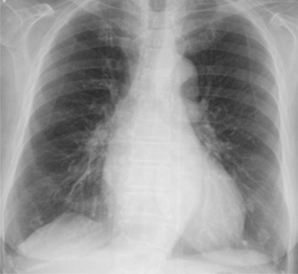
Chest radiograph of 80-year-old woman show subtle infiltrate at the lower zone of left lung.
Fig. 2.

(A) Axial CE CT scan through the lung bases showing the pulmonary lesion (white arrow) in the medial part of the left lower lobe supplied by an anomalous systemic artery (red arrow). (B) Axial lung window CT images through the lung bases showing the pulmonary lesion (black arrow) with adjacent inflammatory consolidation (red arrow). (C) Coronal CT MPR showing the origin of systemic vessel (white arrows) from the lower thoracic descending aorta.
Discussion
Intralobar pulmonary sequestration is a relatively rare congenital anomaly with few reports of initial diagnosis occurring during adulthood. Patients can present with an incidental pulmonary lesion on imaging and be otherwise asymptomatic. More commonly, however, they may manifest with cough, fever, recurrent pneumonia, and chest pain. Few reports have mentioned more severe sequelae such as overlying aspergillosis and even fatal hemoptysis [7], [8], [9]. For these reasons, sequestration has traditionally been treated by definitive resection of the affected lung segment.
Most of the ILSs are located in the medial and posterior basal segments of the left lung. Overall, 98% occur in the lower lobes [1]. Bilateral involvement is uncommon.
In ILS, sequestrations occur within pulmonary visceral pleurae and do not communicate with the bronchial tree. ILS is seen in males and females in equal numbers. The lesions of ILS may be solid, fluid, or hemorrhagic or may contain mucus. Cystic or emphysematous elements may be present, and adjacent atelectasis often exists.
Most lesions appear hypervascular because of abundant systemic vascularization. Super-added infection may lead to some consolidation in adjacent segments, and a chronic inflammatory process may induce localized reactive neovascularization. Mucoid impaction of a bronchus surrounded by a hyperinflated lung is believed to be characteristic of ILS. Associated congenital anomalies are uncommon in ILS.
In ILS, anomalous systemic arterial supply is via the descending thoracic aorta (72%), as seen in our case, via abdominal aorta, celiac axis, or splenic artery (21%), via intercostal artery (3%), and rarely via the subclavian, internal thoracic, and pericardiacophrenic arteries. Venous drainage is usually via the pulmonary veins, but it can also occur through the azygos vein and/or hemiazygos system, portal vein, right atrium, or inferior vena cava.
Achieving a diagnosis of pulmonary sequestration can range in difficulty depending on the type of anomaly and presenting symptoms. CT will typically suffice in most adult cases with some debate still held over the need for angiography [10]. In our experience, CE-MDCT with MPR was clearly sufficient to make the diagnosis plus delineate the anatomic features notable for operative planning.
Plain radiographs of the chest often show a single homogeneous opacity or, less commonly, a cystic mass in the base of one lung that can sometimes suggest the diagnosis of sequestration. [2] Less specific findings include recurrent pneumonia and focal bronchiectatic changes. The principal objective for diagnosis of pulmonary sequestration is to identify the systemic artery supply. With this information, imaging can distinguish sequestration from other causes of lung opacity. Because accessory arteries, pleural investment, and venous drainage are adequately determined intraoperatively, at some institutions only the presence and location of an aberrant systemic artery are considered essential for preoperative assessment for any symptomatic pulmonary sequestration [11].
Imaging strategies for suspected pulmonary sequestration are based on case reports or small series because it is a rare congenital disorder and no study exists that objectively compares imaging techniques for detection, definition, or cost effectiveness [11]. Since the definitive step in the diagnosis of sequestration is the demonstration of the systemic arterial supply, for a long time, diagnosis was made by conventional angiography. More recently, all imaging techniques capable of showing the artery have been implicated in evaluating sequestration. MR imaging and MR angiography can be used together to diagnose pulmonary sequestration in a single noninvasive examination [12]. Nevertheless, MR cannot accurately evaluate lung parenchyma and the airways and must be considered in terms of cost and availability. Other noninvasive techniques for evaluation of sequestration such as scintigraphy are only rarely necessary.
In our case described in this report, CE-MDCT successfully delineated the origin and course of the anomalous systemic artery. Axial images were enough to make the diagnosis but 3D-MPR aided both radiologists and referring clinicians by demonstrating anatomic relationships, particularly for vessels orientated in the z-axis [13]. On the other hand, venous drainage into the lower pulmonary veins was also identified in this case. We have performed 3D-MPR for a better understanding of the anatomy of the abnormal systemic arteries.
In summary, we report a rare case of ILS in elderly woman successfully diagnosed using CE-MDCT. By allowing simultaneous imaging of anomalous vessels and parenchymal lesions in a single examination, CE-MDCT is a particularly efficacious technique and has the potential to become the procedure of choice in the diagnosis and assessment of pulmonary sequestration.
Footnotes
Competing Interests: The authors have declared that no competing interests exist.
References
- 1.Savic B., Birtel F.J., Tholen W., Funke H.D., Knoche R. Lung sequestration: report of seven cases and review of 540 published cases. Thorax. 1979;34:96–101. doi: 10.1136/thx.34.1.96. [DOI] [PMC free article] [PubMed] [Google Scholar]
- 2.Felker R.E., Tankin I.L. Imaging of pulmonary sequestration. AJR Am J Roentgenol. 1990;154:241–249. doi: 10.2214/ajr.154.2.2105007. [DOI] [PubMed] [Google Scholar]
- 3.Nicolette L.A., Kosloske A.M., Bartow S.A., Murphy S. Intralobar pulmonary sequestration: a clinical and pathological spectrum. J Pediatr Surg. 1993;6:802–805. doi: 10.1016/0022-3468(93)90331-e. [DOI] [PubMed] [Google Scholar]
- 4.Frazier A.A., Rosado de Christenson M.L., Stocker J.T., Templeton P.A. Intralobar sequestration: radiologic-pathologic correlation. Radiographics. 1997;17:725–745. doi: 10.1148/radiographics.17.3.9153708. [DOI] [PubMed] [Google Scholar]
- 5.Gustafson R.A., Murray G.F., Warden H.E., Hill R.C., Rozar G.E. Intralobar sequestration. A missed diagnosis. Ann Thorac Surg. 1989;47:841–847. doi: 10.1016/0003-4975(89)90016-7. [DOI] [PubMed] [Google Scholar]
- 6.Petersen G., Martin U., Singhal A., Criner G.J. Intralobar sequestration in the middle-aged and elderly adult: recognition and radiographic evaluation. J Thorac Cardiovasc Surg. 2003;126:2086–2090. doi: 10.1016/s0022-5223(03)01297-2. [DOI] [PubMed] [Google Scholar]
- 7.Morikawa H., Tanaka T., Hamaji M., Ueno Y. A case of aspergillosis associated with intralobar pulmonary sequestration. Asian Cardiovasc Thorac Ann. 2011;19:66–68. doi: 10.1177/0218492310390684. [DOI] [PubMed] [Google Scholar]
- 8.Somja J., De Leval L., Boniver J., Radermecker M.A. Intrapulmonary lung sequestration diagnosed in an adult. Rev Med Liege. 2011;66:7–12. [PubMed] [Google Scholar]
- 9.Rubin E.M., Garcia H., Horowitz M.D., Guerra J.J., Jr. Fatal massive hemoptysis secondary to intralobar sequestration. Chest. 1994;106:954–955. doi: 10.1378/chest.106.3.954. [DOI] [PubMed] [Google Scholar]
- 10.Clements B.S., Warner J.O. Pulmonary sequestration and related congenital bronchopulmonary-vascular malformations: nomenclature and classification based on anatomical and embryological considerations. Thorax. 1987;42:401–408. doi: 10.1136/thx.42.6.401. [DOI] [PMC free article] [PubMed] [Google Scholar]
- 11.Frush D.P., Donnelly L.F. Pulmonary sequestration spectrum: a new spin with helical CT. AJR Am J Roentgenol. 1997;169:679–682. doi: 10.2214/ajr.169.3.9275876. [DOI] [PubMed] [Google Scholar]
- 12.Doyle A.J. Demonstration of blood supply to pulmonary sequestration by MR angiography. AJR Am J Roentgenol. 1992;158:989–990. doi: 10.2214/ajr.158.5.1566704. [DOI] [PubMed] [Google Scholar]
- 13.Johnson P.T., Fishman E.K., Duckwall J.R., Calhoun P.S., Heath D.G. Interactive three-dimensional volume rendering of spiral CT data: current applications in the thorax. Radiographics. 1998;18:165–187. doi: 10.1148/radiographics.18.1.9460115. [DOI] [PubMed] [Google Scholar]