

Abstract
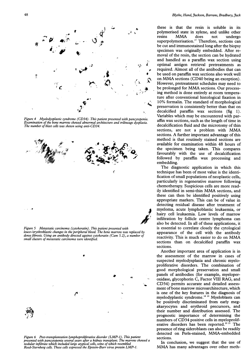
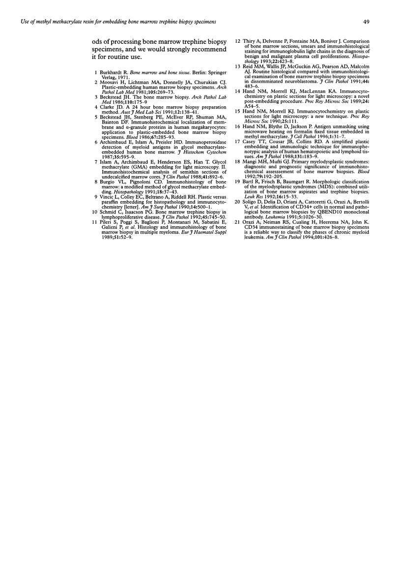
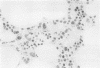
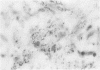
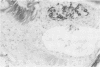
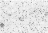
AIMS: To evaluate the use of methyl methacrylate resin as an embedding medium for undecalcified bone marrow trephine biopsy specimens. METHODS: About 2500 undecalcified bone marrow trephine biopsy specimens were processed, and embedded in methyl methacrylate resin. Semithin sections (2-3 microns) were stained by routine tinctorial and immunocytochemical staining methods with a wide range of antibodies using a standard streptavidin biotin horseradish peroxidase technique. Different antigen retrieval pretreatments were evaluated. RESULTS: Bone marrow trephine biopsy specimens are embedded routinely in methyl methacrylate at the Haematological Malignancy Diagnostic Service at The Leeds General Infirmary. Over 50 different primary antibodies are in current use; for the majority of these, microwave antigen retrieval or trypsin digestion, or both, is either essential or greatly enhances the results. CONCLUSIONS: Embedding bone marrow trephine biopsy specimens in methyl methacrylate resin retains morphology and permits reliable, high quality immunocytochemistry. This is particularly desirable for the demonstration of neoplastic cells in regenerative marrow after chemotherapy, and in the detection of residual disease after treatment. The use of methyl methacrylate for routine use on bone marrow trephine biopsy specimens is advocated.
Full text
PDF

Images in this article


Selected References
These references are in PubMed. This may not be the complete list of references from this article.
- Archimbaud E., Islam A., Preisler H. D. Immunoperoxidase detection of myeloid antigens in glycolmethacrylate-embedded human bone marrow. J Histochem Cytochem. 1987 May;35(5):595–599. doi: 10.1177/35.5.2435785. [DOI] [PubMed] [Google Scholar]
- Bartl R., Frisch B., Baumgart R. Morphologic classification of the myelodysplastic syndromes (MDS): combined utilization of bone marrow aspirates and trephine biopsies. Leuk Res. 1992;16(1):15–33. doi: 10.1016/0145-2126(92)90096-p. [DOI] [PubMed] [Google Scholar]
- Beckstead J. H., Stenberg P. E., McEver R. P., Shuman M. A., Bainton D. F. Immunohistochemical localization of membrane and alpha-granule proteins in human megakaryocytes: application to plastic-embedded bone marrow biopsy specimens. Blood. 1986 Feb;67(2):285–293. [PubMed] [Google Scholar]
- Beckstead J. H. The bone marrow biopsy. A diagnostic strategy. Arch Pathol Lab Med. 1986 Mar;110(3):175–179. [PubMed] [Google Scholar]
- Casey T. T., Beckstead J. H. Plastic versus paraffin embedding for histopathology and immunocytochemistry. Am J Surg Pathol. 1990 May;14(5):500–501. doi: 10.1097/00000478-199005000-00013. [DOI] [PubMed] [Google Scholar]
- Casey T. T., Cousar J. B., Collins R. D. A simplified plastic embedding and immunohistologic technique for immunophenotypic analysis of human hematopoietic and lymphoid tissues. Am J Pathol. 1988 May;131(2):183–189. [PMC free article] [PubMed] [Google Scholar]
- Islam A., Archimbaud E., Henderson E. S., Han T. Glycol methacrylate (GMA) embedding for light microscopy. II. Immunohistochemical analysis of semithin sections of undecalcified marrow cores. J Clin Pathol. 1988 Aug;41(8):892–896. doi: 10.1136/jcp.41.8.892. [DOI] [PMC free article] [PubMed] [Google Scholar]
- Mangi M. H., Mufti G. J. Primary myelodysplastic syndromes: diagnostic and prognostic significance of immunohistochemical assessment of bone marrow biopsies. Blood. 1992 Jan 1;79(1):198–205. [PubMed] [Google Scholar]
- Moosavi H., Lichtman M. A., Donnelly J. A., Churukian C. J. Plastic-embedded human marrow biopsy specimens: improved histochemical methods. Arch Pathol Lab Med. 1981 May;105(5):269–273. [PubMed] [Google Scholar]
- Orazi A., Neiman R. S., Cualing H., Heerema N. A., John K. CD34 immunostaining of bone marrow biopsy specimens is a reliable way to classify the phases of chronic myeloid leukemia. Am J Clin Pathol. 1994 Apr;101(4):426–428. doi: 10.1093/ajcp/101.4.426. [DOI] [PubMed] [Google Scholar]
- Reid M. M., Wallis J. P., McGuckin A. G., Pearson A. D., Malcolm A. J. Routine histological compared with immunohistological examination of bone marrow trephine biopsy specimens in disseminated neuroblastoma. J Clin Pathol. 1991 Jun;44(6):483–486. doi: 10.1136/jcp.44.6.483. [DOI] [PMC free article] [PubMed] [Google Scholar]
- Schmid C., Isaacson P. G. Bone marrow trephine biopsy in lymphoproliferative disease. J Clin Pathol. 1992 Sep;45(9):745–750. doi: 10.1136/jcp.45.9.745. [DOI] [PMC free article] [PubMed] [Google Scholar]
- Soligo D., Delia D., Oriani A., Cattoretti G., Orazi A., Bertolli V., Quirici N., Deliliers G. L. Identification of CD34+ cells in normal and pathological bone marrow biopsies by QBEND10 monoclonal antibody. Leukemia. 1991 Dec;5(12):1026–1030. [PubMed] [Google Scholar]
- Thiry A., Delvenne P., Fontaine M. A., Boniver J. Comparison of bone marrow sections, smears and immunohistological staining for immunoglobulin light chains in the diagnosis of benign and malignant plasma cell proliferations. Histopathology. 1993 May;22(5):423–428. doi: 10.1111/j.1365-2559.1993.tb00155.x. [DOI] [PubMed] [Google Scholar]