Abstract
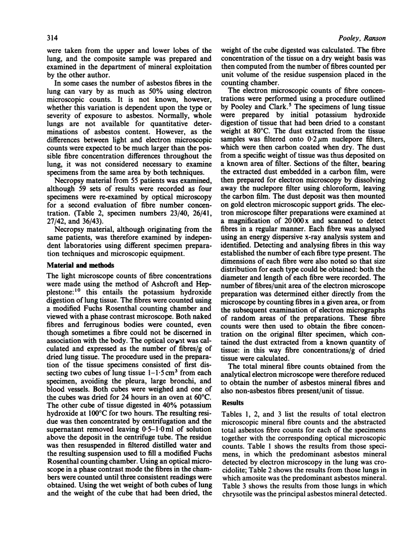
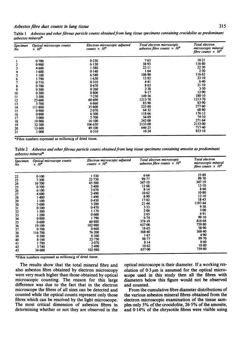
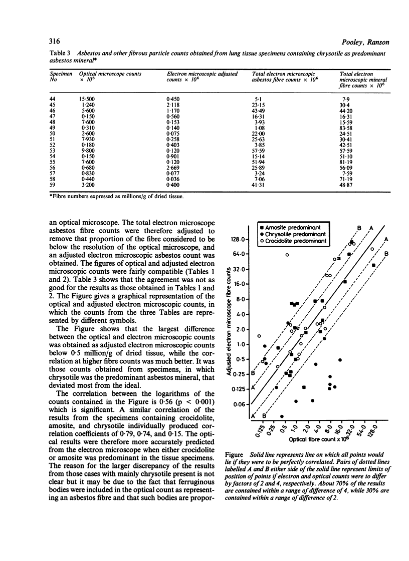
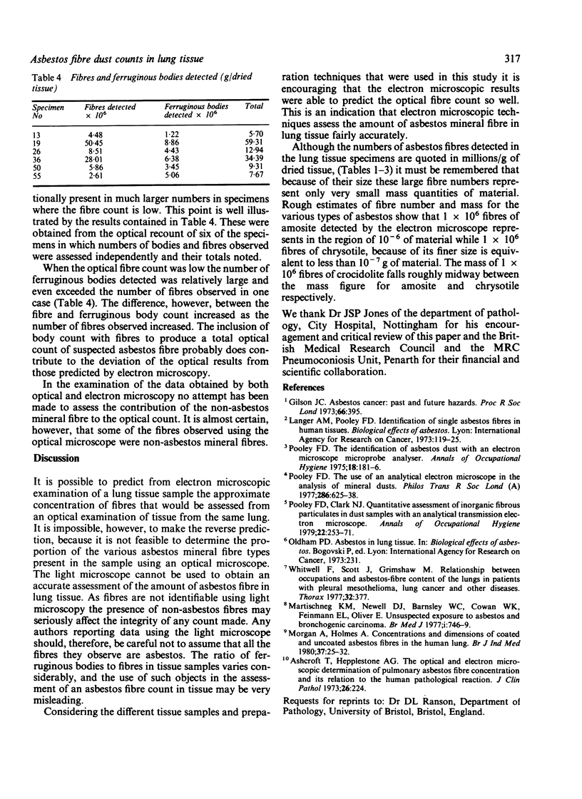
The efficiency of the light microscope with that of the electron microscope in detecting asbestos fibres in human lung tissue was computed. Necropsy material from 55 patients who had died from asbestos related diseases was analysed independently by phase contrast microscopy and electron microscopy. As expected the number of fibres identified using electron microscopy was higher than that identified by light microscopy. By adjusting the electron allow for the limited resolving power of the light microscope, however, a significant correlation of the number of fibres identified using the two methods was obtained. The best correlation was found with specimens containing crocidolite (correlation coefficient 0.79) and amosite (correlation coefficient 0.74), while chrysotile gave a much lower correlation (correlation coefficient 0.15). The cumulated fibre diameter distribution obtained using the electron microscope suggests that the light microscope is able to visualise only 5% of crocidolite, 26.5% of amosite, and 0.14% of chrysotile present in lung tissue. Therefore, although it is possible, using the electron microscope, to predict the asbestos fibre count that would be obtained by light microscopy, the reserve prediction cannot be made: it is impossible to determine the proportion of the various asbestos mineral types using the light microscope.
Full text
PDF
Selected References
These references are in PubMed. This may not be the complete list of references from this article.
- Ashcroft T., Heppleston A. G. The optical and electron microscopic determination of pulmonary asbestos fibre concentration and its relation to the human pathological reaction. J Clin Pathol. 1973 Mar;26(3):224–234. doi: 10.1136/jcp.26.3.224. [DOI] [PMC free article] [PubMed] [Google Scholar]
- Gilson J. C. Asbestos cancer: past and future hazards. Proc R Soc Med. 1973 Apr;66(4):395–403. [PMC free article] [PubMed] [Google Scholar]
- Martischnig K. M., Newell D. J., Barnsley W. C., Cowan W. K., Feinmann E. L., Oliver E. Unsuspected exposure to asbestos and bronchogenic carcinoma. Br Med J. 1977 Mar 19;1(6063):746–749. doi: 10.1136/bmj.1.6063.746. [DOI] [PMC free article] [PubMed] [Google Scholar]
- Morgan A., Holmes A. Concentrations and dimensions of coated and uncoated asbestos fibres in the human lung. Br J Ind Med. 1980 Feb;37(1):25–32. doi: 10.1136/oem.37.1.25. [DOI] [PMC free article] [PubMed] [Google Scholar]
- Pooley F. D., Clark N. J. Quantitative assessment of inorganic fibrous particulates in dust samples with an analytical transmission electron microscope. Ann Occup Hyg. 1979;22(3):253–271. doi: 10.1093/annhyg/22.3.253. [DOI] [PubMed] [Google Scholar]
- Pooley F. D. The identification of asbestos dust with an electron microscope microprobe analyser. Ann Occup Hyg. 1975 Dec;18(3):181–186. doi: 10.1093/annhyg/18.3.181. [DOI] [PubMed] [Google Scholar]
- Whitwell F., Scott J., Grimshaw M. Relationship between occupations and asbestos-fibre content of the lungs in patients with pleural mesothelioma, lung cancer, and other diseases. Thorax. 1977 Aug;32(4):377–386. doi: 10.1136/thx.32.4.377. [DOI] [PMC free article] [PubMed] [Google Scholar]


