Abstract
Tumor angiogenesis plays very important roles for tumorigenesis, tumor development, metastasis, and prognosis. Targeting T1/T2 dual modality magnetic resonance (MR) imaging of the tumor vascular endothelial cells (TVECs) with MR molecular probes can greatly improve diagnostic sensitivity and specificity, as well as helping to make an early diagnosis of tumor at the preclinical stage. In this study, a new T1 and T2 dual modality nanoprobe was successfully fabricated. The prepared nanoprobe comprise peptides CL 1555, poly(ε-caprolactone)-block-poly(ethylene glycol) amphiphilic copolymer shell, and dozens of manganese ferrite (MnFe2O4) nanoparticle core. The results showed that the hydrophobic MnFe2O4 nanoparticles were of uniform spheroidal appearance and narrow size distribution. Due to the self-assembled nanomicelles structure, the prepared probes were of high relaxivity of 281.7 mM−1 s−1, which was much higher than that of MnFe2O4 nanoparticles (67.5 mM 1 s−1). After being grafted with the targeted CD105 peptide CL 1555, the nanomicelles can combine TVECs specifically and make the labeled TVECs dark in T2-weighted MR imaging. With the passage on, the Mn2+ ions were released from MnFe2O4 and the size decreased gradually, making the signal intensity of the second and third passage of labeled TVECs increased in T1-weighted MR imaging. Our results demonstrate that CL-poly(ethylene glycol)-MnFe2O4 can conjugate TVECs and induce dark and bright contrast in MR imaging, and act as a novel molecular probe for T1- and T2-enhanced MR imaging of tumor angiogenesis.
Keywords: CL 1555, CL-PEG-MnFe2O4, TVECs, CD105, tumor angio genesis
Introduction
Malignant tumor has been ranked the second leading cause of human mortality worldwide, accounting for 8.2 million cancer deaths in 2012.1 The lethality of cancer is mainly due to early metastasis and diagnosis at advanced stages. The common routes of metastasis of malignant tumors are hematogenous metastasis, lymphatic metastasis, implantation metastasis, and local infiltration, among which the blood metastases are the most important routes.2,3 In 1971, Folkman reported that the occurrence and development of a tumor were angiogenesis-dependent and tumor angiogenesis accelerated tumor growth and infiltration.4 So, the tumor blood vessels became the focus of cancer research, making vascular endothelial cells the leading targets for cancer research. In recent years, with the discovery of a series of markers of vascular endothelial cells, the study of tumor blood vessels has expanded into the molecular level and the relationship between tumor blood vessels and tumor metastasis was confirmed through these molecular markers.5–8 Endoglin, also called CD105, is a moiety of the transforming growth factor-beta receptor complex and regulates angiogenesis by participating in signal transduction of the transforming growth factor-beta receptor. It has been reported that CD105 is overexpressed in neovascularization of regenerated, inflammatory, and tumor tissues and its expression is positively correlated with the cancer angiogenesis.7–9 Therefore, CD105 is an ideal biomarker for targeting tumor vascular endothelial cells (TVECs).
Magnetic resonance imaging (MRI) is one of the best noninvasive methods used in clinical medicine today because of its superb spatial resolution, soft-tissue contrast resolution, lack of radiation exposure, and multiparameter and multi-sequence imaging.10–12 However, MRI is less sensitive than positron emission tomography and fluorescence imaging, making it not suitable for small lesion monitoring or molecule tracing. The application of magnetic contrast agents (CAs) markedly enhances the sensitivity of MRI.13–15 Usually, MR CAs can be divided into two categories: one is positive CAs, which can mainly reduce longitudinal relaxation (T1) time and induce hyperintensity in T1-weighted imaging; the other is negative CAs which can mainly shorten transverse relaxation (T2) time and provide negative contrast in T2-weighted imaging.16 There have been many types of CAs available for clinical MRI, including gadolinium- or manganese-based chelates as T1 CAs and iron oxide (Fe3O4)-based nanoparticles (NPs) as T2 CAs.17–19 However, both kinds of CAs have limitations. Because of magnetic susceptibility artifacts and their positive or negative contrast effect, the tissues labeled with gadolinium-based complex or Fe3O4 NPs may not be clearly distinguishable from the hyperintensity or low level MR signal arising from adjacent tissues.20,21 Meanwhile, it has been reported that some gadolinium-based CAs can result in nephrogenic systemic fibrosis for patients with severe renal disease.22 Manganese ferrite (MnFe2O4) NPs have been proven to be of higher saturation magnetization and transverse relaxivity (r2) than other ferrite NPs, including cobalt ferrite (CoFe2O4), Fe3O4, and nickel ferrite (NiFe2O4).23–25 Additionally, multiple MnFe2O4 NPs encapsulated inside one nanomicelle (NM) can result in much stronger r2 than a single NP at the same metal concentration, making them excellent candidates for high sensitive T2-enhanced MRI.26,27 The MnFe2O4 NPs within cells will be gradually broken down in lysosomes and then release paramagnetic Mn2+. At the mean time, the size of MnFe2O4 NPs decreased correspondingly. The above two effects can significantly shorten the T1 relaxation time of the surrounding protons and induce hyperintensity in T1-weighted imaging.19,28–31 So, the MnFe2O4 NPs can act as both T1 and T2 CAs in MRI. As a negative CA, MnFe2O4 NPs can cause dark contrast in T2 and T2*-weighted imaging (WI). Upon internalization by cells and localization in lysosomes, released Mn2+ ions and NPs with smaller size can act as strong T1 MRI CAs, making the dark areas in T2/T2*-WI bright in T1-weighted imaging,30,32 to improve the accuracy of diagnosis of MRI greatly.
Bi33 identified a peptide with high binding affinity and selectivity toward CD105 and demonstrated that the selected peptide could act as a CD105 specific ligand. The peptide called CL 1555 (sequence: AHKHVHHVPVRL) was selected from a phage displayed library.34 CL 1555 was condensed with only a dozen of amino acids, resulting in a simpler steric configuration, better diffusion and penetration in vivo, and less immunogenicity compared with anti-CD105 antibody. Because of small steric hindrance, the peptide had a high grafting rate when it was connected to the functional group on the surface of the NPs, which will improve the probe affinity, connective stability, and specificity with the targeted molecule.35 So, CL 1555 is an ideal ligand to CD105 for targeted diagnosis and therapy.
In previous studies, we conjugated anti-CD105 antibody with stabilized immunoliposome (SL)-encapsulated gadolinium-diethylenetriaminepentaacetic acid (Gd-DTPA) and thiol-PEG-carboxyl-stabilized Fe2O3/Au NPs to evaluate tumor angiogenesis on T1- or T2-enhanced MRI, respectively.36,37 The results showed that CD105-Gd-SLs and hybrid-PEG-CD105 could be utilized to detect subcutaneous glioma and breast cancer angiogenesis in tumor-bearing rats. Based on the previous work, this research focuses on synthesizing and characterizing a molecular probe CL-poly(ethylene glycol) (PEG)-MnFe2O4 based on the CD105 specific ligand CL 1555 and MnFe2O4 NMs to detect tumor angiogenesis using T1- and T2-enhanced MRI.
Materials and methods
Materials
Iron acetylacetonate, manganese acetylacetonate, oleic acid, oleylamine, benzyl ether, hexane, ε-caprolactone, tetrahydrofuran (THF), N-(3-dimethylaminopropyl)-Nese acetylacetonate, N-hydroxysuccinimide, stannous octoate, tetramethylammonium hydroxide (TMAH), heparin, L-glutamine, reactive oxygen species (ROS) assay kit, and sodium pyruvate were all purchased from Sigma-Aldrich Co. (St Louis, MO, USA). PEG with a terminal hydroxyl and carboxylic acid functional groups (COOH-PEG-OH) was purchased from JenKem Technology (Beijing, People’s Republic of China). 1,2-Hexadecanediol was obtained from TCI (Shanghai) Development Co., Ltd (Shanghai, People’s Republic of China). Medium 199, Roswell Park Memorial Institute 1640 medium, fetal bovine serum, penicillin/streptomycin, and nonessential amino acid were purchased from Hyclone, Logan, UT, USA. Count Kit-8 (CCK-8) was obtained from Beyotime Biotechnology Company, Beijing, People’s Republic of China. Endothelial cell growth supplement was purchased from Sciencell, CA, Carlsbad, USA. Six-well plates were obtained from Corning Incorporated, Corning, NY, USA.
Synthesis of MnFe2O4 NPs
According to a published procedure,38 iron acetylacetonate (2 mmol), manganese acetylacetonate (1 mmol), 1,2-hexadecanediol (10 mmol), oleic acid (6 mmol), and oleylamine (6 mmol) were mixed in 20 mL benzyl ether under dry and deoxidized argon atmosphere. Then, the mixture was successively heated to 200°C for 2 hours and refluxed at 300°C for 1 hour. After cooling to room temperature, the mixture was treated with ethanol and then centrifuged several times. Finally, the product was dispersed in anhydrous hexane for storage. In order to transfer the prepared NPs into water, hydrophobic MnFe2O4 NPs were treated with TMAH.39 Briefly, 10 mL of NPs suspension in hexane was added into 20 mL of ethanol and the NPs were collected with a permanent magnet. After decanting the solvent, the NPs were redispersed in 25 mL of TMAH aqueous solution (10%, w/v) and sonicated for 10 minutes. After centrifugation, the hydrophobic surfactant (oleic acid/oleylamine) on the surface of NPs was replaced with TMAH, making them bear negative surface charges and be stable in water. Finally, the surface ligand-exchanged NPs (TMAH-MnFe2O4) were suspended in 10 mL of deionized water.
Synthesis of CL-PEG-MnFe2O4
Amphiphilic block copolymer poly(ε-caprolactone)-block-PEG-COOH (PCL-b-PEG-COOH) was synthesized using ε-caprolactone, COOH-PEG-OH, and stannous octoate by ring opening polymerization.40 The resulting copolymers were dissolved in THF and precipitated in excess amount of diethyl ether. The precipitates were then dried in vacuum oven. The dried MnFe2O4 NPs and prepared copolymer were redispersed in THF with a mass ratio of 2:1. Afterwards, the ultrapure water was poured into the mixture under ultrasonication. The oil–water mixture was then dialyzed in a dialysis bag with a molecular weight cutoff of 14,000 overnight to remove the residual THF and the remaining products were PEG-b-PCL-MnFe2O4 NMs. CL 1555 was purchased from ChinaPeptides Co., Ltd (Shanghai, People’s Republic of China) and coupled to terminal carboxylic acid functional amphiphilic block copolymer PCL-b-PEG-COOH through N-(3-dimethylaminopropyl)-Nese acetylacetonate/N-hydroxy succinimide chemistry following a previous method.40,41 Finally, the TVECs targeting nanoprobe CL-PEG-MnFe2O4 NMs were obtained.
NP characterization
The morphology, size, and size distribution of MnFe2O4 and PEG-b-PCL-MnFe2O4 were measured using transmission electron microscopy (TEM) (JEM-2100F, JEOL, Tokyo, Japan). The diameter in dispersion and polydispersity index were determined through the dynamic light scattering technique (Nano zs90, Malvern Instruments, Malvern, UK). The Fe and Mn elemental contents of MnFe2O4 and PEG-b-PCL-MnFe2O4 were quantified by energy dispersive spectrometer analysis in TEM (JEM-2100F) and inductive-coupled plasma optical emission spectrometer. The coercivity, magnetization, and hysteresis loop of MnFe2O4 were evaluated using vibrating sample magnetometer (ADE Technologies, Lowell, MA, USA). Serial metal (Fe + Mn) concentrations (0, 0.01, 0.02, 0.03, 0.04, 0.06, 0.08, 0.1, 0.2, 0.4, 0.6, and 0.8 mM) of both TMAH-MnFe2O4 and PEG-b-PCL-MnFe2O4 were scanned in a head coil using a 3.0 T clinical MR scanner (Signa HDx, GE healthcare, Little Chalfont, UK). The scanning parameters were as follows: matrix 256×256, field of view 16×16 cm, interlayer spacing 0.4 mm, fast spin echo (FSE) T2-weighted imaging (repetition time [TR] 2000 ms and echo time [TE] 43.7 ms), gradient echo [GRE] T2*WI (TR 400 ms, TE 12.0 ms, and flip angle 30°), and 16 echo T2 mapping (TR 1025 ms and TE 2.4–60.5 ms). The r2 was calculated through the curve fitting of the 1/T2 relaxation time (s−1) versus the concentration (mM). The region of interest was 5 mm2.
Cells culture
Human umbilical vein endothelial cells (HUVECs) were routinely harvested by digesting human umbilical veins with type-I collagenase as previously described.42 With approval from the Medical Ethics Committee of Xinqiao Hospital of Third Military Medical University and written patient consent, the human umbilical cords were obtained from the Department of Obstetrics and Gynecology of Xinqiao Hospital. The specificity and purity of the isolated cells were evaluated using immunofluorescence staining and flow cytometry. HUVECs were cultured in medium 199 supplemented with 20% fetal bovine serum, 1% endothelial cell growth supplement, 0.05 mg/mL of heparin, 2 mM of l-glutamine, and 100 U/mL of penicillin/streptomycin. The third to tenth passages were used for the following cocultivation experiments. Human breast cancer cells MDA-MB-231 were purchased from American Type Culture Collection (ATCC, Manassas, VA, USA) and cultured in Roswell Park Memorial Institute 1640 medium containing 10% fetal bovine serum, 1 mM sodium pyruvate, 1% nonessential amino acid, 2 mM of l-glutamine, and 100 U/mL of penicillin/streptomycin. All the cells were incubated in a humidified incubator (Thermo Fisher Scientific, Waltham, MA, USA) with 5% CO2 at 37°C. To simulate the microenvironment of cancer, HUVECs were cocultured with MDA-MB-231 in a transwell coculture system (Merck Millipore, Billerica, MA, USA). HUVECs were seeded in transwell inserts with 0.1 µm pore at a 1:5 ratio to tumor cells and were added into 6-well plates with MDA-MB-231cultured in them. HUVECs and MDA-MB-231 were all cultured overnight for adherence before cocultivation and maintained in cocultural system for a further 48 hours. The collected cells were called TVECs.
Cytotoxicity analysis
To analyze the biocompatibility of CL-PEG-MnFe2O4, we used a Cell Count Kit-8 (CCK-8) to test the cytotoxicity of CL-PEG-MnFe2O4 on TVECs in comparison with a molecular manganese agent. Briefly, cells were seeded in 96-well plates at a density of 5×10³ cells/well and cultured overnight. Then, fresh medium containing CL-PEG-MnFe2O4 with serial metal (Fe + Mn) concentrations (0, 0.01, 0.05, 0.09, 0.23, 0.45, 0.9, 1.8, and 4.5 mM) and MnCl2 with serial Mn concentrations (0, 0.01, 0.05, 0.09, 0.23, 0.45, 0.9, 1.8, and 4.5 mM) was added to replace the previous medium and incubated for a further 24 hours, respectively. Each concentration of the samples was repeated five times. Because the CCK-8 assay relies on the optical density of orange formazan and might be affected by the NPs, the medium containing CL-PEG-MnFe2O4 and MnCl2 was displaced by the mixture containing 100 µL of fresh medium and 10 µL of 2-(2-methoxy-4-nitrophenyl)-3-(4-nitrophenyl)-5-(2,4-disulfophenyl)-2H-tetrazolium after incubation for corresponding time. Following 1.5 hours of coculture, the medium containing formazan was transferred into a new 96-well plate with a permanent magnet under the plate to minimize the influence of NPs on the absorbance. A spectral scanning multimode reader (Varioskan Flash, Thermo Scientific) was used to determine the optical density at a wavelength of 490 nm. In addition, for the cytotoxicity assessment of CL-PEG-MnFe2O4 on TVECs, the level of intracellular ROS was quantified using a ROS assay kit (Sigma-Aldrich Co.). TVECs were labeled with CL-PEG-MnFe2O4 of serial metal concentrations (0, 0.01, 0.05, 0.09, 0.23, 0.45, 0.9, 1.8, and 4.5 mM) for 24 hours. The labeled cells were then incubated with 2,7-dichlorofluorescin diacetate and the fluorescent intensity was measured by flow cytometry (Moflo XDP, Beckman Coulter, Brea, CA, USA) with excitation and emission wavelengths of 488 and 525 nm, respectively.
Labeling of TVECs
To evaluate the targeted labeling efficiency of CL-PEG-MnFe2O4, the TVECs were cocultured with CL-PEG-MnFe2O4, PEG-b-PCL-MnFe2O4, and the mixture of CL-PEG-MnFe2O4 and CL 1555 peptides in a ratio of 1:100 (for competition binding experiment) at the same metal concentration of 0.09 mM for 2 hours. Cells incubated with cell medium without any NPs were used as blank control. Prussian blue staining and MRI were used to determine the location within the cells and the labeling rate. For Prussian blue staining, the labeled cells were fixed with 4% paraformaldehyde, incubated with Prussian blue staining solution (containing equal volumes of 2% hydrochloric acid and 2% potassium ferrocyanide) for 30 minutes and stained with neutral red solution for 10 minutes. The images were obtained using an inverted fluorescence microscope (DMIRB, Leica Microsystems, Wetzlar, Germany). For MRI, cells were harvested by trypsinization and resuspended in 0.5 mL of 1% agarose gel in Eppendorf tubes. All the cells were scanned in a head coil using a 3.0 T clinical MR scanner (Signa HDx) following the previous scanning parameters.
Subculture of the labeled cells and their MRI
The TVECs were seeded into cell culture flask with 25 cm2 effective area at a density of 4×10⁴ cells/cm2. After adherence, the cells were incubated with CL-PEG-MnFe2O4 with metal concentration of 0.18 mM for 2 hours. Then, the culture medium with NPs was displaced with fresh medium and the cells were incubated till 80% confluency was reached and tagged as passage one (P1). Half of labeled TVECs of P1 were passaged in a 1:2 ratio and tagged as passage two (P2). The remaining cells were resuspended in 1% agarose gel and scanned in a head coil using a 3.0 T clinical MR scanner according to the parameters described in the section of NP characterization. Similarly, the labeled TVECs were continuously passaged to passage four and each passage was correspondingly tagged as P3 and P4 and each passage cell was treated similarly to P1.
Statistics
All results were expressed as the mean ± standard deviation. Statistical analysis was carried out using SPSS 13.0 software (SPSS Inc., Chicago, IL, USA). The statistical comparisons were performed using the Student’s t-test and one-way analysis of variance; a P-value <0.05 indicated a significant difference.
Results
TEM analysis
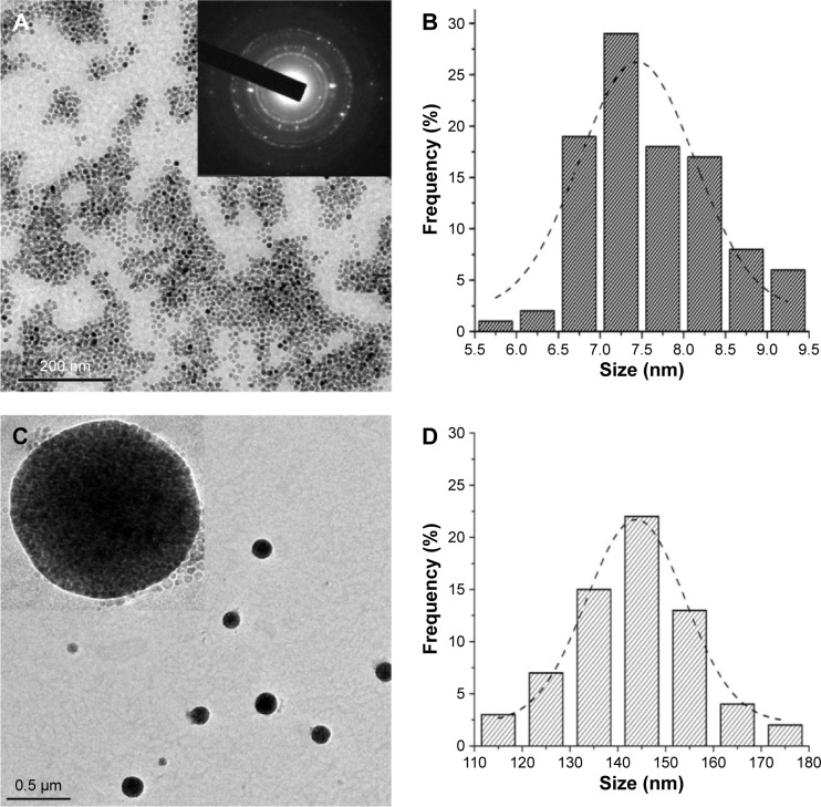
MnFe2O4 NPs synthesized from thermal decomposition of iron acetylacetonate and manganese acetylacetonate in a 2:1 ratio were of spheroidal appearance and narrow size distribution (Figure 1A). The results from Image Pro Plus 6.0 showed that the average size was 7.6±1.0 nm (Figure 1B), as counted with 300 NPs that were randomly selected. After being enveloped with amphiphilic block copolymer PEG-b-PCL, dozens of hydrophobic MnFe2O4 NPs assembled together (Figure 1C). The diameter of PEG-b-PCL-MnFe2O4 was 146.7±25.9 nm according to the TEM photomicrograph (Figure 1D). Figure 2A and B show a high resolution TEM image of a single MnFe2O4 NP. The spacings between the lattice fringes were measured to be around 0.301 and 0.257 nm, which correspond to the planes of (220) and (311) of bulk MnFe2O4 very well. Moreover, the measured interplanar spacings based on the diffraction rings in the selected area electron diffraction (inset of Figure 1A) were perfectly in agreement with the respective hkl indexes of bulk MnFe2O4 from the Joint Committee on Powder Diffraction Standards (JCPDS) database, indicating that the synthetic NPs are MnFe2O4 nanocrystals. The chemical composition of the nanocrystals was confirmed using the energy dispersive spectrometer measurement. The Mn, Fe, and O peaks indicated the presence of Mn, Fe, and O in the NP (Figure 2C); meanwhile, the Cu signal is derived from the TEM grid. The atomic ratio of Fe to Mn was around 2:1, agreeing well with the molar ratio of Fe and Mn in MnFe2O4.
Figure 1.
TEM characterization of MnFe2O4 NPs and PEG-b-PCL-MnFe2CO4 NMs.
Notes: MnFe2O4 NPs were of spheroidal appearance and narrow size distribution (A) and the average size was 7.6±1.0 nm (B). The diameter of PEG-b-PCL-MnFe2O4 NMs was 146.7±25.9 nm (C and D). Inset of (A) is SAED of MnFe2O4 NPs. Inset of (C) is an enlarged TEM of a single PEG-b-PCL-MnFe2O4 NM; magnification ×200,000.
Abbreviations: NMs, nanomicelles; NPs, nanoparticles; PEG-b-PCL, polyethylene glycol-block-poly(ε-caprolactone); SAED, selected area electron diffraction; TEM, transmission electron microscopy.
Figure 2.
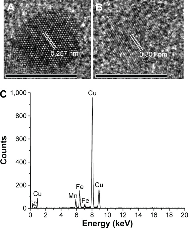
HRTEM and EDS characterization of MnFe2O4 NPs.
Notes: HRTEM image of a single MnFe2O4 NP showed that the spacing between the lattice fringes of planes (220) and (311) was around 0.257 nm (A) and 0.301 nm (B). The chemical composition of the nanocrystals was confirmed using EDS measurement. EDS (C) of the prepared NPs indicated the presence of Mn, Fe, and O, and the atomic ratio of Fe to Mn was around 2:1. Scale bar =10 nm.
Abbreviations: EDS, energy dispersive spectrometer; HRTEM, high resolution transmission electron microscopy; NPs, nanoparticles.
Dynamic light scattering and inductive-coupled plasma optical emission spectrometer measurement
According to the results of dynamic light scattering, the hydrodynamic diameter of MnFe2O4 dispersed in hexane was 10.3±1.2 nm (Figure 3A), which was slightly larger than the size measured by TEM. This might be due to the coating of oleic acid or oleylamine on the outer surface of the NP that could affect the light scattering while it could not be detected under the TEM. Similarly, because of the hydrodynamic size effect and electronic permeability of the PEG-b-PCL, the zeta sizes of PEG-b-PCL-MnFe2O4 and CL-PEG-MnFe2O4 were 162.6±28.9 nm (Figure 3B) and 183.4±26.5 nm (Figure 3C), which were larger than those obtained from TEM observations. Inductive-coupled plasma optical emission spectrometer analysis showed that Fe and Mn elemental contents of MnFe2O4 were 363.5 and 167.9 mg/L, which matched the result of energy dispersive spectrometer analysis and revealed that the molar ratio of Fe and Mn of MnFe2O4 was ~2:1, indicating that the prepared NPs were MnFe2O4.
Figure 3.
Hydrodynamic diameter of MnFe2O4 NPs and NMs.
Notes: The hydrodynamic diameter of MnFe2O4 dispersed in hexane was 10.3±1.2 nm (A) and that of PEG-b-PCL-MnFe2O4 and CL-PEG-b-PCL-MnFe2O4 was 162.6±28.9 nm (B) and 183.4±26.5 (C), respectively.
Abbreviations: NMs, nanomicelles; NPs, nanoparticles; PEG-b-PCL, polyethylene glycol-block-poly(ε-caprolactone).
Magnetization and T2 relaxivity
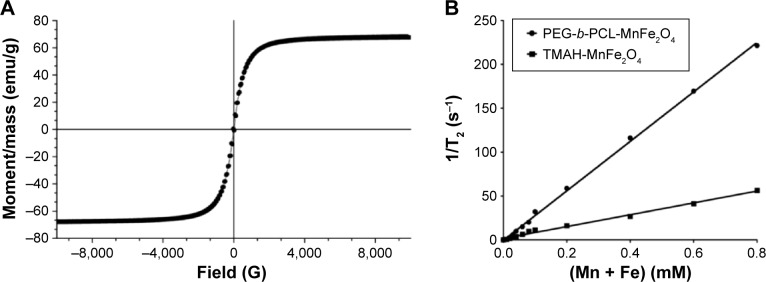
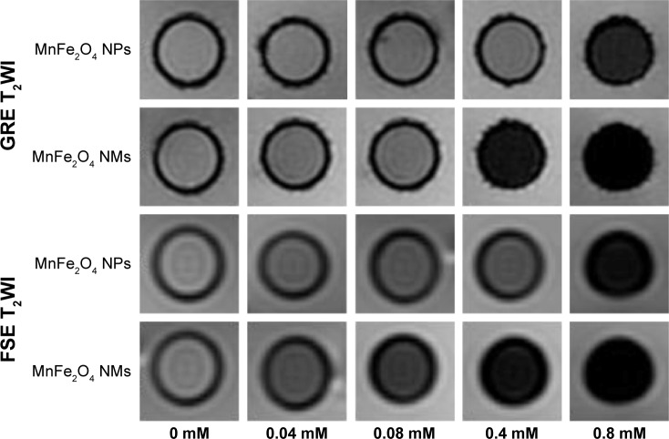
To understand the magnetic property of the prepared MnFe2O4 NPs, 46.8 mg nanopowder was tested using a vibrating sample magnetometer. The magnetization rose nonlinearly with the increase of applied magnetic field (Figure 4A). The prepared NPs showed no remaining net magnetization in the absence of an external field, which indicated that the MnFe2O4 NPs were superparamagnetic at 300 K. Under a powerful magnetic field, NPs will reach their saturation magnetization, which was 68.2 emu/g. Figure 5 shows that TMAH-MnFe2O4 NPs and PEG-b-PCL-MnFe2O4 NMs both exhibited a concentration-dependent signal drop in the GRE T2*WI and FSE T2-weighted imaging. Their signal intensities decreased gradually with the increase of the metal ion concentration. However, PEG-b-PCL-MnFe2O4 NMs induced greater hypointensity at an identical concentration compared with TMAH-MnFe2O4 NPs. The linear fitting of concentration and 1/T2 showed that the r2 of PEG-b-PCL-MnFe2O4 NMs was 281.7 mM−1 s−1, which was ~4.2 times higher than that of TMAH-MnFe2O4 4 NPs (67.5 mM− s−1) (Figure 4B).
Figure 4.
Hysteresis loop and T2 relaxivity.
Notes: The hysteresis loop of MnFe2O4 NPs (A) showed that the magnetization rose nonlinearly with the increase of applied magnetic field and no remaining net magnetization in the absence of an external field, indicating that the MnFe2O4 NPs are superparamagnetic at 300 K. The MnFe2O4 NPs saturation magnetization was 68.2 emu/g (A). The linear fitting of concentration and 1/T2 showed that the r2 of PEG-b-PCL-MnFe2O4 NMs was 281.7 mM−1 s−1, which was ~4.2 times higher than that of TMAH-MnFe2O4 NPs (67.5 mM−1 s−1) (B).
Abbreviations: NMs, nanomicelles; NPs, nanoparticles; PEG-b-PCL, polyethylene glycol-block-poly(ε-caprolactone); r2, transverse relaxivity; T2, transverse relaxation time; TMAH, tetramethylammonium hydroxide.
Figure 5.
MRI of MnFe2O4 TMAH-MnFe2O4 NPs and PEG-b-PCL-MnFe2O4 NMs.
Notes: TMAH-MnFe2O4 NPs and PEG-b-PCL-MnFe2O4 NMs both exhibited a concentration-dependent signal drop in the GRE T2WI and FSE T2WI. The signal intensity decreased gradually with the increase of the metal ions concentration, while NMs induced greater hypointensity at an identical concentration compared with NPs.
Abbreviations: MRI, magnetic resonance imaging; NMs, nanomicelles; NPs, nanoparticles; PEG-b-PCL, polyethylene glycol-block-poly(ε-caprolactone); TMAH, tetramethylammonium hydroxide; T2WI, T2-weighted imaging; GRE, gradient echo; FSE, fast spin echo.
Cytotoxicity analysis
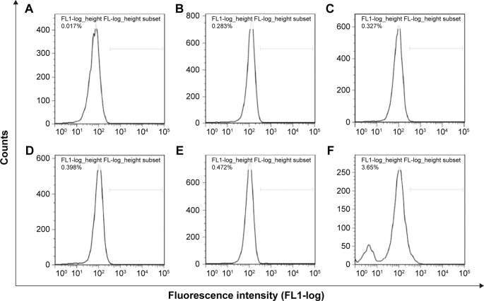
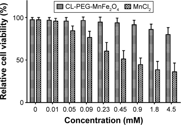
The relative cell viability (RCV) of each treatment group to control group was used to determine the cytotoxicity of CL-PEG-MnFe2O4 and MnCl2 with different concentrations on TVECs. Figure 6 shows that the RCV of TVECs labeled with CL-PEG-MnFe2O4 within 0.9 mM decreased only 7%–9% compared to controls. As the concentration increased, the RCV decreased correspondingly, and a decrease of ~19% in viability of TVECs incubated with 4.5 mM CL-PEG-MnFe2O4 solution was measured. On the contrary, a notable decrease of ~15% in viability was observed in TVECs incubated with only 0.05 mM MnCl2 solution. When the TVECs were incubated with MnCl2 solution as high as 4.5 mM, there were only 36.2% cells that survived. The CCK-8 results demonstrated that the CL-PEG-MnFe2O4 showed no acute toxicity to TVECs even at high concentrations, indicating their much better biocompatibility than molecular manganese agents. The ROS level of TVECs labeled with 0, 0.05, 0.23, 0.45, 0.9, and 4.5 mM CL-PEG-MnFe2O4 solutions are shown in Figure 7. It was shown that the induction of ROS depended on the concentration and an insignificant ROS level elevation was detected in the cells exposed to CL-PEG-MnFe2O4 within 0.9 mM. However, a 3.18% higher intracellular ROS level was measured in the cells incubated with 4.5 mM CL-PEG-MnFe2O4 solution compared with 0.9 mM CL-PEG-MnFe2O4.
Figure 6.
Cytotoxicity analysis.
Notes: The RCV of TVECs labeled with CL-PEG-MnFe2O4 within 0.9 mM decreased only 7%–9% compared to controls. As the concentration increased, the RCV decreased correspondingly, and a significant decrease of ~19% in viability of TVECs incubated with 4.5 mM CL-PEG-MnFe2O4 solution was measured. On the contrary, a notable decrease of ~15% in viability of TVECs incubated with only 0.05 mM MnCl2 solution was observed. When the TVECs were incubated with MnCl2 solution as high as 4.5 mM, there were only 36.2% cells that survived.
Abbreviations: NMs, nanomicelles; PEG, poly(ethylene glycol); RCV, relative cell viability; TVECs, tumor vascular endothelial cells.
Figure 7.
ROS of TVECs labeled with CL-PEG-MnFe2O4.
Notes: The ROS levels of TVECs labeled with 0.05 (B), 0.23 (C), 0.45 (D), and 0.9 (E) mM CL-PEG-MnFe2O4 solutions were very low and depicted an insignificant elevation of ROS level compared with the control (A). However, a 3.18% higher intracellular ROS level was measured in the cells incubated with 4.5 mM CL-PEG-MnFe2O4 solution compared with 0.9 mM CL-PEG-MnFe2O4 (F).
Abbreviations: PEG, poly(ethylene glycol); ROS, reactive oxygen species; TVECs, tumor vascular endothelial cells.
Labeling of TVECs with CL-PEG-MnFe2O4
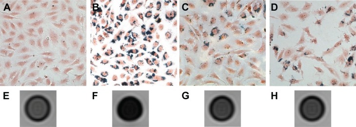
As the ferric ferrocyanide, which is a kind of dark blue pigment (also called Prussian blue), can be produced through the reaction of iron with potassium ferrocyanide within the acidic solution, the uptake of the CL-PEG-MnFe2O4, PEG-b-PCL-MnFe2O4, and the mixture of CL-PEG-MnFe2O4 and CL 1555 peptides can be observed under optical microscope after Prussian blue staining. Figure 8A–D are the results of Prussian blue staining of control group (Figure 8A) and TVECs labeled with CL-PEG-MnFe2O4 (Figure 8B), PEG-b-PCL-MnFe2O4 (Figure 8C) and the mixture of CL-PEG-MnFe2O4 and CL 1555 peptides (Figure 8D), which clearly shows a higher uptake of CL-PEG-MnFe2O4 than PEG-b-PCL-MnFe2O4 into TVECs. Blocking CD105 with the free CL 1555 peptides effectively reduced the amount of blue granules in the cytoplasm of TVECs, indicating that the internalization of CL-PEG-MnFe2O4 was specifically mediated by CL 1555 peptides. Figure 8E–H shows the MR images of the four groups of cells. Compared with the control, the cells cocultured with CL-PEG-MnFe2O4 showed a noticeable signal intensity drop and its T2 relaxation time was 38.3±3.5 ms. However, the cells treated with PEG-b-PCL-MnFe2O4 and the mixture of CL-PEG-MnFe2O4 and CL 1555 peptides showed a similar signal intensity and T2 relaxation time to the control.
Figure 8.
In vitro labeling of TVECs with CL-PEG-MnFe2O4.
Notes: Prussian blue staining (200×) of control group (A), TVECs labeled with CL-PEG-MnFe2O4 (B), PEG-b-PCL-MnFe2O4 (C) and the mixture of CL-PEG-MnFe2O4 and CL 1555 peptides (D). It showed that the uptake of CL-PEG-MnFe2O4 was high, while few PEG-b-PCL-MnFe2O4 was engulfed into TVECs. Blocking CD105 with the free CL 1555 peptides also effectively reduced the amount of blue granules in the cytoplasm of TVECs. (E–H) showed the T2-weighted MR images of the four groups of cells. Correspondingly, the cells co-cultured with CL-PEG-MnFe2O4 (F) showed a more noticeable signal intensity drop than that with PEG-b-PCL-MnFe2O4 (G) and blocking with the free CL 1555 peptides (H).
Abbreviations: MR, magnetic resonance; PEG-b-PCL, polyethylene glycol-block-poly(ε-caprolactone); TVECs, tumor vascular endothelial cells.
Subculture of the labeled cells and their MRI
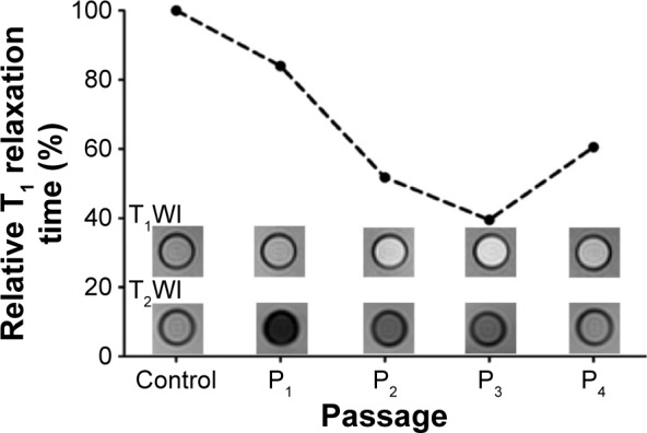
The TVECs labeled with CL-PEG-MnFe2O4 were subcultured from P1 to P4 and each generation was evaluated with a 3.0 T MRI system. As shown in Figure 9, the signal intensity from P1 to P4 in T2-weighted images gradually increased and P4 had almost equal signal intensity with control group. However, the signal intensity firstly increased and then decreased in T1-weighted images. The labeled cells of P1 presented low signal intensity similar to the control and those of P2, P3, and P4 were hyper-intense. The T1 relaxation time from P1 to P4 decreased firstly and then increased back accordingly (Figure 9).
Figure 9.
MRI of subcultured labeled TVECs.
Notes: MRI showed that the signal intensity from P1 to P4 in T2WI gradually increased and P4 had almost equal signal intensity with control group. In T1WI, the signal intensity from P1 to P3 gradually increased and that of P4 came down to the control. The plot showed that the T1 relaxation time from P1 to P4 decreased firstly and then increased back.
Abbreviations: MRI, magnetic resonance imaging; T1WI, T1-weighted imaging; T2WI, T2-weighted imaging; TVECs, tumor vascular endothelial cells.
Discussion
Angiogenesis, the formation of new blood vessels, is a requirement for tumor growth and metastasis.3,4,43 Moreover, the tumor microvascular density was positively correlated with the trend of tumor metastasis and high microvascular density always leads to poor prognosis.44,45 Commonly, the angiogenesis is estimated with histopathological examination. However, histopathological examination for angiogenesis does not demonstrate functionality within the vessels sampled and is inherent invasive and suffers from sampling bias.46,47 Molecular imaging targeting vascular endothelial cells can offer this in a noninvasive way.46,48–50 Due to a relatively good spatial resolution, good soft-tissue contrast, absence of ionizing radiation, and limited side effects, MRI has been proposed to be a good candidate for angiogenesis imaging.51–53
Endoglin, also called CD105, is a 180 kDa transmembrane protein and consists of a homodimer with disulfide links, and it has been found that its expression is usually low in resting endothelial cells and becomes highly expressed once neoangiogenesis begins.7,9,54 The important role that CD105 plays in tumor angiogenesis makes it an ideal candidate for tumor targeting imaging. In previous studies, we conjugated anti-CD105 antibody with SL-encapsulated Gd-DTPA and thiol-PEG-carboxyl-stabilized Fe2O3/Au NPs to evaluate tumor angiogenesis on T1- or T2-enhanced MRI, respectively.36,37 The results showed that CD105-Gd-SLs and hybrid-PEG-CD105 could be utilized to detect subcutaneous glioma and breast cancer angiogenesis in tumor-bearing rats. However, because of the larger molecular weight of anti-CD105 antibody, the probes have low connection efficiency and consequently have not enough affinity for the targeted TVECs. In order to improve the affinity of the probes, we employed a peptide – CL 1555 – which consisted of only 12 amino acids as the targeting molecule in the present study. CL 1555 was selected from a phage displayed library and was demonstrated to be of high affinity and selectivity toward CD105.33,34 Compared with anti-CD105 antibody, CL 1555 had a lower molecular weight and much smaller steric hindrance, resulting in a high grafting rate on the surface of the NMs. The results of our study showed that compared with PEG-MnFe2O4, CL-PEG-MnFe2O4 binds to the surface of TVECs much better and markedly shortens T2 of the labeled TVCEs in T2-weighted images. In a competition binding experiment, the binding of CL-PEG-MnFe2O4 and TVECs was blocked by free CL 1555 peptides, which revealed that the immobilization CL-PEG-MnFe2O4 on the surface of TVECs was mediated by CL 1555. So, it was undoubtedly confirmed that CL 1555 was an ideal ligand to CD105 for targeted diagnosis and therapy.
Because of good biocompatibility, superparamagnetic Fe3O4 NPs have been widely used as a negative CA for enhanced MRI.37,55,56 However, the r2 of nanoscale Fe3O4 was low and not enough for cell imaging and neovessel visualization in the early stage. MnFe2O4 NPs have been proven to be of a higher saturation magnetization (Mp) and r2 than other ferrite NPs, including CoFe2O4, Fe3O4, and NiFe2O4.17,57 Lee et al25 fabricated a series of ferrite NPs, including CoFe2O4, Fe3O4, MnFe2O4, and NiFe2O4, and investigated their magnetic properties. They found that these ferrite NPs with a size of 12 nm all showed superparamagnetism. However, these NPs with an identical size have different Mp and MnFe2O4 exhibited a much higher Mp than CoFe2O4, Fe3O4, and NiFe2O4. So, we employed MnFe2O4 NPs as CAs for TVECs MR molecular imaging in the present study in order to improve the sensitivity of imaging. Besides the Mp, the particle size can also influence the r2 value. It has been reported that a different relation between r2 value and particle size follows when the size is within different regimes. When the particles are small, the diffusional motion of water molecules surrounding these NPs is fast enough to average out the magnetic fields induced by these magnetic NPs. This regime is termed as motional averaging and the r2 value is proportional to Mp2τd, where τd signifies the duration when water protons are under the influence of a magnetic NP and is proportional to r2/D (D is the diffusion coefficient of water protons). Because τd increases with the increase of particle size, the averaging effect diminishes and the magnetic NPs appear as randomly distributed and stationary objects to water protons. In this regime, called static dephasing, the r2 value is only proportional to Mp. So, increasing the size of individual particle can enhance the r2 value by motional averaging relaxation.17,26,58–61 However, this approach often posed technical difficulties because further increasing the size of the single NP will transfer the magnetic NPs from superparamagnetic to becoming ferromagnetic or ferrimagnetic. Then, the large, single core magnetic NPs will tend to aggregation in suspension because of the magnetic force between each other. An alternative route is to embed multiple magnetic NPs into a single multicore micelle.27,62–64 When the NPs aggregate, the size of the single multicore micelle will be increased into static dephasing regime and their r2 value will be further increased through the static dephasing relaxation mechanism. In this work, we first synthesized the hydrophobic MnFe2O4 NPs with a size of ~7.5 nm, which was in the regime of motional averaging. The Mp of the prepared NPs was 68.2 emu/g. Afterward, we transformed the MnFe2O4 NPs from hydrophobic to hydrophilic using amphiphilic block copolymer PEG-b-PCL. The MnFe2O4 NPs modified with PEG-b-PCL then self-assembled into clusters and their size increased to ~146.7 nm, which was in the regime of static dephasing. After linear fitting of concentration and 1/T2, we found that the clusters consisting of dozens of MnFe2O4 NPs were of much higher r2 of 281.7 mM−1 (Mn + Fe) s−1 than that of single NPs (67.5 mM−1 s−1).
Because these nanoprobes were fabricated for biomedical application, we must evaluate their cytotoxicity first. The in vitro cytotoxicity of CL-PEG-MnFe2O4 NMs toward TVECs was evaluated using CCK-8 analysis in comparison with molecular manganese agents. The results showed that the viability of TVECs labeled with CL-PEG-MnFe2O4 within 0.9 mM decreased only 7%–9% compared to controls. As the concentration increased, the RCV decreased correspondingly, and a decrease of ~19% in viability of TVECs incubated with 4.5 mM CL-PEG-MnFe2O4 solution was measured. On the contrary, a notable decrease of ~15% in viability was observed in TVECs incubated with only 0.05 mM MnCl2 solution. When the TVECs were incubated with MnCl2 solution as high as 4.5 mM, there were only 36.2% cells that survived. So, a conclusion can be easily drawn that the CL-PEG-MnFe2O4 showed no acute toxicity to TVECs even at relatively high concentrations, indicating their much better biocompatibility than MnCl2. Several factors may contribute to the low cytotoxicity of CL-PEG-MnFe2O4 NMs. It has been demonstrated that several factors are responsible for the cytotoxicity of NPs, including ROS production and toxic ion leaching.65,66 The ROS induction has been posted as one of the main explanations for these toxic effects and induces various deleterious effects, including cell membrane damage, DNA and cytoskeleton injury, autophagy, and apoptosis.42 Here, the results of ROS assay depicted that an insignificant ROS level elevation was detected in the cells exposed to CL-PEG-MnFe2O4 within 0.9 mM and only a 3.18% higher intracellular ROS level was measured in the cells incubated with 4.5 mM CL-PEG-MnFe2O4 solution compared with that incubated with 0.9 mM CL-PEG-MnFe2O4, which might explain the CCK-8 results well. Moreover, the ferrite magnetic NPs have been proven to have intrinsic peroxidase-like activity, which can decrease the amount of intracellular H2O2 to promote cell proliferation. Regarding the ion leaching, in contrast to MnCl2 solution, Mn ions leaching from Mn-based NPs have been demonstrated to be slower and Mn-based NPs showed no significant toxic effects on cells at a very high dosage of 200 µg/mL in some previous studies.30,31 Actually, to avoid high cytotoxicity of Mn, some researchers employed Mn-based NPs as a sustained release delivery of Mn ions for neuroimaging instead of MnCl2 solution.30,31
Because of clearance and degradation of NPs from the labeled cells, it was reasonable that the signal intensity from P1 to P4 in T2-weighted MR images gradually increased. However, it was very interesting that the signal intensity of these cells firstly increased and then decreased in T1-weighted MR images. We hypothesized that T1 enhancement of the labeled cells resulted from the divalent manganese ions and the smaller size MnFe2O4 NPs. It has been observed that the internalized nano- and microparticles are exclusively located within endosomes and finally in the lysosomes.30,67 Due to the abundance of proteolytic enzymes, chelating agents, and an acidic environment, the internalized particles will finally be degraded. For MnFe2O4 NPs, they were dissolved gradually and metallic ions Mn2+ and Fe3+ slowly released. Divalent manganese ions have five unpaired electrons and are paramagnetic, which have been regarded as strong T1 MRI CAs.31 Additionally, the reduction of NP size accompanied with the degradation of MnFe2O4 may also contribute to the high signal intensity of P3 in T1-weighted MR images. It is known that the surface atom ratio is inversely proportional to the particle size and large particles exhibit lower surface Mn2+ ratio, leading to a small longitudinal relaxivity.28 When the internalized MnFe2O4 was degraded, its size decreased gradually. Accordingly, the surface Mn2+ ratio of MnFe2O4 increased rapidly, resulting in a continuous growth of longitudinal relaxivity. When the size is 2.2 nm, the longitudinal relaxivity of MnFe2O4 is 6.61 mM−1s−1, which is higher than that of the clinically used Gd-DTPA agent (r =4.8 mM−1s−1).29
Conclusion
In this study, we have successfully prepared an angiogenesis-targeting nanoprobe of high sensitivity for T1 and T2 MRI. The nanoprobe consists of MnFe2O4 NMs, which act as an MRI CA, and a peptide named CL 1555 for active targeting. These nanoprobes could specifically target TVECs with CD105 expression. The labeled TVECs could be imaged in T2-weighted MRI and also in T1-weighted MRI with the passage of labeled cells and the degradation of the internalized NPs. Based on these results, the prepared CL-PEG-MnFe2O4 nanoprobe could act as a promising CA for early detection of tumor angiogenesis using targeting T1- and T2-enhanced MRI.
Acknowledgments
We are very grateful to the National Natural Science Foundation of China (81401466 and 81501521) and General project of the frontier and application basic research plan of Chongqing (cstc2015jcyjA1338) for financial support.
Footnotes
Disclosure
The authors report no conflicts of interest in this work.
References
- 1.Torre LA, Bray F, Siegel RL, Ferlay J, Lortet-Tieulent J, Jemal A. Global cancer statistics, 2012. CA Cancer J Clin. 2015;65(2):87–108. doi: 10.3322/caac.21262. [DOI] [PubMed] [Google Scholar]
- 2.Morgan-Parkes JH. Metastases: mechanisms, pathways, and cascades. AJR Am J Roentgenol. 1995;164(5):1075–1082. doi: 10.2214/ajr.164.5.7717206. [DOI] [PubMed] [Google Scholar]
- 3.Blood CH, Zetter BR. Tumor interactions with the vasculature: angiogenesis and tumor metastasis. Biochim Biophys Acta. 1990;1032(1):89–118. doi: 10.1016/0304-419x(90)90014-r. [DOI] [PubMed] [Google Scholar]
- 4.Folkman J, Merler E, Abernathy C, Williams G. Isolation of a tumor factor responsible for angiogenesis. J Exp Med. 1971;133(2):275–288. doi: 10.1084/jem.133.2.275. [DOI] [PMC free article] [PubMed] [Google Scholar]
- 5.Erber R, Thurnher A, Katsen AD, et al. Combined inhibition of VEGF and PDGF signaling enforces tumor vessel regression by interfering with pericyte-mediated endothelial cell survival mechanisms. FASEB J. 2004;18(2):338–340. doi: 10.1096/fj.03-0271fje. [DOI] [PubMed] [Google Scholar]
- 6.Brooks PC, Montgomery AM, Rosenfeld M, et al. Integrin αvβ3 antagonists promote tumor regression by inducing apoptosis of angiogenic blood vessels. Cell. 1994;79(7):1157–1164. doi: 10.1016/0092-8674(94)90007-8. [DOI] [PubMed] [Google Scholar]
- 7.Fonsatti E, Altomonte M, Nicotra MR, Natali PG, Maio M. Endoglin (CD105): a powerful therapeutic target on tumor-associated angiogenetic blood vessels. Oncogene. 2003;22(42):6557–6563. doi: 10.1038/sj.onc.1206813. [DOI] [PubMed] [Google Scholar]
- 8.Seon BK. Expression of endoglin (CD105) in tumor blood vessels. Int J Cancer. 2002;99(2):310–311. doi: 10.1002/ijc.10378. [DOI] [PubMed] [Google Scholar]
- 9.Duff SE, Li C, Garland JM, Kumar S. CD105 is important for angiogenesis: evidence and potential applications. FASEB J. 2003;17(9):984–992. doi: 10.1096/fj.02-0634rev. [DOI] [PubMed] [Google Scholar]
- 10.Edelman RR, Warach S. Magnetic resonance imaging. N Engl J Med. 1993;328(10):708–716. doi: 10.1056/NEJM199303113281008. [DOI] [PubMed] [Google Scholar]
- 11.Pichler BJ, Kolb A, Nägele T, Schlemmer H-P. PET/MRI: paving the way for the next generation of clinical multimodality imaging applications. J Nucl Med. 2010;51(3):333–336. doi: 10.2967/jnumed.109.061853. [DOI] [PubMed] [Google Scholar]
- 12.Thompson WO, Thaete FL, Fu FH, Dye SF. Tibial meniscal dynamics using three-dimensional reconstruction of magnetic resonance images. Am J Sports Med. 1991;19(3):210–216. doi: 10.1177/036354659101900302. [DOI] [PubMed] [Google Scholar]
- 13.Weissleder R, Mahmood U. Molecular imaging 1. Radiology. 2001;219(2):316–333. doi: 10.1148/radiology.219.2.r01ma19316. [DOI] [PubMed] [Google Scholar]
- 14.Satoh Y, Ichikawa T, Motosugi U, et al. Diagnosis of peritoneal dissemination: comparison of 18F-FDG PET/CT, diffusion-weighted MRI, and contrast-enhanced MDCT. AMJ Am J Roentgenol. 2011;196(2):447–453. doi: 10.2214/AJR.10.4687. [DOI] [PubMed] [Google Scholar]
- 15.Hu B, Dai F, Fan Z, Ma G, Tang Q, Zhang X. Nanotheranostics: Congo Red/Rutin-MNPs with enhanced magnetic resonance imaging and H2O2-responsive therapy of Alzheimer’s disease in APPswe/PS1dE9 transgenic mice. Adv Mater. 2015;27(37):5499–5505. doi: 10.1002/adma.201502227. [DOI] [PubMed] [Google Scholar]
- 16.Geraldes CF, Laurent S. Classification and basic properties of contrast agents for magnetic resonance imaging. Contrast Media Mol Imaging. 2009;4(1):1–23. doi: 10.1002/cmmi.265. [DOI] [PubMed] [Google Scholar]
- 17.Tromsdorf UI, Bigall NC, Kaul MG, et al. Size and surface effects on the MRI relaxivity of manganese ferrite nanoparticle contrast agents. Nano Lett. 2007;7(8):2422–2427. doi: 10.1021/nl071099b. [DOI] [PubMed] [Google Scholar]
- 18.Hong S, Chang Y, Rhee I. Chitosan-coated ferrite (Fe3O4) nanoparticles as a T2 contrast agent for magnetic resonance imaging. J Korean Phys Soc. 2010;56(3):868–873. [Google Scholar]
- 19.Leal MP, Rivera-Fernández S, Franco JM, Pozo D, Jesús M, García- Martín ML. Long-circulating PEGylated manganese ferrite nanoparticles for MRI-based molecular imaging. Nanoscale. 2015;7(5):2050–2059. doi: 10.1039/c4nr05781c. [DOI] [PubMed] [Google Scholar]
- 20.Bae KH, Kim YB, Lee Y, Hwang J, Park H, Park TG. Bioinspired synthesis and characterization of gadolinium-labeled magnetite nanoparticles for dual contrast T1-and T2-weighted magnetic resonance imaging. Bioconjug Chem. 2010;21(3):505–512. doi: 10.1021/bc900424u. [DOI] [PubMed] [Google Scholar]
- 21.Hu F, Jia Q, Li Y, Gao M. Facile synthesis of ultrasmall PEGylated iron oxide nanoparticles for dual-contrast T1-and T2-weighted magnetic resonance imaging. Nanotechnology. 2011;22(24):245604. doi: 10.1088/0957-4484/22/24/245604. [DOI] [PubMed] [Google Scholar]
- 22.High WA, Ayers RA, Cowper SE. Gadolinium is quantifiable within the tissue of patients with nephrogenic systemic fibrosis. J Am Acad Dermatol. 2007;56(4):710–712. doi: 10.1016/j.jaad.2007.01.022. [DOI] [PubMed] [Google Scholar]
- 23.Song Q, Zhang ZJ. Controlled synthesis and magnetic properties of bimagnetic spinel ferrite CoFe2O4 and MnFe2O4 nanocrystals with core–shell architecture. J Am Chem Soc. 2012;134(24):10182–10190. doi: 10.1021/ja302856z. [DOI] [PubMed] [Google Scholar]
- 24.Xu C, Sun S. New forms of superparamagnetic nanoparticles for biomedical applications. Adv Drug Deliv Rev. 2013;65(5):732–743. doi: 10.1016/j.addr.2012.10.008. [DOI] [PubMed] [Google Scholar]
- 25.Lee JH, Huh YM, Jun YW, et al. Artificially engineered magnetic nanoparticles for ultra-sensitive molecular imaging. Nat Med. 2007;13(1):95–99. doi: 10.1038/nm1467. [DOI] [PubMed] [Google Scholar]
- 26.Lee H, Shin TH, Cheon J, Weissleder R. Recent developments in magnetic diagnostic systems. Chem Rev. 2015;115(19):10690–10724. doi: 10.1021/cr500698d. [DOI] [PMC free article] [PubMed] [Google Scholar]
- 27.Yoon T-J, Lee H, Shao H, Hilderbrand SA, Weissleder R. Multicore assemblies potentiate magnetic properties of biomagnetic nanoparticles. Adv Mater. 2011;23(41):4793–4797. doi: 10.1002/adma.201102948. [DOI] [PMC free article] [PubMed] [Google Scholar]
- 28.Niu D, Luo X, Li Y, Liu X, Wang X, Shi J. Manganese-loaded dual-mesoporous silica spheres for efficient T1-and T2-weighted dual mode magnetic resonance imaging. ACS Appl Mater Interfaces. 2013;5(20):9942–9948. doi: 10.1021/am401856w. [DOI] [PubMed] [Google Scholar]
- 29.Li Z, Wang SX, Sun Q, et al. Ultrasmall manganese ferrite nanoparticles as positive contrast agent for magnetic resonance imaging. Adv Healthc Mater. 2013;2(7):958–964. doi: 10.1002/adhm.201200340. [DOI] [PubMed] [Google Scholar]
- 30.Shapiro EM, Koretsky AP. Convertible manganese contrast for molecular and cellular MRI. Magn Reson Med. 2008;60(2):265–269. doi: 10.1002/mrm.21631. [DOI] [PMC free article] [PubMed] [Google Scholar]
- 31.Chen W, Lu F, Chen CC, et al. Manganese-enhanced MRI of rat brain based on slow cerebral delivery of manganese(II) with silica-encapsulated MnxFe1–xO nanoparticles. NMR Biomed. 2013;26(9):1176–1185. doi: 10.1002/nbm.2932. [DOI] [PubMed] [Google Scholar]
- 32.Chen F-Y, Gu Z-J, Wan H-P, Xu X-Z, Tang Q. Manganese nanosystem for new generation of MRI contrast agent. Rev Nanosci Nanotechnol. 2015;4(2):81–91. [Google Scholar]
- 33.Bi X, Liang Z, Shi L. Determination of functional affinity of rhEndoglin conjugated eptide. J Third Mil Med Univ. 2007;29(8):685–687. [Google Scholar]
- 34.Bi X, Liang Z, Shi L. Screening and function analysis of targeted short binding peptides of endoglin. J Chongqing Medical Univ. 2007;32(3):232–235. [Google Scholar]
- 35.Ghosh P, Han G, De M, Kim CK, Rotello VM. Gold nanoparticles in delivery applications. Adv Drug Deliv Rev. 2008;60(11):1307–1315. doi: 10.1016/j.addr.2008.03.016. [DOI] [PubMed] [Google Scholar]
- 36.Zhang D, Feng XY, Henning TD, et al. MR imaging of tumor angiogenesis using sterically stabilized Gd-DTPA liposomes targeted to CD105. Eur J Radiol. 2009;70(1):180–189. doi: 10.1016/j.ejrad.2008.04.022. [DOI] [PubMed] [Google Scholar]
- 37.Zhang S, Gong M, Zhang D, Yang H, Gao F, Zou L. Thiol-PEG-carboxyl-stabilized Fe2O3/Au nanoparticles targeted to CD105: Synthesis, characterization and application in MR imaging of tumor angiogenesis. Eur J Radiol. 2014;83(7):1190–1198. doi: 10.1016/j.ejrad.2014.03.034. [DOI] [PubMed] [Google Scholar]
- 38.Sun S, Zeng H, Robinson DB, et al. Monodisperse MFe2O4 (M = Fe, Co, Mn) nanoparticles. J Am Chem Soc. 2004;126(1):273–279. doi: 10.1021/ja0380852. [DOI] [PubMed] [Google Scholar]
- 39.Lim J, Eggeman A, Lanni F, Tilton RD, Majetich SA. Synthesis and single-particle optical detection of low-polydispersity plasmonic-superparamagnetic nanoparticles. Adv Mater. 2008;20(9):1721–1726. [Google Scholar]
- 40.Farokhzad OC, Jon S, Khademhosseini A, Tran TN, Lavan DA, Langer R. Nanoparticle-aptamer bioconjugates: a new approach for targeting prostate cancer cells. Cancer Res. 2004;64(21):7668–7672. doi: 10.1158/0008-5472.CAN-04-2550. [DOI] [PubMed] [Google Scholar]
- 41.Farokhzad OC, Cheng J, Teply BA, et al. Targeted nanoparticle-aptamer bioconjugates for cancer chemotherapy in vivo. Proc Natl Acad Sci U S A. 2006;103(16):6315–6320. doi: 10.1073/pnas.0601755103. [DOI] [PMC free article] [PubMed] [Google Scholar]
- 42.Gong M, Yang H, Zhang S, et al. Superparamagnetic core/shell GoldMag nanoparticles: size-, concentration- and time-dependent cellular nanotoxicity on human umbilical vein endothelial cells and the suitable conditions for magnetic resonance imaging. J Nanobiotechnology. 2015;13:24. doi: 10.1186/s12951-015-0080-x. [DOI] [PMC free article] [PubMed] [Google Scholar]
- 43.O’Reilly MS, Boehm T, Shing Y, et al. Endostatin: An endogenous inhibitor of angiogenesis and tumor growth. Cell. 1997;88(2):277–285. doi: 10.1016/s0092-8674(00)81848-6. [DOI] [PubMed] [Google Scholar]
- 44.Jackson A, Kassner A, Annesley-Williams D, Reid H, Zhu X-P, Li K-L. Abnormalities in the recirculation phase of contrast agent bolus passage in cerebral gliomas: comparison with relative blood volume and tumor grade. Am J Neuroradiol. 2002;23(1):7–14. [PMC free article] [PubMed] [Google Scholar]
- 45.Strohmeyer D, Rössing C, Strauss F, Bauerfeind A, Kaufmann O, Loening S. Tumor angiogenesis is associated with progression after radical prostatectomy in pT2/pT3 prostate cancer. Prostate. 2000;42(1):26–33. doi: 10.1002/(sici)1097-0045(20000101)42:1<26::aid-pros4>3.0.co;2-6. [DOI] [PubMed] [Google Scholar]
- 46.Barrett T, Kobayashi H, Brechbiel M, Choyke PL. Macromolecular MRI contrast agents for imaging tumor angiogenesis. Eur J Radiol. 2006;60(3):353–366. doi: 10.1016/j.ejrad.2006.06.025. [DOI] [PubMed] [Google Scholar]
- 47.Barrett T, Brechbiel M, Bernardo M, Choyke PL. MRI of tumor angiogenesis. J Magn Reson Imaging. 2007;26(2):235–249. doi: 10.1002/jmri.20991. [DOI] [PubMed] [Google Scholar]
- 48.Samei E, Saunders RS, Badea CT, et al. Micro-CT imaging of breast tumors in rodents using a liposomal, nanoparticle contrast agent. Int J Nanomedicine. 2009;4:277–282. [PMC free article] [PubMed] [Google Scholar]
- 49.Liu Y, Yang Y, Zhang C. A concise review of magnetic resonance molecular imaging of tumor angiogenesis by targeting integrin αvβ3 with magnetic probes. Int J Nanomedicine. 2013;8:1083–1093. doi: 10.2147/IJN.S39880. [DOI] [PMC free article] [PubMed] [Google Scholar]
- 50.Key J, Leary JF. Nanoparticles for multimodal in vivo imaging in nanomedicine. Int J Nanomedicine. 2014;9:711–726. doi: 10.2147/IJN.S53717. [DOI] [PMC free article] [PubMed] [Google Scholar]
- 51.Wong EC. Vessel-encoded arterial spin-labeling using pseudocontinuous tagging. Magn Reson Med. 2007;58(6):1086–1091. doi: 10.1002/mrm.21293. [DOI] [PubMed] [Google Scholar]
- 52.Li SP, Padhani AR, Makris A. Dynamic contrast-enhanced magnetic resonance imaging and blood oxygenation level-dependent magnetic resonance imaging for the assessment of changes in tumor biology with treatment. J Natl Cancer Inst Monogr. 2010;2011(43):103–107. doi: 10.1093/jncimonographs/lgr031. [DOI] [PubMed] [Google Scholar]
- 53.Hayes C, Padhani AR, Leach MO. Assessing changes in tumour vascular function using dynamic contrast-enhanced magnetic resonance imaging. NMR Biomed. 2002;15(2):154–163. doi: 10.1002/nbm.756. [DOI] [PubMed] [Google Scholar]
- 54.Clara CA, Marie SK, de Almeida JR, et al. Angiogenesis and expression of PDGF-C, VEGF, CD105 and HIF-1α in human glioblastoma. Neuropathology. 2014;34(4):343–352. doi: 10.1111/neup.12111. [DOI] [PubMed] [Google Scholar]
- 55.Xie J, Xu C, Kohler N, Hou Y, Sun S. Controlled PEGylation of monodisperse Fe3O4 nanoparticles for reduced non-specific uptake by macrophage cells. Adv Mater. 2007;19(20):3163–3166. [Google Scholar]
- 56.Ling D, Lee N, Hyeon T. Chemical synthesis and assembly of uniformly sized iron oxide nanoparticles for medical applications. Acc Chem Res. 2015;48(5):1276–1285. doi: 10.1021/acs.accounts.5b00038. [DOI] [PubMed] [Google Scholar]
- 57.Mazarío E, Sánchez-Marcos J, Menéndez N, et al. High specific absorption rate and transverse relaxivity effects in manganese ferrite nanoparticles obtained by an electrochemical route. J Phys Chem C. 2015;119(12):6828–6834. [Google Scholar]
- 58.Chouly C, Pouliquen D, Lucet I, Jeune J, Jallet P. Development of superparamagnetic nanoparticles for MRI: effect of particle size, charge and surface nature on biodistribution. J Microencapsul. 1996;13(3):245–255. doi: 10.3109/02652049609026013. [DOI] [PubMed] [Google Scholar]
- 59.Duan H, Kuang M, Wang X, Wang YA, Mao H, Nie S. Reexamining the effects of particle size and surface chemistry on the magnetic properties of iron oxide nanocrystals: new insights into spin disorder and proton relaxivity. J Phys Chem C. 2008;112(22):8127–8131. [Google Scholar]
- 60.Yablonskiy DA, Haacke EM. Theory of NMR signal behavior in magnetically inhomogeneous tissues: the static dephasing regime. Magn Reson Med. 1994;32(6):749–763. doi: 10.1002/mrm.1910320610. [DOI] [PubMed] [Google Scholar]
- 61.Brooks RA. T2-shortening by strongly magnetized spheres: A chemical exchange model. Magn Reson Med. 2002;47(2):388–391. doi: 10.1002/mrm.10064. [DOI] [PubMed] [Google Scholar]
- 62.Lu J, Ma S, Sun J, et al. Manganese ferrite nanoparticle micellar nanocomposites as MRI contrast agent for liver imaging. Biomaterials. 2009;30(15):2919–2928. doi: 10.1016/j.biomaterials.2009.02.001. [DOI] [PubMed] [Google Scholar]
- 63.Ge J, Hu Y, Biasini M, Beyermann WP, Yin Y. Superparamagnetic magnetite colloidal nanocrystal clusters. Angew Chem Int Ed Engl. 2007;46(23):4342–4345. doi: 10.1002/anie.200700197. [DOI] [PubMed] [Google Scholar]
- 64.Roca AG, Veintemillas-Verdaguer S, Port M, Robic C, Serna CJ, Morales MP. Effect of nanoparticle and aggregate size on the relaxometric properties of MR contrast agents based on high quality magnetite nanoparticles. J Phys Chem B. 2009;113(19):7033–7039. doi: 10.1021/jp807820s. [DOI] [PubMed] [Google Scholar]
- 65.Nel A, Xia T, Mädler L, Li N. Toxic potential of materials at the nanolevel. Science. 2006;311(5761):622–627. doi: 10.1126/science.1114397. [DOI] [PubMed] [Google Scholar]
- 66.Soenen SJ, Manshian B, Montenegro JM, et al. Cytotoxic effects of gold nanoparticles: a multiparametric study. ACS Nano. 2012;6(7):5767–5783. doi: 10.1021/nn301714n. [DOI] [PubMed] [Google Scholar]
- 67.Arbab AS, Wilson LB, Ashari P, Jordan EK, Lewis BK, Frank JA. A model of lysosomal metabolism of dextran coated superparamagnetic iron oxide (SPIO) nanoparticles: implications for cellular magnetic resonance imaging. NMR Biomed. 2005;18(6):383–389. doi: 10.1002/nbm.970. [DOI] [PubMed] [Google Scholar]


