

Abstract
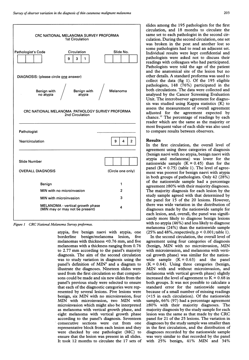
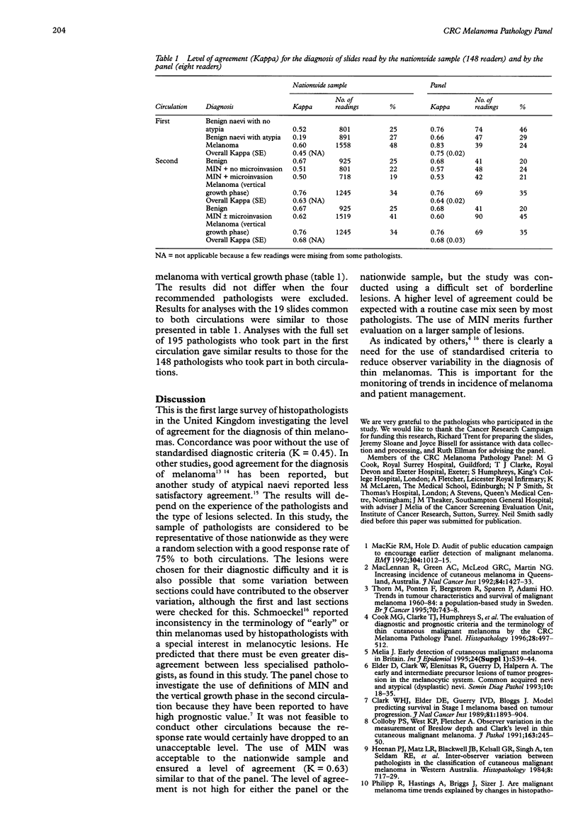
AIM: To investigate observer variation in the diagnosis of thin cutaneous malignant melanoma and related lesions in a nationwide sample of histopathologists in the UK. METHODS: Out of a random sample of 195 pathologists, 148 (76%) participated in two circulations, the first with 20 slides and the second with 25 slides. The results were compared with those for the Cancer Research Campaign (CRC) Melanoma Pathology Panel, consisting of seven histopathologists and one dermatopathologist, which had developed and evaluated diagnostic criteria. RESULTS: In the first circulation, when no standardised diagnostic criteria were used, a fair level of agreement was achieved for an overall diagnosis using the categories benign naevi with no atypia, benign naevi with atypia and melanoma (Kappa = 0.45). This was low compared with the agreement of the panel who used agreed criteria (Kappa = 0.75). Moreover, participants in the nationwide survey were more likely to diagnose melanoma and less likely to diagnose benign naevi without atypia than the panel. In the second circulation, when diagnostic criteria and diagrams were used, there was a higher level of agreement for overall diagnosis using the categories benign, melanocytic intraepidermal neoplasia (MIN) with or without microinvasion and melanoma with vertical growth phase, and was the same as that achieved by the panel using the same criteria (Kappa = 0.68). CONCLUSIONS: As the incidence rate of thin melanomas has been increasing in the UK, it is important that standardised diagnostic criteria are used to ensure accurate reporting of incidence and correct management of patients. The use of MIN and the vertical growth phase seemed to be generally acceptable.
Full text
PDF
Selected References
These references are in PubMed. This may not be the complete list of references from this article.
- Clark W. H., Jr, Elder D. E., Guerry D., 4th, Braitman L. E., Trock B. J., Schultz D., Synnestvedt M., Halpern A. C. Model predicting survival in stage I melanoma based on tumor progression. J Natl Cancer Inst. 1989 Dec 20;81(24):1893–1904. doi: 10.1093/jnci/81.24.1893. [DOI] [PubMed] [Google Scholar]
- Colloby P. S., West K. P., Fletcher A. Observer variation in the measurement of Breslow depth and Clark's level in thin cutaneous malignant melanoma. J Pathol. 1991 Mar;163(3):245–250. doi: 10.1002/path.1711630310. [DOI] [PubMed] [Google Scholar]
- Cook M. G., Clarke T. J., Humphreys S., Fletcher A., McLaren K. M., Smith N. P., Stevens A., Theaker J. M., Melia J. The evaluation of diagnostic and prognostic criteria and the terminology of thin cutaneous malignant melanoma by the CRC Melanoma Pathology Panel. Histopathology. 1996 Jun;28(6):497–512. doi: 10.1046/j.1365-2559.1996.d01-464.x. [DOI] [PubMed] [Google Scholar]
- Duray P. H., DerSimonian R., Barnhill R., Stenn K., Ernstoff M. S., Fine J., Kirkwood J. M. An analysis of interobserver recognition of the histopathologic features of dysplastic nevi from a mixed group of nevomelanocytic lesions. J Am Acad Dermatol. 1992 Nov;27(5 Pt 1):741–749. doi: 10.1016/0190-9622(92)70248-e. [DOI] [PubMed] [Google Scholar]
- Elder D. E., Clark W. H., Jr, Elenitsas R., Guerry D., 4th, Halpern A. C. The early and intermediate precursor lesions of tumor progression in the melanocytic system: common acquired nevi and atypical (dysplastic) nevi. Semin Diagn Pathol. 1993 Feb;10(1):18–35. [PubMed] [Google Scholar]
- Hastrup N., Clemmensen O. J., Spaun E., Søndergaard K. Dysplastic naevus: histological criteria and their inter-observer reproducibility. Histopathology. 1994 Jun;24(6):503–509. doi: 10.1111/j.1365-2559.1994.tb00567.x. [DOI] [PubMed] [Google Scholar]
- Heenan P. J., Matz L. R., Blackwell J. B., Kelsall G. R., Singh A., ten Seldam R. E., Holman C. D. Inter-observer variation between pathologists in the classification of cutaneous malignant melanoma in western Australia. Histopathology. 1984 Sep;8(5):717–729. doi: 10.1111/j.1365-2559.1984.tb02388.x. [DOI] [PubMed] [Google Scholar]
- MacKie R. M., Hole D. Audit of public education campaign to encourage earlier detection of malignant melanoma. BMJ. 1992 Apr 18;304(6833):1012–1015. doi: 10.1136/bmj.304.6833.1012. [DOI] [PMC free article] [PubMed] [Google Scholar]
- MacLennan R., Green A. C., McLeod G. R., Martin N. G. Increasing incidence of cutaneous melanoma in Queensland, Australia. J Natl Cancer Inst. 1992 Sep 16;84(18):1427–1432. doi: 10.1093/jnci/84.18.1427. [DOI] [PubMed] [Google Scholar]
- Melia J. Early detection of cutaneous malignant melanoma in Britain. Int J Epidemiol. 1995;24 (Suppl 1):S39–S44. doi: 10.1093/ije/24.supplement_1.s39. [DOI] [PubMed] [Google Scholar]
- Philipp R., Hastings A., Briggs J., Sizer J. Are malignant melanoma time trends explained by changes in histopathological criteria for classifying pigmented skin lesions? J Epidemiol Community Health. 1988 Mar;42(1):14–16. doi: 10.1136/jech.42.1.14. [DOI] [PMC free article] [PubMed] [Google Scholar]
- Schmoeckel C. How consistent are dermatopathologists in reading early malignant melanomas and lesions "precursor" to them? An international survey. Am J Dermatopathol. 1984 Summer;6 (Suppl):13–24. [PubMed] [Google Scholar]
- Thörn M., Pontén F., Bergström R., Sparén P., Adami H. O. Trends in tumour characteristics and survival of malignant melanoma 1960-84: a population-based study in Sweden. Br J Cancer. 1994 Oct;70(4):743–748. doi: 10.1038/bjc.1994.388. [DOI] [PMC free article] [PubMed] [Google Scholar]
- de Wit P. E., van't Hof-Grootenboer B., Ruiter D. J., Bondi R., Bröcker E. B., Cesarini J. P., Hastrup N., Hou-Jensen K., MacKie R. M., Scheffer E. Validity of the histopathological criteria used for diagnosing dysplastic naevi. An interobserver study by the pathology subgroup of the EORTC Malignant Melanoma Cooperative Group. Eur J Cancer. 1993;29A(6):831–839. doi: 10.1016/s0959-8049(05)80419-8. [DOI] [PubMed] [Google Scholar]
- van der Esch E. P., Muir C. S., Nectoux J., Macfarlane G., Maisonneuve P., Bharucha H., Briggs J., Cooke R. A., Dempster A. G., Essex W. B. Temporal change in diagnostic criteria as a cause of the increase of malignant melanoma over time is unlikely. Int J Cancer. 1991 Feb 20;47(4):483–489. doi: 10.1002/ijc.2910470402. [DOI] [PubMed] [Google Scholar]