

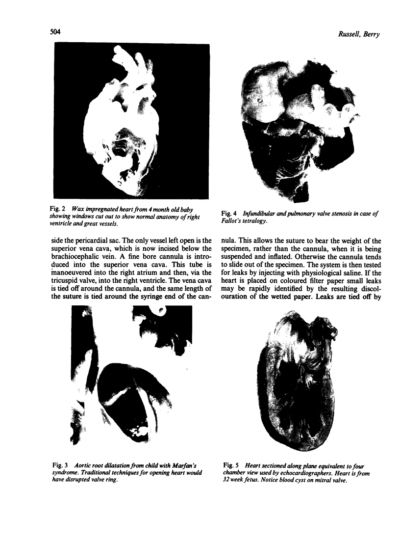
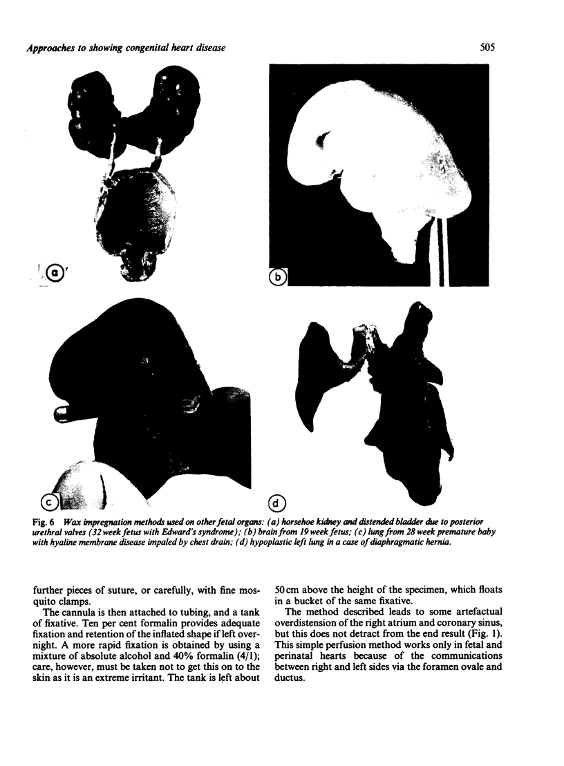
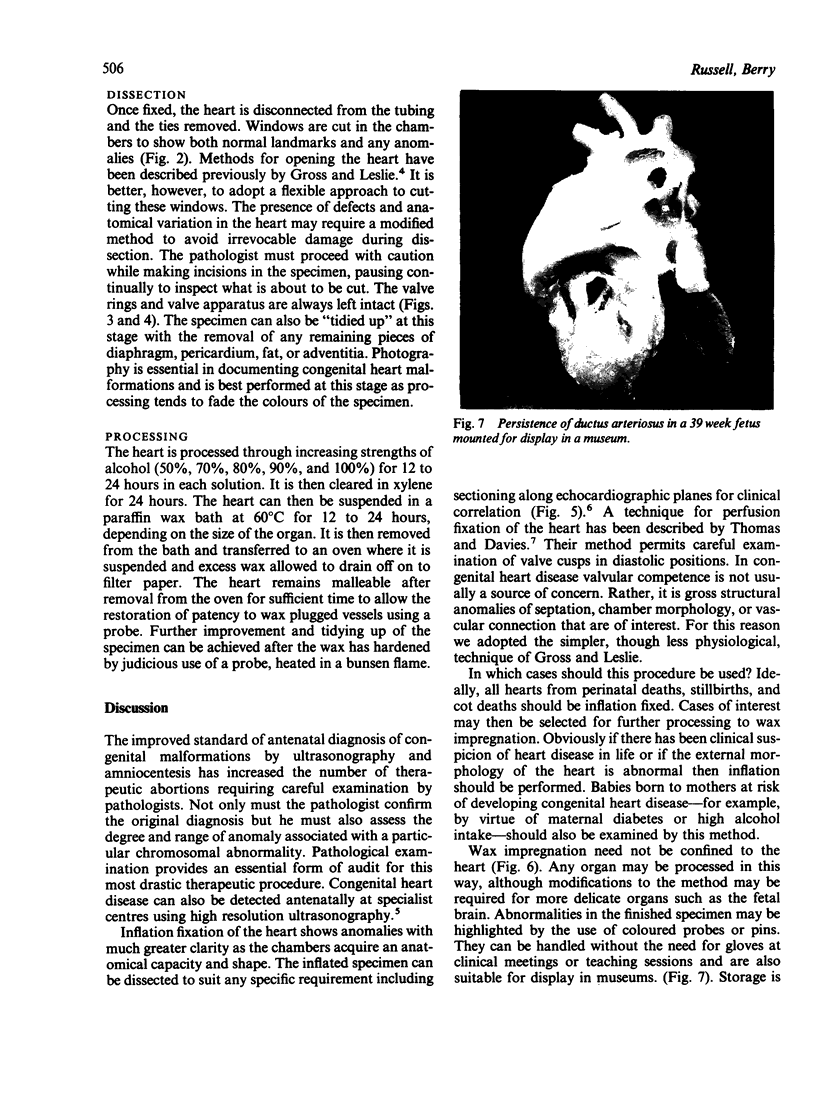
Abstract
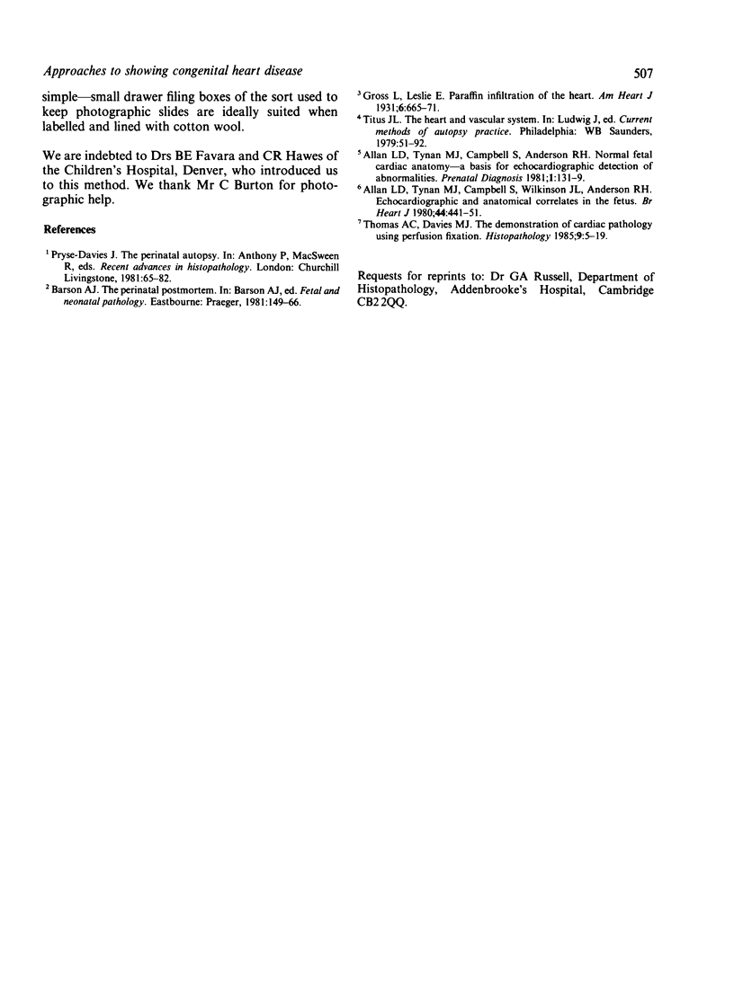
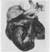
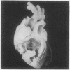
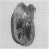
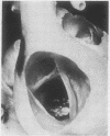
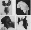
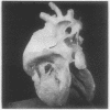
Advances in antenatal screening techniques have increased the interest in obtaining a detailed pathological correlation with the ultrasonographic findings obtained before death. As a consequence, inadequacies in traditional methods used by pathologists to display congenital malformations have been brought to light. We describe a simple technique of inflation and wax impregnation for the permanent proof of congenital heart defects that can be used in routine perinatal necropsies.
Full text
PDF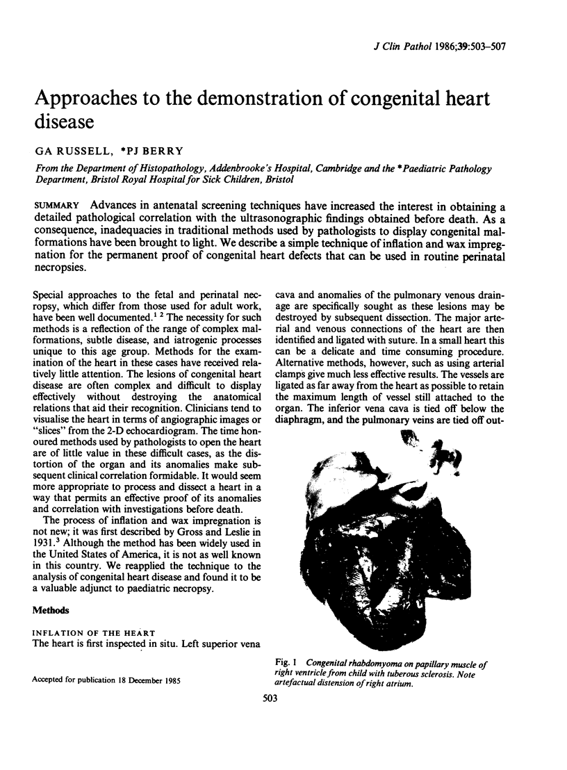
Images in this article
Selected References
These references are in PubMed. This may not be the complete list of references from this article.
- Allan L. D., Tynan M., Campbell S., Anderson R. H. Normal fetal cardiac anatomy--a basis for the echocardiographic detection of abnormalities. Prenat Diagn. 1981 Apr;1(2):131–139. doi: 10.1002/pd.1970010208. [DOI] [PubMed] [Google Scholar]
- Thomas A. C., Davies M. J. The demonstration of cardiac pathology using perfusion-fixation. Histopathology. 1985 Jan;9(1):5–19. doi: 10.1111/j.1365-2559.1985.tb02967.x. [DOI] [PubMed] [Google Scholar]