The presence of hemichorea associated with a lesion in the cortex is exceedingly rare. We report a case of a 62-year-old female who developed delayed left hemichorea after an ischemic infarct in the right temporal–occipital lobe. This case is unique because of the location of infarct and expands our knowledge of hemichorea arising after an ischemic stroke.
A 62-year-old female patient was initially admitted to the hospital after waking up that morning on the floor beside her bed, with a severe right temporal headache. She denied any associated photophobia, phonophobia, weakness, facial droop, slurred speech, dizziness, double vision, nausea or vomiting. The patient had a remote history of sarcoidosis (not under immunosuppression) and was being treated for hypertension. She denied use of alcohol, tobacco or illicit drugs. She had no family history of any neurodegenerative disease. During neurological examination, the patient was fully conscious. Left homonymous hemianopia with normal extraocular movements was noted. Muscle strength was preserved in all four limbs and plantar responses were flexor. A slight decrease in tactile and thermal sensations was found on the left side, but vibration and joint position were intact.
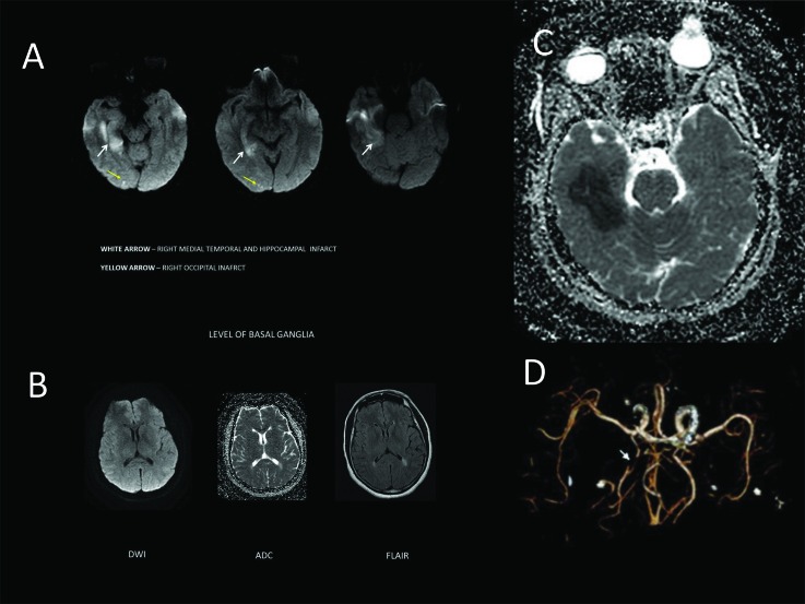
Laboratory findings, including levels of electrolytes and blood sugar, were within the normal limits. Magnetic resonance imaging (MRI) of the brain revealed an acute ischemic stroke involving predominantly the right mesial-temporal and hippocampal cortical regions, and minimally the occipital cortex. The basal ganglia, thalamus, and subthalamic nucleus (STN) were entirely spared. Computed tomography angiogram of the head and neck revealed focal occlusion in the right posterior cerebral artery (PCA) (P2 segment) (Figure 1). The patient was prescribed a treatment with 325 mg of aspirin, 75 mg of clopidogrel, and 80 mg of atorvastatin in addition to an anti-hypertensive regimen, and discharged home.
Figure 1. Neuroimaging studies: Axial Diffusion weighted imaging (DWI). (A) shows restricted diffusion in the right temporal lobe and scattered hyperintensity in right occipital lobe. Axial Apparent diffussion coefficient (ADC) (C) shows hypointensity in the same area. Axial ADC, DWI, and Fluid attenuation inversion recovery (FLAIR) (B) do not show any evidence of infarct in the basal ganglia or thalamus. Computed tomography angiogram of neck (D) shows occlusion of the right posterior cerebral artery.
Three months after hospital discharge, the patient presented for an outpatient disability assessment at the neurology clinic. At this time, she reported involuntary movements on the left side, involving preferentially her leg than her arm, which developed 1 week after the stroke and persisted until the last follow-up. Upon examination, these movements involving left upper and lower extremities were irregular, involuntary, purposeless, non-rhythmic, and rapid (Video 1). The patient was not aware of whether these movements persisted in her sleep. She was offered tetrabenazine to relieve the movements, but she chose to remain off treatment.
Video 1. Left-sided hemichorea. The video demonstrates irregular, involuntary, purposeless, non-rhythmic, rapid, and partially suppressible movements in the left upper and lower extremity. There were no abnormal movements in the face and it is not shown.
This patient presented with a delayed onset of hemichorea after an ischemic infarct in the right PCA, involving the temporal and occipital lobes. Few cases of hemichorea with PCA stenosis leading to an infarct in the thalamus1,2 have been reported, but, in this case, there was no evidence of infarct in the thalamus or the STN. A possible cause of hemichorea in this case may have been ischemia in the STN, as this region is supplied by penetrating branches of the PCA, and ischemic changes were not visible on brain MRI. In the Lausanne stroke registry,2 hyperkinetic movement disorders were seen in only 1% of the patients following stroke, and none of them presented with a cortical location of the stroke. In older patients, chorea is the commonest movement disorder after stroke, but dystonia is common in younger age groups.3 The pathophysiology underlying the delayed onset of movement disorders after central nervous system (CNS) injury is not well known. It has been hypothesized that such symptoms may be explained by the changes in CNS plasticity after injury and the re-establishment of functional synapses between the injured CNS neurons and their target cells.4 STN is involved in a minority of the cases of hemichorea, and the lesions are mostly observed in basal ganglia, thalamus, or cortex.5 It is advisable to look for proprioceptive loss, as stroke in the parietal lobe can lead to pseudoathetosis.6 Hemichorea arising after cortical stroke has better prognosis than when it follows other types of stroke.1 Perfusion imaging of the brain performed in cases of cortical hemichorea has rendered conflicting results. A case of temporal–parietal lobe infarct showed reduced perfusion in the ipsilateral basal ganglion, and a series of three patients with cortical strokes showed no perfusion changes in the basal ganglia or thalamus.7,8 It is possible that some cases of hemichorea result from an interruption of cortical projections to the basal ganglia, but others arise following an interruption of inter-cortical network leading to sensorimotor disintegration.8 Current management of hemichorea involves the use of antidopaminergic agents or benzodiazepines.5 As antidopaminergic agents can lead to parkinsonism or tardive dyskinesia, alternative agents such as topiramate may be useful.9 In cases of medically refractory hemichorea, deep brain stimulation should be considered given that long-term follow-up has shown good results.10
Footnotes
Funding: None.
Financial Disclosures: None.
Conflict of Interest: All patients that appear on video have provided written informed consent; authorization for the videotaping and for publication of the videotape was provided.
Ethics Statement: Not applicable for this category of article.
References
- 1.Chung SJ, Im JH, Lee MC, Kim JS. Hemichorea after stroke: clinical-radiological correlation. J Neurol. 2004;251:725–729. doi: 10.1007/s00415-004-0412-5. doi: http://dx.doi.org/10.1007/s00415-004-0412-5. [DOI] [PubMed] [Google Scholar]
- 2.Ghika-Schmid F, Ghika J, Regli F, Bogousslavsky J. Hyperkinetic movement disorders during and after acute stroke: the Lausanne Stroke Registry. J Neurol Sci. 1997;146:109–116. doi: 10.1016/s0022-510x(96)00290-0. doi: http://dx.doi.org/10.1016/S0022-510X(96)00290-0. [DOI] [PubMed] [Google Scholar]
- 3.Alarcón F, Zijlmans JC, Dueñas G, Cevallos N. Post-stroke movement disorders: report of 56 patients. J Neurol Neurosurg Psychiatry. 2004;75:1568–1574. doi: 10.1136/jnnp.2003.011874. doi: http://dx.doi.org/10.1136/jnnp.2003.011874. [DOI] [PMC free article] [PubMed] [Google Scholar]
- 4.Scott BL, Jankovic J. Delayed-onset progressive movement disorders after static brain lesions. Neurology. 1996;46:68–74. doi: 10.1212/wnl.46.1.68. doi: http://dx.doi.org/10.1212/WNL.46.1.68. [DOI] [PubMed] [Google Scholar]
- 5.Postuma RB, Lang AE. Hemiballism: revisiting a classic disorder. Lancet Neurol. 2003;2:661–668. doi: 10.1016/s1474-4422(03)00554-4. doi: http://dx.doi.org/10.1016/S1474-4422(03)00554-4. [DOI] [PubMed] [Google Scholar]
- 6.Mehanna R, Jankovic J. Movement disorders in cerebrovascular disease. Lancet Neurol. 2013;12:597–608. doi: 10.1016/S1474-4422(13)70057-7. doi: http://dx.doi.org/10.1016/S1474-4422(13)70057-7. [DOI] [PubMed] [Google Scholar]
- 7.Murakami T, Wada T, Sasaki I, et al. Hemichorea-hemiballism in a patient with temporal-parietal lobe infarction appearing after reperfusion by recombinant tissue plasminogen activator. Mov Disord Clin Pract. 2015;2:426–428. doi: 10.1002/mdc3.12198. doi: http://dx.doi.org/10.1002/mdc3.12198. [DOI] [PMC free article] [PubMed] [Google Scholar]
- 8.Hwang KJ, Hong IK, Ahn TB, Yi SH, Lee D, Kim DY. Cortical hemichorea-hemiballism. J Neurol. 2013;260:2986–2992. doi: 10.1007/s00415-013-7096-7. doi: http://dx.doi.org/10.1007/s00415-013-7096-7. [DOI] [PubMed] [Google Scholar]
- 9.Kim JA, Jung S, Kim MJ, Kwon SB, Hwang SH, Kwon KH. A case of vascular hemichorea responding to topiramate. J Mov Disord. 2009;2:80–81. doi: 10.14802/jmd.09021. doi: http://dx.doi.org/10.14802/jmd.09021. [DOI] [PMC free article] [PubMed] [Google Scholar]
- 10.Hasegawa H, Mundil N, Samuel M, Jarosz J, Ashkan K. The treatment of persistent vascular hemidystonia-hemiballismus with unilateral GPi deep brain stimulation. Mov Disord. 2009;24:1697–1698. doi: 10.1002/mds.22598. doi: http://dx.doi.org/10.1002/mds.22598. [DOI] [PubMed] [Google Scholar]


