Abstract
Introduction
We developed a device (FreeO2) that automatically adjusts the oxygen flow rates based on patients’ needs, in order to limit hyperoxia and hypoxemia and to automatically wean them from oxygen.
Objective
The aim of this study was to evaluate the feasibility of using FreeO2 in patients hospitalized in the respiratory ward for an acute exacerbation of COPD.
Methods
We conducted a randomized controlled trial comparing FreeO2 vs manual oxygen titration in the respiratory ward of a university hospital. We measured the perception of appropriateness of oxygen titration and monitoring in both groups by nurses and attending physicians using a Likert scale. We evaluated the time in the target range of oxygen saturation (SpO2) as defined for each patient by the attending physician, the time with severe desaturation (SpO2 <85%), and the time with hyperoxia (SpO2 >5% above the target). We also recorded length of stay, intensive care unit admissions, and readmission rate. Fifty patients were randomized (25 patients in both groups; mean age: 72±8 years; mean forced expiratory volume in 1 second: 1.00±0.49 L; and mean initial O2 flow 2.0±1.0 L/min).
Results
Nurses and attending physicians felt that oxygen titration and monitoring were equally appropriate with both O2 administration systems. The percentage of time within the SpO2 target was significantly higher with FreeO2, and the time with severe desaturation and hyperoxia was significantly reduced with FreeO2. Time from study inclusion to hospital discharge was 5.8±4.4 days with FreeO2 and 8.4±6.0 days with usual oxygen administration (P=0.051).
Conclusion
FreeO2 was deemed as an appropriate oxygen administration system by nurses and physicians of a respiratory unit. This system maintained SpO2 at the target level better than did manual titration and reduced periods of desaturation and hyperoxia. Our results also suggest that FreeO2 has the potential to reduce the hospital length of stay.
Keywords: oxygen inhalation therapy, technological innovations, hypoxia, hyperoxia, closed-loop
Video abstract
Introduction
Patients with COPD who are hospitalized for an acute exacerbation often receive oxygen therapy as a part of their treatment. Despite current recommendations,1 oxygen therapy is often suboptimally adjusted during acute exacerbation of COPD, with episodes of both hypoxemia and hyperoxia reported.2 Hyperoxia may be particularly problematic in COPD because of its association with hypercapnia,3–7 especially in the acute phase of exacerbations,3,4,8 where it may be associated with adverse clinical outcomes, including respiratory acidosis and even increased mortality.4,8,9 Continuous adjustment of oxygen flows is time consuming, and clinicians are more concerned with oxygen desaturation than with hyperoxia, potentially leading to the utilization of higher than required oxygen flows for longer than necessary.
Closed-loop adjustment of oxygen administration based on pulsed oxygen saturation (SpO2) may optimize oxygen therapy and improve patients’ safety.10 We recently developed a closed-loop device, FreeO2, that automatically adjusts the oxygen flows administered to spontaneously breathing patients to maintain SpO2 in a predefined target set by physicians and to progressively and automatically wean patients from oxygen.11 This pilot trial was intended to test the acceptability of FreeO2 by nurses and attending physicians and the potential clinical impact of using such a device in patients hospitalized in the respiratory ward for acute exacerbation of COPD. Our hypotheses were that 1) nurses and physicians would conclude that FreeO2 is an acceptable oxygen administration device, 2) time spent in the target SpO2 zone would be increased by automatic O2 flow adjustments in comparison to manual O2 titration, and 3) weaning time from oxygen would be shortened by the automatic O2 adjustment device (FreeO2).
Methods
Participants
We conducted a randomized trial (Clinical Trial Registration: NCT01393015) comparing manual oxygen titration and automated oxygen adjustment with FreeO2 in patients hospitalized for an acute exacerbation of COPD in whom oxygen therapy was prescribed by the attending physician based on the documentation of resting hypoxemia (SpO2 <90%). We included patients aged ≥40 years with a past or current smoking history of at least 10 pack-years. Patients had to maintain an SpO2 of ≥92% with supplemental oxygen at a maximum flow rate of 8 L/min. We excluded patients admitted for >24 hours, patients infected with multidrug-resistant bacteria, patients on intermittent noninvasive ventilation (including CPAP for obstructive sleep apnea), and patients with cognitive impairment precluding informed consent or collaboration. The study was conducted at the Institut Universitaire de Cardiologie et de Pneumologie de Québec. The study was approved by the Institutional Ethics Committee (# 20694), and a signed consent was obtained from each participant.
FreeO2 system
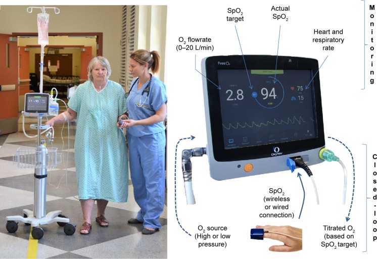
The FreeO2 system automatically adjusts oxygen flow rates administered via nasal cannula or nonocclusive mask, using a closed-loop algorithm based on physiological data.11 It also provides continuous monitoring of respiratory parameters in spontaneously breathing patients.11 The main parameter that is monitored within this closed-loop system is SpO2 that continuously feeds the algorithm at a rate of 1 value/s. A proportional integral controller adjusts the oxygen flow delivered by a mass-flow controller from 0 L/min to 20 L/min (flow accuracy ±0.1 L/min), with the aim of maintaining SpO2 at a predefined target. The system was developed by the authors (FL and ELH) in collaboration with the Department of Electronic and Informatics Engineering, Laval University, Quebec (Figure 1).
Figure 1.
Main features of the FreeO2: oxygen automated titration and weaning with SpO2–O2 flow closed-loop and monitoring.
Notes: The FreeO2 adjusts the oxygen flow rate from 0 L/min to 20 L/min (flow accuracy ±0.1 L/min) every second, based on the patient’s needs. A proportional integral controller adjusts the oxygen flow based on the difference between the SpO2 target set by clinicians and the continuously measured SpO2. Several cardiorespiratory parameters are continuously recorded (O2 flow rate, SpO2, respiratory rate, and heart rate), and trends for these parameters are available for clinicians. Both heart rate and respiratory rate are derived from the pulse oximeter plethysmographic wave form. The version used in the study was a previous version of the device with similar technical features.
Protocol
Randomization was conducted with sealed envelopes after inclusion of the patients in the study. Randomization sequences were externally generated using www.random.org. Patients were allocated to either 1) automated oxygen titration by FreeO2 with continuous monitoring of physiological parameters, including SpO2 and oxygen flow rates (FreeO2 arm), at bedside and remotely at the nursing station or 2) manual oxygen administration with bedside titration from usual oxygen flow-meter (rotameter) and usual oxygen monitoring with bedside pulsed oximetry (control arm). In the control group, the nurses manually adjusted the oxygen flows guided by local protocols and by SpO2 target provided by the attending physician before randomization. SpO2 monitoring was conducted as per the local practice (ie, approximately three times per day). However, there were no specific training to the oxygen management guidelines and no recording of the compliance to these guidelines. In the FreeO2 arm, oxygen was titrated automatically every second to maintain SpO2 in the target zone set by the attending physician before randomization.
The principal investigators of the study did not manage the patients included in the study and were not involved in the treatment and discharge decisions.
Measurements
At baseline, demographic data, arterial or capillary blood gases, oxygen flow rate, and respiratory rate were recorded. Pulmonary function tests (forced expiratory volume in 1 second and forced vital capacity) were conducted using standardized postbronchodilator spirometry. These pulmonary parameters were recorded during a stable period of COPD within 1 year before or after exacerbation and during hospitalization.
In order to evaluate whether FreeO2 could be used in daily practice and to quantify its acceptance and ease of use by caregivers (primary outcomes), the nurses and physicians’ perception of the appropriateness of the oxygen management was monitored by direct interview when available on a daily basis using a 10-point Likert scale (from 0: not appropriate at all to 10: perfectly appropriate). The secondary outcomes were 1) time spent within the SpO2 target (±2%), 2) time with severe desaturation (SpO2 <85%), 3) time with hyperoxia (SpO2 >5% above the target), 4) total duration of oxygen therapy, and 5) hospital length of stay.
Patients in both groups had continuous monitoring of SpO2, respiratory rate, heart rate, and end-tidal CO2 with the FreeO2 system set in the “FreeO2 mode” (automated closed-loop adjustment of oxygen and continuous recording) or in the “recording mode” (continuous recording of physiological data without administration of oxygen by the device for those in the control group). Capillary blood gases were collected every day during the first week or up to discharge.
Statistical analyses
We could not calculate a priori a sample size based on the primary outcome as we did not have any data to estimate how the system would be accepted by the caregivers. Nevertheless, we chose to include 50 patients to obtain sufficient exposure and experience with FreeO2. Data were expressed using mean with standard deviation or median with interquartile ranges for continuous variables or proportions for categorical data, respectively. Categorical variables were compared between groups using the chi-square or Fisher’s exact tests, and continuous variables were compared using one-way analyses of variance. Wilcoxon rank sum tests (nonparametric tests) were performed for data that did not fulfill the normality or variance assumptions after transformation. When different conclusions between analyses from one-way analysis of variance and nonparametric tests occurred, the latest approach was retained. The results were considered significant with P-values ≤0.05. All analyses were conducted using the statistical package SAS, Version 9.4 (SAS Institute Inc., Cary, NC, USA).
Results
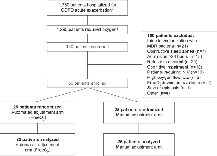
We randomized 25 patients with COPD in each group (total of 50 patients) from August 2011 to February 2015. The flow chart of patient participation in the study is shown in Figure 2. Patients’ baseline characteristics are shown in Table 1. Overall, baseline characteristics were similar between the two study groups. The mean age was 72±9 years, and the mean forced expiratory volume in 1 second was 1.00±0.49 L, representing 37%±22% predicted. Twenty-three patients (46%) were women.
Figure 2.
Flow chart of the study.
Note: *Based on administrative records of the institution.
Abbreviations: MDR, multidrug resistant; NIV, noninvasive ventilation.
Table 1.
Patient characteristics
| Characteristics | FreeO2 patients (n=25) | Control patients (n=25) |
|---|---|---|
| Age (years) | 71±8 | 73±8 |
| Men, n (%) | 15 (60) | 12 (48) |
| ED admission to inclusion (days) | 0.6±0.7 | 0.6±1.1 |
| Spirometry during stable statea | ||
| FEV1 (L) | 1.1±0.3 | 1.1±0.4 |
| FEV1 (% predicted) | 40±11 | 51±20 |
| FEV1/FVC (%) | 41±0.11 | 47±20 |
| Spirometry during hospital stayb | ||
| FEV1 (L) | 1.0±0.5 | 1.0±0.5 |
| FEV1 (% predicted) | 36±19 | 38±24 |
| FEV1/FVC (%) | 45±12 | 49±13 |
| Physiologic data at study inclusion | ||
| O2 flow (L/min) | 2.0±1.0 | 2.0±1.0 |
| pH | 7.40±0.05 | 7.39±0.05 |
| PaCO2 (mmHg) | 46±12 | 46±11 |
| HCO3− (mmol/L) | 28±5 | 27±5 |
| SpO2 (%) | 93±2 | 93±2 |
| Body temperature (°C) | 36.6±0.6 | 36.7±0.6 |
| Respiratory rate (breaths/min) | 22±3 | 21±2 |
Notes: Data expressed as mean ± SD, unless otherwise specified.
Forced spirometry performed within 1 year before or after hospitalization, during a stable state (value available for 62% of the patients).
Forced spirometry performed during the hospital stay (available for all patients).
Abbreviations: ED, emergency department; FEV1, forced expiratory volume in 1 second; FVC, forced vital capacity; SD, standard deviation.
Primary outcomes
There were 924 evaluations of the oxygen adjustment and monitoring by nurses and 96 evaluations by physicians. Nurses and physicians considered FreeO2 adjustments and monitoring to be at least as appropriate and as acceptable as manual oxygen management. Only monitoring was deemed slightly better with FreeO2 by the physicians, but the difference was not statistically significant (Table 2). We did not directly evaluate patient’s tolerance of the system, but 80% of the patients completed the study in both groups. The main reason to prematurely stop the study was related to the difficulty in continuously wearing a pulse oximeter and the reduced mobility (70% of the reasons to stop); this occurred similarly in both groups.
Table 2.
Evaluation of the nurses’ and physicians’ perception of the appropriateness of the oxygen therapy management
| FreeO2 (n=25) | Control (n=25) | P-value | |
|---|---|---|---|
| Nurses | |||
| Oxygen titration | 8.9±1.5 | 8.8±1.8 | 0.46 |
| Oxygen monitoring | 8.9±1.4 | 8.7±2.0 | 0.19 |
| Physicians | |||
| Oxygen titration | 8.2±2.2 | 7.8±2.1 | 0.48 |
| Oxygen monitoring | 8.2±2.2 | 6.7±3.2 | 0.07 |
Notes: Data expressed as mean ± standard deviation, unless otherwise specified. Based on a 10-point Likert scale (from 0, not appropriate at all, to 10, perfectly appropriate).
Secondary outcomes
Oxygenation and blood gases
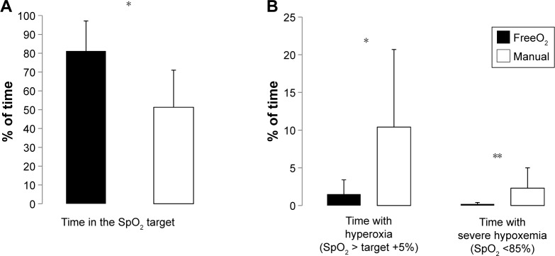
SpO2 targets chosen by physicians were similar in both groups, averaging 90.0% and 90.1% for FreeO2 and manual O2 adjustment, respectively. The mean SpO2 during the study was 90.9±1.2 in the FreeO2 group and 91.9±1.2 in the manual adjustment group (P=0.009). The proportion of time within SpO2 target was 81.2%±19.9% with FreeO2 vs 51.3%±19.7% with manual O2 adjustments (P<0.001). The percentage of time with severe desaturation (SpO2 <85%) and with hyperoxia (SpO2 >5% above the target) was significantly lower with FreeO2 in comparison with manual oxygen adjustment (Figure 3 and Table 3). There was no significant difference in blood gases measured on day 3 and day 7 between the two groups.
Figure 3.
Percentage of time in the SpO2 target (A), with hyperoxia and with severe hypoxemia (B) with FreeO2 (black bars) and with manual adjustment (white bars).
Notes: *P<0.001. **P=0.01.
Table 3.
Oxygenation and capillary blood gases
| FreeO2 | Control | P-value | |
|---|---|---|---|
| SpO2 target defined by the physicians (%)a | 90.0±1.2 | 90.1±1.0 | 0.89 |
| Mean SpO2 (%) | 90.9±1.2 | 91.9±1.2 | 0.009 |
| % of time in the SpO2 target ±2% | 81.2±15.9 | 51.3±19.7 | <0.001 |
| % of time with hyperoxiab | 1.5±1.9 | 10.4±10.3 | <0.001 |
| % of time with hypoxemiac | 0.2±0.2 | 2.3±2.7 | 0.001 |
| Mean O2 flow (L/min)d | 0.7±0.7 | 1.2±1.0 | 0.06 |
| % of time with SpO2 signal available | 90.4±7.6 | 82.4±12.7 | 0.01 |
| Day 3e | |||
| pH | 7.42±0.03 | 7.41±0.05 | 0.33 |
| PaCO2 | 45±7 | 46±10 | 0.62 |
| HCO3− | 29±4 | 29±5 | 0.94 |
| O2 flow (L/min) | 1.0±1.1 | 1.6±1.4 | 0.21 |
| Day 7f | |||
| pH | 7.43±0.02 | 7.42±0.06 | 0.46 |
| PaCO2 | 40±4 | 47±13 | 0.09 |
| HCO3− | 27±2 | 30±6 | 0.13 |
| O2 flow (L/min) | 0.6±0.4 | 1.2±1.0 | 0.47 |
Notes: Data expressed as mean ± standard deviation, unless otherwise specified.
At study entry.
Hyperoxia was defined by SpO2 >5% above the target.
Hypoxemia was defined by SpO2 <85%.
During the time of recording.
Data available (n=25 in both groups).
Data available (n=12 in the FreeO2 arm and n=15 in the control group).
Clinical outcomes
Duration of oxygen administration was reduced by 1.8 days with FreeO2, but this difference did not reach statistical significance (Table 4). Time from randomization to hospital discharge was reduced by 2.6 days with FreeO2 (P=0.051). There was no difference between the two groups in the requirement for noninvasive ventilation during hospitalization, need of transfer to the intensive care unit, or death. The readmission rates at 30, 60, and 180 days were also similar in the two groups.
Table 4.
Clinical outcome
| Outcome | FreeO2 patients (n=25) | Control patients (n=25) | P-value |
|---|---|---|---|
| Duration of O2 administration (days) | 4.0±2.1 | 5.8±9.9 | 0.14 |
| Length of hospital stay | |||
| Randomization to hospital discharge (days) | 5.8±4.4 | 8.4±6.0 | 0.051 |
| Admission to hospital discharge (days) | 6.7±4.3 | 9.5±6.0 | 0.053 |
| Complications | |||
| NIV (n) | 0 | 1 | 0.48 |
| ICU transfer (n) | 0 | 1 | 0.48 |
| Death (n) | 1 | 1 | 1 |
| Readmission rate | |||
| 30 days (n) | 6 | 6 | 1 |
| 60 days (n) | 6 | 9 | 0.54 |
| 180 days (n) | 10 | 13 | 0.57 |
Note: Data expressed as mean ± standard deviation, unless otherwise specified.
Abbreviations: ICU, intensive care unit; NIV, noninvasive ventilation.
Safety
There was no safety issue as we did not record any oxygen delivery interruption with FreeO2. Few technical issues occurred with end-tidal CO2 monitoring, but none concerned the oxygen delivery valve, and this was not associated with safety issues.
Discussion
Our pilot trial demonstrates the feasibility of providing oxygen therapy with FreeO2 to patients hospitalized for an acute exacerbation of COPD, even during prolonged administration of oxygen (up to 8 consecutive days). The device was well accepted by nurses and physicians. FreeO2 maintained SpO2 within the prespecified target better than manual oxygen administration. It also reduced the periods of hyperoxia and desaturation. In addition, our results suggest that FreeO2 may have the potential to reduce hospital length of stay, although this pilot observation requires formal testing in a proper clinical trial.
Our study is the first to investigate the potential effects of a continuous and an automated oxygen titration and weaning system on physiological and clinical outcomes in patients hospitalized for an acute exacerbation of COPD. Several studies evaluated similar systems in patients with COPD in other settings, such as home care12 or during rehabilitation,13,14 and in other populations, including neonates15,16 and in patients with acute respiratory failure in the emergency department.17 Automated therapy has always proved effective in safely providing oxygen in these settings and populations, and our results are consistent with this notion.
The introduction of new devices in clinical practice may not be well accepted, and barriers to technology acceptance should not be overlooked.18 The primary study outcome was selected accordingly. FreeO2 was deemed at least as adequate for oxygen administration and monitoring as usual oxygen administration. Also, most patients tolerated the instrument, as 80% completed the study in both groups. Few patients did not complete the study due to reduced mobility related to the continuous oximetry recording. Implementation of an oximeter with wireless communication features may further improve the acceptance of O2 automated systems in the future.
We were successful in continuously recording respiratory parameters with identical oximeters in both groups with second-by-second data available 90.4% and 82.4% of the time in the FreeO2 and control groups, respectively. As expected and in line with other evaluations of automated oxygen titration,10,14,17 oxygenation parameters were improved in the FreeO2 group, with more time spent in the targeted range of SpO2 and less time with oxygen desaturation or hyperoxia. This may be clinically relevant when considering that hyperoxia may induce hypercapnia3–7 with potentially severe consequences.4,8,9 Hyperoxia is also of concern in patients with COPD who frequently have coronary artery disease,19 as it may increase coronary artery resistance.20,21 On the other hand, periods of oxygen desaturations may be responsible for arythmias and myocardial ischemia,22,23 and they should also be avoided.
In the control group, patients were in the SpO2 target 50% of the time, which may not be compared with other data, as these are the first data with continuous recording of SpO2 in this situation to our knowledge. In the recent review of Cousin et al,24 the mean rate of accurate prescription of oxygen therapy in eleven studies was 17.4% before and 51.2% after implementation of specific interventions.
One strength of our study is that we could evaluate an automated oxygen therapy during prolonged duration (ie, up to 8 days), compared with previous studies where automated O2 titration was provided for a duration of <1 hour11,13,14 and up to 3 hours.17 An important result of our trial is the trend toward a reduction in duration of oxygen therapy and hospital length of stay with automated oxygen titration, although we cannot ascertain whether this effect was related to closer O2 monitoring and surveillance as a part of the FreeO2 protocol or whether this was due to the FreeO2 device itself. Although the trial was not primarily designed to demonstrate an impact on these outcomes, this finding is in agreement with the hypothesis that unnecessarily high flows and prolonged oxygen therapy are common25 and that automated weaning may be useful to abbreviate oxygen therapy in hospitalized patients with COPD. Length of stay must be interpreted in comparison to local statistics obtained off-protocol. In 2010, the mean length of stay for acute exacerbation of COPD in our institution was 9.8±8.1 days (unpublished data), which is in-line with the length of stay in the control group of the present study. Reducing the length of hospital stay by 31% as found in this study would be expected to have major logistic and financial impacts on our health care setting. This study was underpowered to conclude on the hospital length of stay, and larger studies will be required to confirm this potential benefit of automated oxygen titration.
Our study has potential limitations that should be considered for proper interpretation. First, this was a pilot study with a small sample size. A more complete evaluation of relevant clinical outcomes, such as length of stay and cost effectiveness, will likely require more patients. Second, the recruitment rate was low as we recruited only 50 patients in 3 years. The main reason was the limited resources during prolonged periods that did not allow continuous screening of potential study participants. There were >500 COPD hospitalized every year during the study period, and several of these would have been eligible to the trial. Third, the study was not blinded. Nurses had to be aware of the patients’ allocation as they were in charge of the manual titration in the control group, and remote monitoring of cardiorespiratory parameters at the nursing station was available only in the FreeO2 arm. To limit the impact of this potential source of bias, study investigators were not involved in any treatment or discharge decisions.
Conclusion
This study demonstrates the feasibility of using automated oxygen titration and weaning as well as remote monitoring with the FreeO2 system in patients hospitalized for acute exacerbation of COPD. The system was well accepted by both nurses and physicians. Automated oxygen titration provided benefits in terms of safety (ie, reduction in the time with severe desaturation and hyperoxia) and may have contributed to a trend toward the reduced hospital length of stay, a potentially important finding, if these benefits are confirmed in subsequent studies specifically designed to demonstrate these effects.
Acknowledgments
Our research laboratory has been funded by the Canadian Foundation for Innovation (Leaders Opportunity Funds) to develop automated systems for respiratory support. Part of this funding was used to make FreeO2 prototypes. Funding sources had no role in study design, in the collection, analysis, and interpretation of data, in the writing of the report, and in the decision to submit the article for publication. OxyNov provided the FreeO2 device prototypes. This study was supported by the Groupe de recherche en santé respiratoire de l’Université Laval. This study was presented in part at the 2015 International Conference of the American Thoracic Society, May 15–20, Denver, USA.
Footnotes
Author contributions
All authors contributed toward data analysis, drafting and critically revising the paper, gave final approval of the version to be published, and agree to be accountable for all aspects of the work.
Disclosure
The Fond de Recherche en Santé du Québec contributes to FL’s salary for research activities (clinical research scholar) and to the research assistant’s salary (clinical research grant). FL and ELH are the coinventors of the FreeO2 system and made the first prototypes with the engineering department of Laval University. FL and ELH are the cofounders of a Laval University spin-off research and development company (OxyNov) to develop automated systems for respiratory support. FM holds a GlaxoSmithKline/Canadian Institutes of Health Research chair on COPD at Laval University. FM and YL participate in Innovair, a company that owns shares in OxyNov, the owner of the FreeO2 device. PAB and MR have no financial interests that may be relevant to the submitted work. The authors report no other conflicts of interest in this work.
References
- 1.Pauwels RA, Buist AS, Calverley PM, Jenkins CR, Hurd SS, GOLD Scientific Committee Global strategy for the diagnosis, management, and prevention of chronic obstructive pulmonary disease. NHLBI/WHO Global Initiative for chronic obstructive lung disease (GOLD) Workshop summary. Am J Respir Crit Care Med. 2001;163(5):1256–1276. doi: 10.1164/ajrccm.163.5.2101039. [DOI] [PubMed] [Google Scholar]
- 2.Hale KE, Gavin C, O’Driscoll BR. Audit of oxygen use in emergency ambulances and in a hospital emergency department. Emerg Med J. 2008;25(11):773–776. doi: 10.1136/emj.2008.059287. [DOI] [PubMed] [Google Scholar]
- 3.Aubier M, Murciano D, Fournier M, Milic-Emili J, Pariente R, Derenne JP. Central respiratory drive in acute respiratory failure of patients with chronic obstructive pulmonary disease. Am Rev Respir Dis. 1980;122(2):191–199. doi: 10.1164/arrd.1980.122.2.191. [DOI] [PubMed] [Google Scholar]
- 4.Austin MA, Wills KE, Blizzard L, Walters EH, Wood-Baker R. Effect of high flow oxygen on mortality in chronic obstructive pulmonary disease patients in prehospital setting: randomised controlled trial. BMJ. 2010;341:c5462. doi: 10.1136/bmj.c5462. [DOI] [PMC free article] [PubMed] [Google Scholar]
- 5.Beasley R, Patel M, Perrin K, O’Driscoll BR. High-concentration oxygen therapy in COPD. Lancet. 2011;378(9795):969–970. doi: 10.1016/S0140-6736(11)61431-1. [DOI] [PubMed] [Google Scholar]
- 6.Sassoon CS, Hassell KT, Mahutte CK. Hyperoxic-induced hypercapnia in stable chronic obstructive pulmonary disease. Am Rev Respir Dis. 1987;135(4):907–911. doi: 10.1164/arrd.1987.135.4.907. [DOI] [PubMed] [Google Scholar]
- 7.Mithoefer JC, Karetzky MS, Mead GD. Oxygen therapy in respiratory failure. N Engl J Med. 1967;277(18):947–949. doi: 10.1056/NEJM196711022771802. [DOI] [PubMed] [Google Scholar]
- 8.Plant PK, Owen JL, Elliott MW. One year period prevalence study of respiratory acidosis in acute exacerbations of COPD: implications for the provision of non-invasive ventilation and oxygen administration. Thorax. 2000;55(7):550–554. doi: 10.1136/thorax.55.7.550. [DOI] [PMC free article] [PubMed] [Google Scholar]
- 9.Warren PM, Flenley DC, Millar JS, Avery A. Respiratory failure revisited: acute exacerbations of chronic bronchitis between 1961–68 and 1970–76. Lancet. 1980;1(8166):467–470. doi: 10.1016/s0140-6736(80)91008-9. [DOI] [PubMed] [Google Scholar]
- 10.Claure N, Bancalari E. Automated closed loop control of inspired oxygen concentration. Respir Care. 2013;58(1):151–161. doi: 10.4187/respcare.01955. [DOI] [PubMed] [Google Scholar]
- 11.Lellouche F, L’Her E. Automated oxygen flow titration to maintain constant oxygenation. Respir Care. 2012;57(8):1254–1262. doi: 10.4187/respcare.01343. [DOI] [PubMed] [Google Scholar]
- 12.Rice KL, Schmidt MF, Buan JS, Lebahn F, Schwarzock TK. A portable, closed-loop oxygen conserving device for stable COPD patients: comparison with fixed dose delivery systems. Respir Care. 2011;56(12):1901–1905. [PubMed] [Google Scholar]
- 13.Cirio S, Nava S. Pilot study of a new device to titrate oxygen flow in hypoxic patients on long-term oxygen therapy. Respir Care. 2011;56(4):429–434. doi: 10.4187/respcare.00983. [DOI] [PubMed] [Google Scholar]
- 14.Lellouche F, Maltais F, Bouchard PA, Brouillard C, L’Her E. FreeO2: closed-loop adaptation of oxygen flow based on SpO2. Evaluation in COPD patients during effort. Respir Care. 2016 [Google Scholar]
- 15.Claure N, D’Ugard C, Bancalari E. Automated adjustment of inspired oxygen in preterm infants with frequent fluctuations in oxygenation: a pilot clinical trial. J Pediatr. 2009;155(5):640-5.e1–640-5.e2. doi: 10.1016/j.jpeds.2009.04.057. [DOI] [PubMed] [Google Scholar]
- 16.Claure N, Gerhardt T, Everett R, Musante G, Herrera C, Bancalari E. Closed-loop controlled inspired oxygen concentration for mechanically ventilated very low birth weight infants with frequent episodes of hypoxemia. Pediatrics. 2001;107(5):1120–1124. doi: 10.1542/peds.107.5.1120. [DOI] [PubMed] [Google Scholar]
- 17.L’Her E, Dias P, Gouillou M, et al. Automatisation of oxygen titration in patients with acute respiratory distress at the emergency department. A multicentric international randomized controlled study. Am J Respir Critic Care Med. 2015;191:A6329. [Google Scholar]
- 18.Boonstra A, Broekhuis M. Barriers to the acceptance of electronic medical records by physicians from systematic review to taxonomy and interventions. BMC Health Serv Res. 2010;10:231. doi: 10.1186/1472-6963-10-231. [DOI] [PMC free article] [PubMed] [Google Scholar]
- 19.Vanfleteren LE, Franssen FM, Uszko-Lencer NH, et al. Frequency and relevance of ischemic electrocardiographic findings in patients with chronic obstructive pulmonary disease. Am J Cardiol. 2011;108(11):1669–1674. doi: 10.1016/j.amjcard.2011.07.027. [DOI] [PubMed] [Google Scholar]
- 20.Farquhar H, Weatherall M, Wijesinghe M, et al. Systematic review of studies of the effect of hyperoxia on coronary blood flow. Am Heart J. 2009;158(3):371–377. doi: 10.1016/j.ahj.2009.05.037. [DOI] [PubMed] [Google Scholar]
- 21.McNulty PH, King N, Scott S, et al. Effects of supplemental oxygen administration on coronary blood flow in patients undergoing cardiac catheterization. Am J Physiol Heart Circ Physiol. 2005;288(3):H1057–H1062. doi: 10.1152/ajpheart.00625.2004. [DOI] [PubMed] [Google Scholar]
- 22.Galatius-Jensen S, Hansen J, Rasmussen V, Bildsoe J, Therboe M, Rosenberg J. Nocturnal hypoxaemia after myocardial infarction: association with nocturnal myocardial ischaemia and arrhythmias. Br Heart J. 1994;72(1):23–30. doi: 10.1136/hrt.72.1.23. [DOI] [PMC free article] [PubMed] [Google Scholar]
- 23.Gogenur I, Rosenberg-Adamsen S, Lie C, Carstensen M, Rasmussen V, Rosenberg J. Relationship between nocturnal hypoxaemia, tachycardia and myocardial ischaemia after major abdominal surgery. Br J Anaesth. 2004;93(3):333–338. doi: 10.1093/bja/aeh208. [DOI] [PubMed] [Google Scholar]
- 24.Cousins JL, Wark PA, McDonald VM. Acute oxygen therapy: a review of prescribing and delivery practices. Int J Chron Obstruct Pulmon Dis. 2016;11:1067–1075. doi: 10.2147/COPD.S103607. [DOI] [PMC free article] [PubMed] [Google Scholar]
- 25.Brill SE, Wedzicha JA. Oxygen therapy in acute exacerbations of chronic obstructive pulmonary disease. Int J Chron Obstruct Pulmon Dis. 2014;9:1241–1252. doi: 10.2147/COPD.S41476. [DOI] [PMC free article] [PubMed] [Google Scholar]


