

Abstract
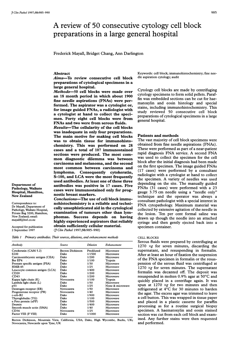
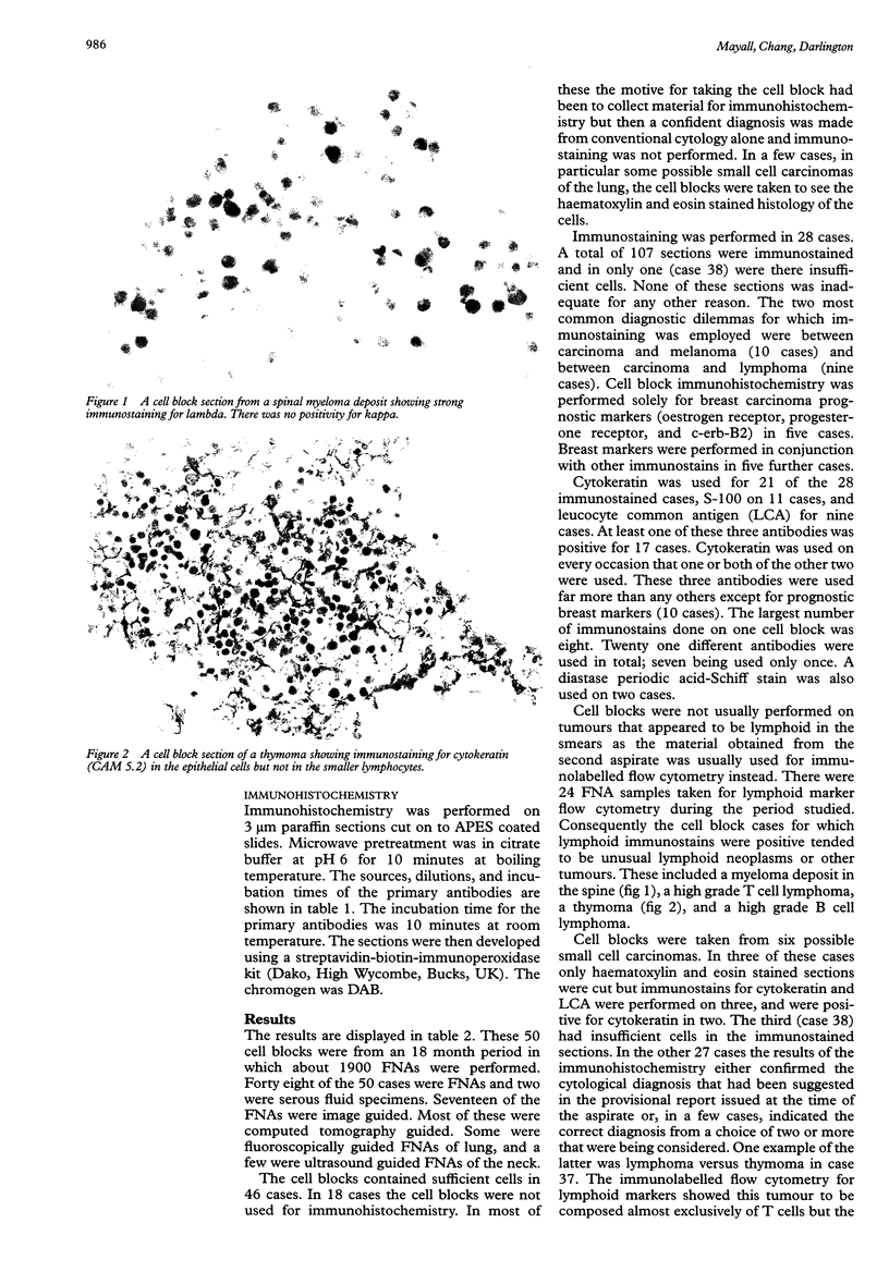
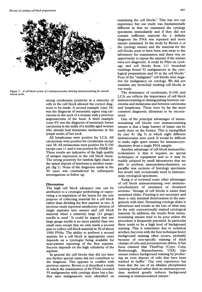
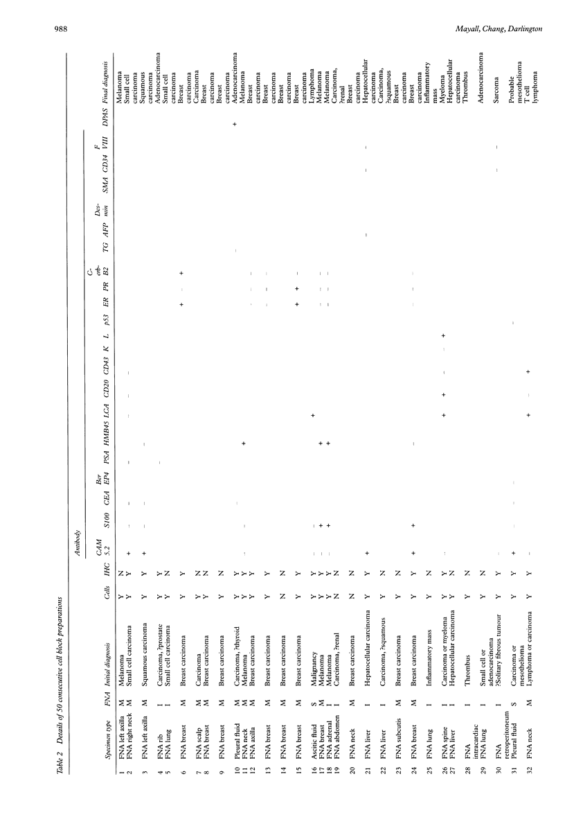
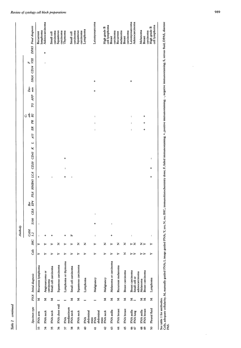
AIMS: To review consecutive cell block preparations of cytological specimens in a large general hospital. METHODS: 50 cell blocks were made over an 18 month period in which about 1900 fine needle aspirations (FNAs) were performed. The aspirator was a cytologist or, for image guided FNAs, a radiologist with a cytologist at hand to collect the specimen. Forty eight cell blocks were from FNAs and two were from serous fluids. RESULTS: The cellularity of the cell blocks was inadequate in only four preparations. The main motive for making cell blocks was to obtain tissue for immunohistochemistry. This was performed on 28 cases and a total of 107 immunostained sections were produced. The most common diagnostic dilemma was between carcinoma and melanoma, and the second most common between carcinoma and lymphoma. Consequently cytokeratin, S-100, and LCA were the most frequently used antibodies. At least one of these three antibodies was positive in 17 cases. Five cases were immunostained only for prognostic breast markers. CONCLUSIONS: The use of cell block immunohistochemistry is a reliable and technically unsophisticated aid in the cytological examination of tumours other than lymphomas. Success depends on having highly experienced aspirators that reliably obtain sufficiently cellular material.
Full text
PDF
Images in this article
Selected References
These references are in PubMed. This may not be the complete list of references from this article.
- Brown K. T., Fulbright R. K., Avitabile A. M., Bashist B. Cytologic analysis in fine-needle aspiration biopsy: smears vs cell blocks. AJR Am J Roentgenol. 1993 Sep;161(3):629–631. doi: 10.2214/ajr.161.3.8352121. [DOI] [PubMed] [Google Scholar]
- Chernoff W. G., Lampe H. B., Cramer H., Banerjee D. The potential clinical impact of the fine needle aspiration/flow cytometric diagnosis of malignant lymphoma. J Otolaryngol. 1992 Feb;21 (Suppl 1):1–15. [PubMed] [Google Scholar]
- Flint A. Detection of pulmonary neoplasms by bronchial washings. Are cell blocks a diagnostic aid? Acta Cytol. 1993 Jan-Feb;37(1):21–23. [PubMed] [Google Scholar]
- Hanson C. A. Fine-needle aspiration and immunophenotyping. A role in diagnostic hematopathology? Am J Clin Pathol. 1994 May;101(5):555–556. doi: 10.1093/ajcp/101.5.555. [DOI] [PubMed] [Google Scholar]
- Kung I. T., Chan S. K., Lo E. S. Application of the immunoperoxidase technique to cell block preparations from fine needle aspirates. Acta Cytol. 1990 May-Jun;34(3):297–303. [PubMed] [Google Scholar]
- Leung S. W., Bédard Y. C. Immunocytochemical staining on ThinPrep processed smears. Mod Pathol. 1996 Mar;9(3):304–306. [PubMed] [Google Scholar]
- Zito F. A., Gadaleta C. D., Salvatore C., Filotico R., Labriola A., Marzullo A., Prete F., Marzullo F. A modified cell block technique for fine needle aspiration cytology. Acta Cytol. 1995 Jan-Feb;39(1):93–99. [PubMed] [Google Scholar]