Abstract
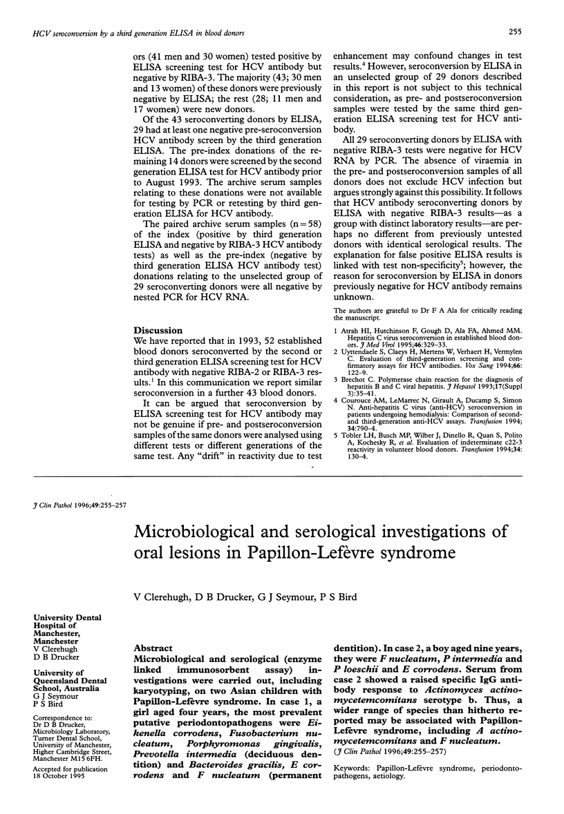
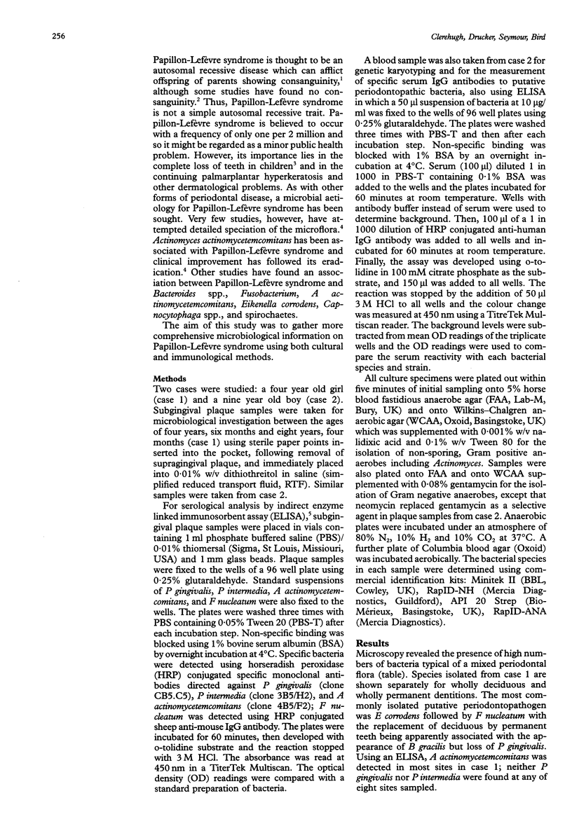
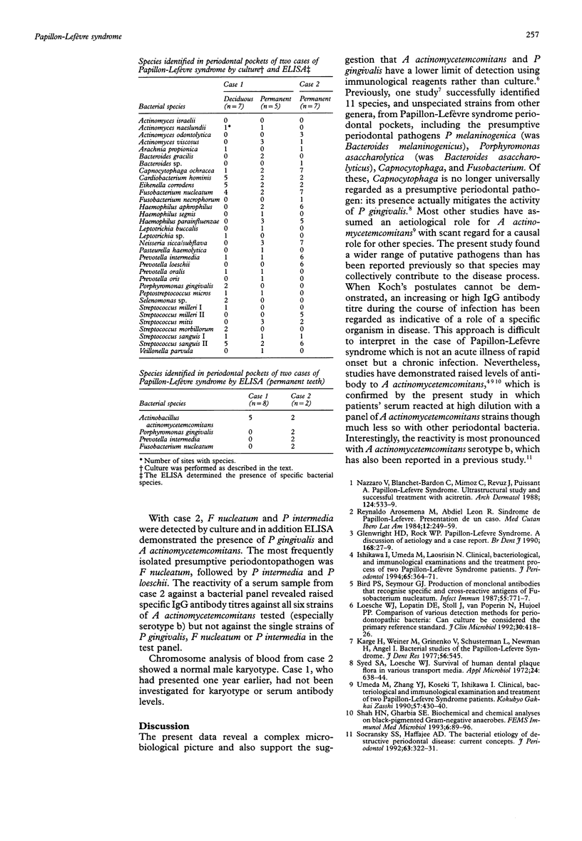
Microbiological and serological (enzyme linked immunosorbent assay) investigations were carried out, including karyotyping, on two Asian children with Papillon-Lefèvre syndrome. In case 1, a girl aged four years, the most prevalent putative periodontopathogens were Eikenella corrodens, Fusobacterium nucleatum, Porphyromonas gingivalis, Prevotella intermedia (deciduous dentition) and Bacteroides gracilis, E corrodens and F nucleatum (permanent dentition). In case 2, a boy aged nine years, they were F nucleatum, P intermedia and P loeschii and E corrodens. Serum from case 2 showed a raised specific IgG antibody response to Actinomyces actino-mycetemcomitans serotype b. Thus, a wider range of species than hitherto reported may be associated with Papillon-Lefèvre syndrome, including A actino-mycetemcomitans and F nucleatum.
Full text
PDF


Selected References
These references are in PubMed. This may not be the complete list of references from this article.
- Bird P. S., Seymour G. J. Production of monoclonal antibodies that recognize specific and cross-reactive antigens of Fusobacterium nucleatum. Infect Immun. 1987 Mar;55(3):771–777. doi: 10.1128/iai.55.3.771-777.1987. [DOI] [PMC free article] [PubMed] [Google Scholar]
- Glenwright H. D., Rock W. P. Papillon-Lefevre syndrome. A discussion of aetiology and a case report. Br Dent J. 1990 Jan 6;168(1):27–29. doi: 10.1038/sj.bdj.4807059. [DOI] [PubMed] [Google Scholar]
- Ishikawa I., Umeda M., Laosrisin N. Clinical, bacteriological, and immunological examinations and the treatment process of two Papillon-Lefèvre syndrome patients. J Periodontol. 1994 Apr;65(4):364–371. doi: 10.1902/jop.1994.65.4.364. [DOI] [PubMed] [Google Scholar]
- Loesche W. J., Lopatin D. E., Stoll J., van Poperin N., Hujoel P. P. Comparison of various detection methods for periodontopathic bacteria: can culture be considered the primary reference standard? J Clin Microbiol. 1992 Feb;30(2):418–426. doi: 10.1128/jcm.30.2.418-426.1992. [DOI] [PMC free article] [PubMed] [Google Scholar]
- Nazzaro V., Blanchet-Bardon C., Mimoz C., Revuz J., Puissant A. Papillon-Lefèvre syndrome. Ultrastructural study and successful treatment with acitretin. Arch Dermatol. 1988 Apr;124(4):533–539. [PubMed] [Google Scholar]
- Newman M., Angel I., Karge H., Weiner M., Grinenko V., Schusterman L. Bacterial studies of the Papillon-Lefévre syndrome. J Dent Res. 1977 May;56(5):545–545. doi: 10.1177/00220345770560052201. [DOI] [PubMed] [Google Scholar]
- Reynaldo Arosemena M., Abdiel León R. Síndrome de Papillon-Lefèvre. Presentación de un caso. Med Cutan Ibero Lat Am. 1984;12(3):245–249. [PubMed] [Google Scholar]
- Shah H. N., Gharbia S. E. Biochemical and chemical analyses of black-pigmented gram-negative anaerobes. FEMS Immunol Med Microbiol. 1993 Mar;6(2-3):89–96. doi: 10.1111/j.1574-695X.1993.tb00308.x. [DOI] [PubMed] [Google Scholar]
- Socransky S. S., Haffajee A. D. The bacterial etiology of destructive periodontal disease: current concepts. J Periodontol. 1992 Apr;63(4 Suppl):322–331. doi: 10.1902/jop.1992.63.4s.322. [DOI] [PubMed] [Google Scholar]
- Syed S. A., Loesche W. J. Survival of human dental plaque flora in various transport media. Appl Microbiol. 1972 Oct;24(4):638–644. doi: 10.1128/am.24.4.638-644.1972. [DOI] [PMC free article] [PubMed] [Google Scholar]
- Umeda M., Zhang Y. J., Koseki T., Ishikawa I. [Clinical, bacteriological, and immunological examination and treatment of two Papillon-Lefevre syndrome patients]. Kokubyo Gakkai Zasshi. 1990 Sep;57(3):430–440. doi: 10.5357/koubyou.57.430. [DOI] [PubMed] [Google Scholar]


