Abstract
Introduction
Basal cell carcinoma (BCC) is an immunogenic neoplasm and the imbalance in Th1/Th2 cytokines expression seems to play the major role in pathogenesis and clinical behaviour of the tumour.
Aim
To investigate the association of soluble interleukin 2α receptor (sIL-2Rα) and interleukin-2 (IL-2) serum concentrations with BCC.
Material and methods
The study involved 110 individuals with BCC and 60 healthy age- and sex-matched volunteers. Serum levels of sIL-2Rα and IL-2 were measured using ELISA test.
Results
We found significantly (p = 0.027) increased sIL-2Rα serum levels in BCC patients, in comparison to healthy controls. Statistically (p = 0.04) higher sIL-2Rα levels were observed in patients with more advanced tumours. Serum levels of sIL-2Rα showed a significant linear (r = 0.24, p = 0.018) correlation with tumour size. The average IL-2 serum levels in BCC patients were statistically (p = 0.039) decreased compared to controls. Significantly (p = 0.0454) lower median IL-2 levels were observed in patients with more advanced tumours. A negative correlation between sIL-2Rα and IL-2 serum concentrations was revealed (r = –0.22; p = 0.027).
Conclusions
Our results testify to the importance of the IL-2/sIL-2Rα signalling pathway in pathogenesis of BCC, suggesting that IL-2 and sIL-2Rα might be considered as potential markers of disease and targets for immunotherapy in BCC patients.
Keywords: basal cell carcinoma, soluble interleukin-2 receptor α, interleukin-2, sIL-2Rα, IL-2
Introduction
Several studies have been focused on understanding the role of the immune system in development and progression of cancer in recent decades. It was shown that various cytokines secreted by neoplastic and host cells play an essential role as mediators and regulators of tumour-host interactions. Many types of malignant solid tumours have been found to be associated with marked alterations of pro- and anti-inflammatory cytokines’ patterns, reflecting the tumours’ biological aggressiveness, clinical advancement and prognosis [1].
Basal cell carcinoma (BCC) is an immunogenic neoplasm. The tumour tissue is infiltrated by regulatory CD4+, CD25+, Foxp3+ T regulatory (Treg) lymphocytes and immature dendritic cells. The immune response seems to play the major role in spontaneous and pharmacologic induced (imiquimod) BCC regression [2–4].
Interleukin 2 (IL-2) plays a central role in the activation of T cell-mediated immune response. It is known as the main cytokine involved in effector T (Teff) and Treg cells’ proliferation and differentiation. In relation to neoplastic process, IL-2 plays an essential role in anti-tumour immunity. It has been reported that transfection of the IL-2 gene into tumour cells may enhance both specific and nonspecific anti-tumour immune responses [5–8].
Interleukin-2 exerts its biological effects via signalling through its receptor system, which is required for T-cell functioning. The high affinity cell-bound IL-2R consists of three polypeptide subunits: IL-2Rα (CD25); IL-2Rβ (CD122) and IL-2Rγ (CD132). IL-2Rβ and IL-2Rγ are constitutively expressed on the surface of T-cells, while IL-2Rα expression increases rapidly after T-cell activation. The IL-2Rα is shed from the cell surface and can be measurable in the serum as a 45 kDa soluble form (sIL-2Rα), thus serving as a surrogate marker for T cell activation [9]. Several types of malignancies, mainly of lymphoproliferative origin, have been found to release large amounts of sIL-2Rα from their unstimulated cells [10, 11]. It is suggested that IL-2Rα may stimulate the proliferation of neoplastic cells and reflect the tumour burden and aggressiveness [9, 12]. Circulating sIL-2Rα is able to bind IL-2 efficiently, competing for the ligand with the cellular receptor on normal lymphocytes, thus blocking anti-tumour immunity dependent on IL-2 [13]. Accordingly, elevated initial levels of serum sIL-2Rα correlating with poorer response to treatment and survival were noted in various malignancies [9].
Aim
To our best knowledge, serum levels of sIL-2Rα have not been explored in patients with BCC yet. Therefore, in this study we analysed the concentrations of the cytokines in a numerous group of BCC patients and a control group to investigate the possible correlations between the analysed markers and some clinical aspects of disease.
Material and methods
Patients and control group
This study group included 110 unrelated patients with BCC (42 women, 68 men; mean age at diagnosis: 68.9 ±11.8 years) and 60 healthy, unrelated age- and sex-matched volunteers (control group). Patients were treated surgically because of primary or recurrent BCC, in the Department of Dermatology, Venereology and Allergology, Medical University of Gdansk, Poland, between 2009 and 2010. Individuals with histopathological features of tumours regression were excluded. The clinical characteristics of the patients are shown in Table 1. None of the subjects were organ transplant recipients, suffered from any systemic inflammatory disease or other malignancy, nor have been treated with immunosuppressive drugs. The patients and controls had no symptoms and signs of infection during and at least 2 weeks before the samples collection.
Table 1.
Characteristics of 110 BCC patients
| Variables | Number (%) |
|---|---|
| Gender: | |
| Males | 68 (62) |
| Females | 42 (38) |
| Tumour size [cm]: | |
| ≤ 1 | 49 (45) |
| > 1 | 61 (55) |
| Recognition: | |
| Primary BCC | 78 (71) |
| Recurrent BCC | 32 (29) |
| Number of tumours: | |
| One tumour | 79 (72) |
| More than one tumour | 31 (28) |
| Location: | |
| Area exposed to UV | 81 (74) |
| Area not exposed to UV | 29 (36) |
Serum IL-2 and sIL-2Rα levels measurement
Serum levels of IL-2 and sIL-2Rα proteins were measured with ELISA test (Human ELISA kit, Diaclone SAS, France), following the manufacturer's instructions. The proteins concentrations were not affected by the age or sex at enrolment either in the BCC patients or in the controls.
Statistical analysis
The Mann-Whitney U-test was used to compare the mean values and the correlation was determined by mean Spearman coefficient values. Analyses were performed with Statistica 10.0 data analysis software system (StatSoft, Inc. 2011). P ≤ 0.05 was considered statistically significant.
Results
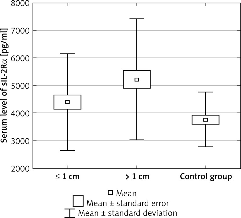
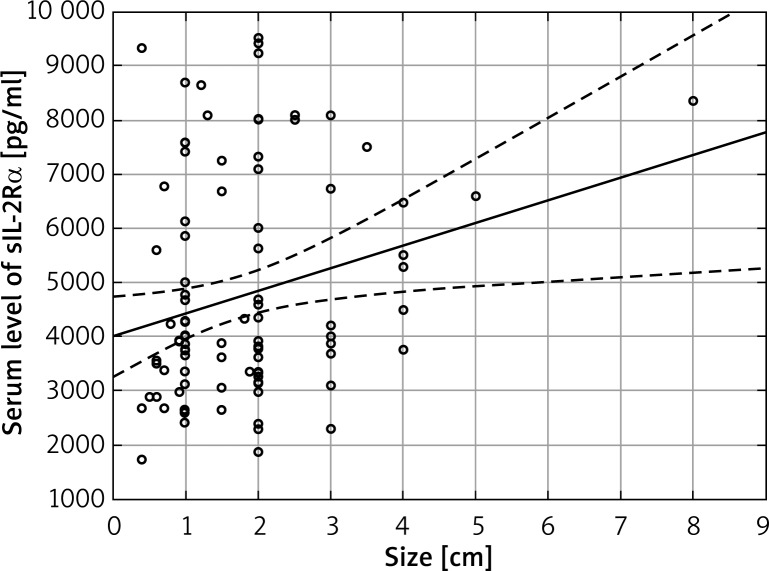
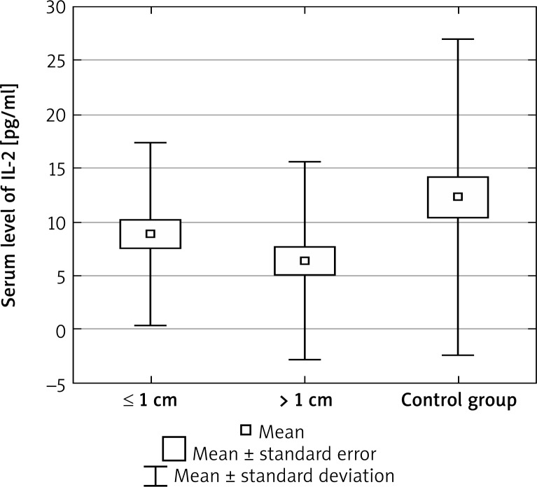
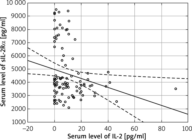
The mean and median sIL-2Rα and IL-2 serum levels in patients and controls are shown in Table 2. We found significantly increased sIL-2Rα serum levels in BCC patients, in comparison to healthy controls (mean: 4855.794 ±2019.533 pg/ml vs. 3764.990 ±998.536 pg/ml; p = 0.027). Moreover, statistically higher sIL-2Rα levels were observed in patients with tumours larger than 1 cm in diameter (mean: 5218.061 ±2165.194 pg/ml vs. 4393.370 ±1752.960 pg/ml; p = 0.04) (Figure 1). Serum levels of sIL-2Rα showed a significant linear correlation with tumour size: r = 0.24, p = 0.018 (Figure 2). The average IL-2 serum levels in BCC patients were statistically decreased as compared to controls (mean: 8.148 ±9.0487 pg/ml vs. 12.308 ±14.726 pg/ml; p = 0.039). We also noted lower mean IL-2 levels in patients with tumours larger than 1 cm (mean: 6.348 ±9.188 pg/ml vs. 8.859 ±8.523 pg/ml; p = 0.0454) (Figure 3). A negative correlation between sIL-2Rα and IL-2 serum concentrations was revealed: r = –0.22; p = 0.027 (Figure 4).
Table 2.
Comparison between serum levels of sIL-2Rα and IL-2 in BCC patients and controls
| Parameter | BCC patients N = 110 |
Controls N = 60 |
P-value |
|---|---|---|---|
| sIL-2Rα serum level [pg/ml] |
Mean: 4855.794 ±2019.533 Median: 3900.450 Range: 2285–9500 |
Mean: 3764.997 ±998.536 Median: 3834.830 Range: 2094–7642 |
0.026817 |
| IL-2 serum level [pg/ml] |
Mean: 8.14791 ±9.04868 Median: 4.885 Range: 0–40.29 |
Mean: 12.30850 ±14.72643 Median: 8.535 Range: 0–90.75 |
0.039 |
Figure 1.
sIL-2Rα serum levels in BCC patients in relation to tumour size and in healthy controls. Statistically higher sIL-2Rα levels in patients with tumours larger than 1 cm in diameter compared to smaller ones (mean: 5218.061 ±2165.194 pg/ml vs. 4393.370 ±1752.960 pg/ml; p = 0.04)
Figure 2.
Linear correlation between serum levels of sIL-2Rα and tumour size (r = 0.24, p = 0.018)
Figure 3.
IL-2 serum levels in BCC patients in relation to tumour size and in healthy controls. Lower mean IL-2 levels in patients with tumours larger than 1 cm compared to smaller ones (mean: 6.348750 ±9.187712 pg/ml vs. 8.859556 ± 8.523332 pg/ml; p = 0.0454)
Figure 4.
Negative linear correlation between sIL-2Rα and IL-2 serum levels in patients with BCC (r = –0.22; p = 0.027)
Discussion
Interactions between the immune system and neoplastic cells play an important role in tumorigenesis. Anti-tumour responses in cancer are generally attributed to the activation of tumour-specific T-cells (CD4+ and CD8+), particularly at the tumours microenvironment. It has been confirmed that IL-2 is able to stimulate NK cells and CD8+ T-cells lysis of tumour targets, which was the reason to consider its role in anti-neoplastic response as a biological medicament [14].
The present study is the first to report the significance of diminished serum IL-2 and elevated sIL-2Rα concentrations in patients with BCC. High sIL-2Rα serum levels were noted in various malignancies and have been correlated with disease progression and poor response to treatment [9, 15–17].
The role of sIL-2Rα was explored in a few cutaneous malignancies, including cutaneous T cell lymphoma, and melanoma, however none of them have concerned BCC patients [18–20]. Ottaiano et al. [19] proved that high or increasing initial serum levels of sIL-2Rα in patients with malignant melanoma correlated with a higher Breslow scale, progression of the tumour and significantly lower 5-year disease-free survival rate.
As we noted, elevated serum levels of sIL-2Rα were present in BCC patients as compared to the healthy controls. Moreover, serum concentration of sIL-2Rα correlated positively with the size of the tumour. Our results are similar to those found in other malignant solid tumours, including melanoma, nasopharyngeal carcinoma, non-small cell lung cancer and, head and neck carcinoma. In these studies elevation of serum sIL-2Rα level was associated with the disease stage and/or progression and has been revealed as a negative prognostic factor [9, 18, 19, 21–23].
The source of the sIL-2Rα in the serum of BCC patients has not been defined. The host and tumour origin should be considered. Serum soluble IL-2Rα has been associated with its cell-bound receptor expression on malignant cells of leukemias and lymphomas thus providing a reliable marker of disease extent and IL-2/IL-2R pathway activity [11]. In cases of malignant solid tumours, the biological source and role of sIL-2Rα is more complex. It seems to be mainly released from activated lymphoid cells, especially those infiltrating neoplastic tissue [9].
The biological function of sIL-2Rα in neoplasms is unclear. Eicher and Waldmann [24] showed that sIL-2Rα on one cell may augment the IL-2 signalling by presenting IL-2 to the sIL-2Rβ/γ on another cell. Thus, the extent of sIL-2Rα expression may reflect the magnitude of clone expansion and the resultant immune response. These findings may partially explain the diminished immunocompetence in patients with the impairment of sIL-2Rα expression and function on effector cells and/or decreased IL-2 production.
Our results seem to reflect the finding of Rubin et al. [13] that high concentrations of circulating sIL-2Rα compete for IL-2 binding with the cellular receptor on normal lymphocytes. This may contribute to diminishing of the IL-2-dependent anti-tumour immunity and result in BCC progression. However, the role of sIL-2Rα in BCC pathogenesis seems to be more complex. Glaser et al. [25] noted that lower levels of CD25, CD3ɛ, CD68 and ICAM-1 mRNA in BCC biopsies at baseline predicted subsequent BCC. Halliday et al. [26] showed a higher CD25 mRNA expression in actively regressing BCC tumours compared with these without evidence of current or past regression.
Our data have revealed that the IL-2 serum levels were significantly lower in BCC patients as compared to controls. There are not many reports concerning this issue; however, our results are consistent with most of the existing studies. Wei et al. [27] had demonstrated significantly lower IL-2 serum concentrations in individuals with nasopharyngeal carcinoma. Li et al. [28] have shown lower levels of Th1 (IL-2, IFN-γ) and higher Th2 cytokines (IL-4, IL-10) in patients with non-small cell lung cancer. Similarly, the BCC microenvironment is dominated by Th2 cytokines (enhanced expression of IL-4, IL-5, IL-6, IL-10 and CCL22) [29].
Moreover, Wong et al. [4] reported that actively regressing BCCs tended to express higher levels of IL-2 and IFN-γ, as detected by RT-PCR within the T cells infiltrating the tumour. This finding suggests that these Th1 cytokines might play a role in promoting BCC regression. Elsässer-Beile et al. [30] have not found differences in IL-2 levels in the whole blood cell cultures between BCC patients and controls. Interestingly, we noted a significantly lower concentration of cytokine in tumours larger than 1 cm. Also, the tendency to a negative linear correlation between IL-2 serum levels and tumour size was noted; however, it was not statistically significant.
These results suggest that diminution of IL-2 response might play a role in BCC pathogenesis and clinical behaviour of the tumour. High sIL-2Rα and low serum IL-2 levels seem to be possible markers of a more aggressive clinical course of BCC. The negative correlation between sIL-2Rα and IL-2 serum levels in patients with BCC is a novel finding and strongly suggests the role of IL-2/sIL-2Rα signalling pathway in pathogenesis of BCC. These phenomena have to be explored in further investigations.
Kaur et al. [2] demonstrated distinct epithelial-stromal-inflammatory patterns of BCC, which correlated with tumour subtypes and their progression. We suppose that subtypes of BCC may be also characterized by distinct serum cytokine patterns. In our opinion, the main limitation of this study was that all BCC patients have been analysed as a homogenous group without division into histopathological subtypes. However, that designed investigation would require a definitely larger study group to make statistical analysis reliable.
Because of BCC immunogenicity, also the role of others cytokines, including IL-6, IL-8 and tumor necrosis factor (TNF)-α, were explored and shown to be involved in BCC pathogenesis [5, 8, 23, 29, 31, 32]. These fields seem to be interesting and novel research is required.
Recently novel therapeutic strategies including immunotherapy have been introduced in oncology. The administration of unmodified and modified IL-2 formulations were approved in metastatic melanoma and renal cell carcinoma treatment [19]. Also in BCC the intralesional IL-2-based immunotherapy (PEG-IL-2) seems to be a promising novel approach. Kaplan and Moy [33] reported 67% of complete responses in patients with BCC treated with perilesional PEG-IL-2 injections. The treatment was found encouraging, safe and well-tolerated. The sIL-2Rα has been also established as a target for immunotherapy of several CD25(+) malignancies [9]. Our results support the thesis that sIL-2Rα and IL-2 can be also considered new therapeutic approaches in BCC treatment.
Conclusions
Ours study has shown for the first time the clinical significance of the measurements of serum levels of IL-2 and sIL-2Rα in BCC patients. Our results testify to the importance of the IL-2/sIL-2Rα signalling pathway in pathogenesis of BCC, suggesting that IL-2 and sIL-2Rα might be considered as potential markers of disease and targets for immunotherapy in BCC patients.
Acknowledgments
Funding source: The study was funded by the Medical University of Gdansk, project no. 02-0066/07.
The study was approved by the local ethics committee of the Medical University of Gdansk.
Conflict of interest
The authors declare no conflict of interest.
References
- 1.Lippitz BE. Cytokine patterns in patients with cancer: a systematic review. Lancet Oncol. 2013;14:e218–28. doi: 10.1016/S1470-2045(12)70582-X. [DOI] [PubMed] [Google Scholar]
- 2.Kaur P, Mulvaney M, Carlson JA. Basal cell carcinoma progression correlates with host immune response and stromal alterations: a histologic analysis. Am J Dermatopathol. 2006;28:293–307. doi: 10.1097/00000372-200608000-00002. [DOI] [PubMed] [Google Scholar]
- 3.Urosevic M, Maier T, Benninghoff B, et al. Mechanisms underlying imiquimod-induced regression of basal cell carcinoma in vivo. Arch Dermatol. 2003;139:1325–32. doi: 10.1001/archderm.139.10.1325. [DOI] [PubMed] [Google Scholar]
- 4.Wong DA, Bishop GA, Lowes MA, et al. Cytokine profiles in spontaneously regressing basal cell carcinomas. Br J Dermatol. 2000;143:91–8. doi: 10.1046/j.1365-2133.2000.03596.x. [DOI] [PubMed] [Google Scholar]
- 5.Masuda A, Arai K, Nishihara D, et al. Clinical significance of serum soluble T cell regulatory molecules in clear cell renal cell carcinoma. Biomed Res Int. 2014;2014:396064. doi: 10.1155/2014/396064. [DOI] [PMC free article] [PubMed] [Google Scholar]
- 6.Okamoto T, Tsuburaya A, Yanoma S, et al. Inhibition of peritoneal metastasis in an animal gastric cancer model by interferon-gamma and interleukin-2. Anticancer Res. 2003;23:149–53. [PubMed] [Google Scholar]
- 7.Sakaguchi S, Yamaguchi T, Nomura T, Ono M. Regulatory T cells and immune tolerance. Cell. 2008;133:775–87. doi: 10.1016/j.cell.2008.05.009. [DOI] [PubMed] [Google Scholar]
- 8.Zhang Y, Luo CL, He BC, et al. Exosomes derived from IL-12-anchored renal cancer cells increase induction of specific antitumor response in vitro: a novel vaccine for renal cell carcinoma. Int J Oncol. 2010;36:133–40. [PubMed] [Google Scholar]
- 9.Bien E, Balcerska A. Serum soluble interleukin 2 receptor alpha in human cancer of adults and children: a review. Biomarkers. 2008;13:1–26. doi: 10.1080/13547500701674063. [DOI] [PubMed] [Google Scholar]
- 10.Waldmann TA. The IL2/IL15 receptor systems: targets for immunotherapy. J Clin Immunol. 2002;22:51–6. doi: 10.1023/a:1014416616687. [DOI] [PubMed] [Google Scholar]
- 11.Zhang Z, Zhang M, Garmestani K, et al. Effective treatment of a murine model of adult T-cell leukemia using 211At-7G7/B6 and its combination with unmodified anti-Tac (daclizumab) directed toward CD25. Blood. 2006;108:1007–12. doi: 10.1182/blood-2005-11-4757. [DOI] [PMC free article] [PubMed] [Google Scholar]
- 12.Waldmann TA. Anti-Tac (daclizumab, Zenapax) in the treatment of leukemia, autoimmune diseases, and in the prevention of allograft rejection: a 25-year personal odyssey. J Clin Immunol. 2007;27:1–18. doi: 10.1007/s10875-006-9060-0. [DOI] [PubMed] [Google Scholar]
- 13.Rubin LA, Kurman CC, Fritz ME, et al. Soluble interleukin 2 receptors are released from activated human lymphoid cells in vitro. J Immunol. 1985;135:3172–7. [PubMed] [Google Scholar]
- 14.Rosalia RA, Arenas-Ramirez N, Bouchaud G, et al. Use of enhanced interleukin-2 formulations for improved immunotherapy against cancer. Curr Opin Chem Biol. 2014;23:39–46. doi: 10.1016/j.cbpa.2014.09.006. [DOI] [PubMed] [Google Scholar]
- 15.Bien E, Balcerska A, Kuchta G. Serum level of soluble interleukin-2 receptor alpha correlates with the clinical course and activity of Wilms’ tumour and soft tissue sarcomas in children. Biomarkers. 2007;12:203–13. doi: 10.1080/13547500601066410. [DOI] [PubMed] [Google Scholar]
- 16.El Houda Agueznay N, Badoual C, Hans S, et al. Soluble interleukin-2 receptor and metalloproteinase-9 expression in head and neck cancer: prognostic value and analysis of their relationships. Clin Exp Immunol. 2007;150:114–23. doi: 10.1111/j.1365-2249.2007.03464.x. [DOI] [PMC free article] [PubMed] [Google Scholar]
- 17.Wang LS, Chow KC, Li WY, et al. Clinical significance of serum soluble interleukin 2 receptor-alpha in esophageal squamous cell carcinoma. Clin Cancer Res. 2000;6:1445–51. [PubMed] [Google Scholar]
- 18.Boyano MD, Garcia-Vázquez MD, López-Michelena T, et al. Soluble interleukin-2 receptor, intercellular adhesion molecule-1 and interleukin-10 serum levels in patients with melanoma. Br J Cancer. 2000;83:847–52. doi: 10.1054/bjoc.2000.1402. [DOI] [PMC free article] [PubMed] [Google Scholar]
- 19.Ottaiano A, Leonardi E, Simeone E, et al. Soluble interleukin-2 receptor in stage I-III melanoma. Cytokine. 2006;33:150–5. doi: 10.1016/j.cyto.2006.01.002. [DOI] [PubMed] [Google Scholar]
- 20.Vonderheid EC, Zhang Q, Lessin SR, et al. Use of serum soluble interleukin-2 receptor levels to monitor the progression of cutaneous T-cell lymphoma. J Am Acad Dermatol. 1998;38:207–20. doi: 10.1016/s0190-9622(98)70597-3. [DOI] [PubMed] [Google Scholar]
- 21.Hsiao SH, Lee MS, Lin HY, et al. Clinical significance of measuring levels of tumor necrosis factor-alpha and soluble interleukin-2 receptor in nasopharyngeal carcinoma. Acta Otolaryngol. 2009;129:1519–23. doi: 10.3109/00016480902849427. [DOI] [PubMed] [Google Scholar]
- 22.Sakata H, Murakami S, Hirayama R. Serum soluble interleukin-2 receptor (IL-2R) and immunohistochemical staining of IL-2R/Tac antigen in colorectal cancer. Int J Clin Oncol. 2002;7:312–7. doi: 10.1007/s101470200046. [DOI] [PubMed] [Google Scholar]
- 23.Tartour E, Mosseri V, Jouffroy T, et al. Serum soluble interleukin-2 receptor concentrations as an independent prognostic marker in head and neck cancer. Lancet. 2001;357:1263–4. doi: 10.1016/s0140-6736(00)04420-2. [DOI] [PubMed] [Google Scholar]
- 24.Eicher DM, Waldmann TA. IL-2R alpha on one cell can present IL-2 to IL-2R beta/gamma(c) on another cell to augment IL-2 signaling. J Immunol. 1998;161:5430–7. [PubMed] [Google Scholar]
- 25.Glaser R, Andridge R, Yang EV, et al. Tumor site immune markers associated with risk for subsequent basal cell carcinomas. PLoS One. 2011;6:e25160. doi: 10.1371/journal.pone.0025160. [DOI] [PMC free article] [PubMed] [Google Scholar]
- 26.Halliday GM, Patel A, Hunt MJ, et al. Spontaneous regression of human melanoma/nonmelanoma skin cancer: association with infiltrating CD4+ T cells. World J Surg. 1995;19:352–8. doi: 10.1007/BF00299157. [DOI] [PubMed] [Google Scholar]
- 27.Wei YS, Lan Y, Zhang L, Wang JC. Association of the interleukin-2 polymorphisms with interleukin-2 serum levels and risk of nasopharyngeal carcinoma. DNA Cell Biol. 2010;29:363–8. doi: 10.1089/dna.2010.1019. [DOI] [PubMed] [Google Scholar]
- 28.Li J, Wang Z, Mao K, Guo X. Clinical significance of serum Thelper 1/Thelper 2 cytokine shift in patients with non-small cell lung cancer. Oncol Lett. 2014;8:1682–6. doi: 10.3892/ol.2014.2391. [DOI] [PMC free article] [PubMed] [Google Scholar]
- 29.Elamin I, Zecević RD, Vojvodić D, et al. Cytokine concentrations in basal cell carcinomas of different histological types and localization. Acta Dermatovenerol Alp Pannonica Adriat. 2008;17:55–9. [PubMed] [Google Scholar]
- 30.Elsässer-Beile U, von Kleist S, Stähle W, et al. Cytokine levels in whole blood cell cultures as parameters of the cellular immunologic activity in patients with malignant melanoma and basal cell carcinoma. Cancer. 1993;71:231–6. doi: 10.1002/1097-0142(19930101)71:1<231::aid-cncr2820710136>3.0.co;2-3. [DOI] [PubMed] [Google Scholar]
- 31.Gambichler T, Skrygan M, Hyun J, et al. Cytokine mRNA expression in basal cell carcinoma. Arch Dermatol Res. 2006;298:139–41. doi: 10.1007/s00403-006-0673-1. [DOI] [PubMed] [Google Scholar]
- 32.Sobjanek M, Zablotna M, Michajlowski I, et al. -308 G/A TNF-alpha gene polymorphism influences on basal cell carcinoma course in Polish population. Arch Med Sci. 2015;11:599–604. doi: 10.5114/aoms.2015.52364. [DOI] [PMC free article] [PubMed] [Google Scholar]
- 33.Kaplan B, Moy RL. Effect of perilesional injections of PEG-interleukin-2 on basal cell carcinoma. Dermatol Surg. 2000;26:1037–104. doi: 10.1046/j.1524-4725.2000.0260111037.x. [DOI] [PubMed] [Google Scholar]


