Abstract
Background:
The latest advancement in adhesive dentistry is the development of self adhering flowable composite resin which incorporates the self-etch adhesion technology to eliminate the steps of etching, rinsing, priming and bonding. Few studies have addressed resin bonding to primary teeth.
Aim:
The aim of this study was to compare the shear bond strength and nanoleakage of conventional and self adhering flowable composites to primary teeth dentin.
Settings and Design:
This study was conducted in the Department of Pedodontics and Preventive Dentistry, I.T.S Dental College, Hospital and Research Centre, Greater Noida; in association with the Department of Mechanical Engineering, I.T.S Engineering College, Greater Noida; and the Advanced Instrumentation Research Facility (AIRF), Jawaharlal Nehru University, New Delhi.
Materials and Methods:
Sixty of the ninety primary teeth were evaluated for shear bond strength and thirty for nanoleakage. The samples were divided into three groups; Group I – Dyad Flow (Kerr), Group II – Fusio Liquid Dentin (Pentron Clinical Technologies) and Group III – G-aenial Universal Flo (GC). Shear bond strength was determined using a universal testing machine. Nanoleakage pattern was observed under scanning electron microscope.
Results:
The shear bond strength of conventional flowable composite was significantly greater than self adhering flowable composite (p<0.05). Nanoleakage scores of both conventional and self adhering flowable composites were comparable.
Conclusions:
Self adhering flowable composites combine properties of composites and self etch adhesives, eliminating the need for separate bond application that simplifies direct restorative procedure. The evolution of self adhering materials could open new horizons for pediatric dentistry.
Key words: nanoleakage, self adhering flowable composite, shear bond strength
Introduction
The current adhesive systems obtain acceptable micromechanical retention between resin and dentin by two different ways. The first method utilizes acid etching for demineralization of subsurface intact dentin and complete removal of smear layer. The second method, called the self-etch approach, integrates usage of monomers that are slightly acidic. This leads to partial demineralization of the smear layer and the underlying dentin, hence incorporating the demineralized remnants of smear layer to be used as bonding substrate. There has been a growing trend to move toward simplified, consolidated bonding systems from the original type of multicomponent systems over the last few years.[1]
The first generation of flowable composites was introduced in 1996, with their major indication of use in Class V restorations.[2] The flowable composite materials contain a lower filler content than their hybrid counterparts (weight: 60–70% vs. 70–80%) which results in reduced elastic modulus and enhanced flow.[3] The introduction of self-adhering flowable composite resin has opened new doors in adhesive dentistry. The incorporation of self-etch technology in the self-adhering flowable composite resin eliminates the cardinal steps of etching, rinsing, priming, and bonding. Subsequently, the use of self-adhering composites (SACs) is easier, simpler, and less time consuming. In addition, a reduction in postoperative sensitivity was also noted.[4]
Although only a few studies have been conducted on primary teeth, the results indicate that primary teeth tend to have a lower bond strength owing to their different physiological, morphological, and chemical properties when compared to permanent teeth.[5] The aim of this in vitro study was to compare and evaluate the shear bond strength and nanoleakage of conventional and self-adhering flowable composites to primary teeth dentin.
Methodology
This study was conducted on 90 primary teeth, of which 60 were evaluated for shear bond strength and 30 teeth were evaluated for nanoleakage. Teeth close to their natural exfoliation, over retained teeth, teeth indicated for serial extraction, and balanced extraction were chosen for the study. Grossly carious teeth, teeth affected due to developmental anomalies, and teeth fractured while extraction were excluded. Protocols in cross-infection control as per occupational safety and health administration regulations were observed.
The samples were divided into three groups on the basis of the composite material used, namely, Group I – Dyad Flow (Kerr, Orange, CA, USA), Group II – Fusio Dentin Liquid (Pentron Clinical, Orange, CA, USA), and Group III – G-aenial Universal Flo (GC, Tokyo, Japan). For shear bond strength evaluation, flat occlusal dentinal surfaces in primary teeth were prepared using a straight fissure bur. Silicon carbide paper was then sequentially used until 600 grit to standardize the smear layer. Teeth were cleansed, rinsed, and dried lightly. Orthodontic elastic with an internal diameter of 2.5 mm and height of approximately 3 mm was seated on the flat dentin surface and filled with the composite resin material to be tested. Composite material was then light-cured incrementally according to the manufacturer's instructions. The orthodontic elastics were removed before testing and specimens were placed in a shear bond testing machine - universal testing machine. The bonding interface was loaded in shear with a device constructed to direct the shearing force using the universal testing machine (Banbros machine, Taiwan) with a crosshead speed of 0.5 mm/min until failure. Shear bond strength was determined in MegaPascals (MPa) by dividing failure load with the cross-sectional (bonded area) of the bonded composite.
For nanoleakage evaluation, standardized Class V (3 mm × 2 mm × 2 mm) cavities were made on the labial surface of each tooth 1 mm above the cementoenamel junction. The cavities were restored with composite resins according to the manufacturer's instructions. A modified silver staining technique was used with basic 50 weight% ammoniacal silver nitrate (pH = 9.5).[6] The teeth were placed in ammoniacal silver nitrate solution in total darkness for 24 h. This was followed by rinsing under running distilled water for 5 min. The teeth were then placed in a photo developing solution for 8 h under fluorescent light. This reduces the diamine silver ions into metallic silver. After removal from the developing solution, the teeth were placed under running distilled water for 5 min. The stained teeth were then sectioned and polished with Sof-lex discs. The polished sections were then rinsed and stored in distilled water. They were then ultrasonically cleaned and air dried. The samples were mounted on aluminum stubs with an adhesive carbon tape and gold-coated (Polaron SC 7640, United Kingdom) to analyze the resin-dentin interfaces by scanning electron microscope (SEM; Carl Zeiss EVO 40, Germany) at 20 Kv under backscattered electron mode. Nanoleakage pattern was observed under SEM and evaluated qualitatively by the use of scores, following an adaptation of the method suggested by Yuan et al.[7]
0 – No leakage
1 – Mild leakage, <25% of the evaluated area
2 – Clear leakage, between 25% and 50% of the evaluated area
3 – Large leakage, more than 50% of the evaluated area.
Collected data were analyzed using Statistical Package for the Social Sciences version 16.5 (IBM Corp., Chicago, Illinois, USA). The tested null hypothesis was that statistically similar bond strengths are achieved by the conventional and new self-adhering flowable composites. The level of statistical significance was set at P = 0.05 and if P < 0.05, the null was rejected. Shear bond strength of all the specimens in the three groups was summarized as mean and standard deviation. Intergroup comparison of shear bond strength was analyzed by one-way analysis of variance test.
Results
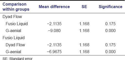
At the end of the study, it was found that the shear bond strength of conventional flowable composite was significantly greater than self-adhering flowable composite. (P < 0.05) [Figure 1 and Table 1]. Among the self-adhering flowable composites, shear bond strength of Fusio Liquid Dentin (Pentron Clinical Technologies) was greater than Dyad Flow (Kerr) although the results were not statistically significant [Table 2]. Nanoleakage scores of both conventional and self-adhering flowable composites were comparable. No correlation was found between bond strength and nanoleakage among different composite materials tested.
Figure 1.
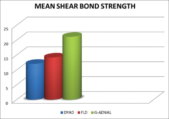
Mean shear bond strength of the three composite materials tested
Table 1.
Shear bond strength of various samples of the three groups tested
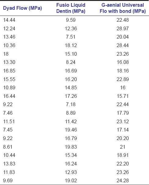
Table 2.
Statistical analysis of shear bond strength
Discussion
The adhesion between composite resin and dental hard tissue is the most important determinant of the bond between tooth and restoration. The advantages of both adhesive and restorative materials are combined in self-adhering flowable composites, hence providing a broader scope to restorative techniques, as it is a direct composite resin restorative material that amalgamates the qualities of adhesive resin and flowable composite resin.[3]
One of the methods to test the adhesion of dental adhesives is by measuring the shear bond strength. The universal testing machine is popularly used for measuring shear bond strength.[8] In vitro shear bond strength tests are valuable and fundamental for analyzing the potential of adhesive systems and association with clinical scenario.[9]
The teeth used in this study were stored in 10% formalin. Lee et al. reported that immersion and storage of teeth in 10% formalin is a suitable option for in vitro dental bonding studies.[10] The storage duration and condition of teeth after extraction may cause changes within dentin and, hence, may influence the adhesion in in vitro bonding studies. Titley et al.[11] stated that postmortem changes could occur in dentin, which in turn could affect the result outcome. However, in another study that compared shear bond strength of dentin to restorative material, no significant differences were observed between the group stored in distilled water for 8 days and the other stored for 6 months.[12]
The shear bond strength of composite bonded to primary teeth dentin is less than that of permanent teeth.[13] A minimum bond strength of 17-20 Mpa is needed to resist contraction forces of resin composite materials for enamel and dentin as confirmed by clinical data.[14]
A comparatively low bond strength of primary teeth as compared to permanent teeth may be attributed to the differences in the chemical and morphological features between them. Calcium level and total available area of solid dentin are the two major criteria which affect the shear bond strength of adhesives as suggested by Bordin-Aykroyd et al.[15] Primary teeth have relatively larger pulp chambers. A lower level of calcium is seen in dentin which lies closer to pulp. This may lower the bond strength in primary teeth as the effective dentin which remains after cavity preparation is the dentin which lies near pulp. Intertubular dentin is the most important site for bonding. However, primary teeth have relatively less intertubular dentin and a comparatively thicker peritubular dentin.[16] The reduced adhesive strength in primary teeth may be due to the thicker interface and the bonding system being incompletely impregnated in the collagen network.[5]
Literature demonstrates that adhesion force between composite resin systems and primary dentin ranges from 5.53 to 70.1 MPa. This wide variation could be attributed to the differences in methods employed, as well as the innate factors related to the tooth and material.[17] Results from studies with permanent teeth were taken as reference due to the lack of studies conducted in primary teeth.
The results of this study showed that the conventional flowable composite had significantly higher bond strength than that of self-adhering flowable composites tested [Table 2]. This result was in accordance with Kerby et al., Bouillaguet et al., and Chuang et al.[8,18,19] The results of the study conducted by Senawongse et al. demonstrated that total-etch systems had a higher bond strength than self-etch systems.[20] Contradictory to this, Kiremitçi et al. concluded that self-etching adhesive systems produced higher bond strength than conventional total-etch systems.[14] However, in a study conducted by Sensi et al., total- and self-etch primer showed comparable dentin bond strength.[21] When compared to phosphoric acid etching, self-etch adhesives had lower bond strength because they produced shorter and thinner resin tags.[22]
In this study, Fusio Liquid Dentin demonstrated higher shear bond strength (14.11 Mpa) than Dyad Flow (12.03 Mpa); however, the results were not statistically significant. The composition of the two tested self-adhering flowable composites differs which may have resulted in the difference in shear bond strength.[23] Dyad Flow (Kerr) contains glycerol phosphate dimethacrylate and a filler content of 70 weight%. Fusio Liquid Dentin (Pentron), on the other hand, contains the functional monomer 4-methacryloxyethyl trimellitic acid (4- MET) and a filler in range of 65 weight%. In this study, Fusio Liquid Dentin was found to be placed more easily onto the tooth surface. The easier handling may have contributed to the better bonding effectiveness of Fusio Liquid Dentin (Pentron) when compared to that of Vertise Flow (Kerr).[24]
Microleakage has been used to express the longevity of bonded restorations for many years. Microleakage may lead to marginal discoloration, postoperative sensitivity, secondary caries, pulpal inflammation, and eventually partial or complete loss of that restoration leading to decrease in longevity.[25] “Nanoleakage” is a type of leakage which occurs within the hybrid layer in nanometer scaled spaces.[26] It causes seepage of oral fluids and bacterial products through the interface which may in turn compromise the stability of the bond between tooth dentin and composite resin. Nanoleakage evaluation is considered as useful determinant of hybrid layer quality and sealability of restorative material.[27]
The term nanoleakage was first quoted as being the impregnation of silver grains within the porosities of the hybrid layer that were not properly filled with adhesive resin. The second mode of nanoleakage, termed as “reticular mode,” has also been described. These delicate, branching channels of nanovoids are thought to be morphological manifestations of the water treeing phenomenon, which is probably a result of aging. Aging is thought to cause polymer deterioration which is water induced.[28] In this study, “spotted” and “reticular” patterns of nanoleakage were observed. The absence of “water-tree” pattern could be attributed to the fact that all the samples were prepared and evaluated under SEM on the same day, hence eliminating the effect of aging. Figures 2–4 depict the scanning electron microscope image of Dyad Flow, Fusio Liquid Dentin and G-aenial Universal Flo respectively.
Figure 2.
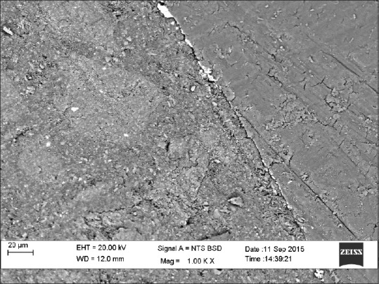
Scanning electron microscope image of Dyad Flow
Figure 4.
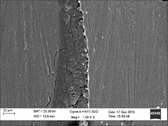
Scanning electron microscope image of G-aenial Universal Flo
Figure 3.
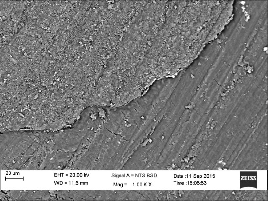
Scanning electron microscope image of Fusio Liquid Dentin
This aim of this study was to evaluate and compare the sealability of self-adhering flowable composite through nanoleakage examination by means of standardized Class V restoration. The results of the study showed that the sealability of self-adhesive flowable composite was comparable to conventional flowable composites. Mobarak and Seyam found that all tested self-adhesive systems showed slight nanoleakage when compared to self-etch adhesives.[29] This could be attributed to the fact that nanoleakage is higher when the hybrid layer is thicker and the demineralized dentin is deeper, resulting in penetration of silver ions into the partially or fully demineralized dentin and hybrid layer.[30] Tay et al.,[6] Hashimoto et al.,[22] and El-Badrawy et al.[31] however showed the presence of a high amount of nanoleakage in self-adhesive systems. The authors asserted that this might be due to the presence of some amount of residual water which is retained as a result of its low vaporization in the presence of HEMA, leading to increased silver uptake. In the presence of water, HEMA copolymerizes with low pH resin monomers and forms homologous hydrogels causing deposition of fine silver at the bonded interface.[32]
Class V clinical trials are very useful for providing a correlation between in vitro and in vivo study conditions.[33] However, till date, only one short-term trial has evaluated the clinical potential of SACs.[34] Hence, before concluding on the efficiency of self-adhering flowable composites in clinical settings, further studies are warranted. Nevertheless, high bond strength without nanoleakage is essentially desirable for a successful restoration.
Conclusion
From the present study, following conclusions were drawn:
Among the self-adhering flowable composites, shear bond strength of Fusio Liquid Dentin (Pentron Clinical Technologies) was greater than Dyad Flow (Kerr) although the results were not statistically significant
Nanoleakage scores of both conventional and self-adhering flowable composites were comparable
There was no correlation between bond strength and nanoleakage among different composite materials tested.
Financial support and sponsorship
Nil.
Conflicts of interest
There are no conflicts of interest.
References
- 1.Vashisth P, Mittal M, Goswami M, Chaudhary S, Dwivedi S. Bond strength and interfacial morphology of different dentin adhesives in primary teeth. J Dent (Tehran) 2014;11:179–87. [PMC free article] [PubMed] [Google Scholar]
- 2.Bayne SC, Thompson JY, Swift EJ, Jr, Stamatiades P, Wilkerson M. A characterization of first-generation flowable composites. Am Dent Assoc. 1998;129:567–77. doi: 10.14219/jada.archive.1998.0274. [DOI] [PubMed] [Google Scholar]
- 3.Bektas OO, Eren D, Akin EG, Akin H. Evaluation of a self-adhering flowable composite in terms of micro-shear bond strength and microleakage. Acta Odontol Scand. 2013;71:541–6. doi: 10.3109/00016357.2012.696697. [DOI] [PubMed] [Google Scholar]
- 4.Ferracane JL. Resin composite - State of the art. Dent Mater. 2011;27:29–38. doi: 10.1016/j.dental.2010.10.020. [DOI] [PubMed] [Google Scholar]
- 5.Nör JE, Feigal RJ, Dennison JB, Edwards CA. Dentin bonding: SEM comparison of the resin-dentin interface in primary and permanent teeth. J Dent Res. 1996;75:1396–403. doi: 10.1177/00220345960750061101. [DOI] [PubMed] [Google Scholar]
- 6.Tay FR, Pashley DH, Yoshiyama M. Two modes of nanoleakage expression in single-step adhesives. J Dent Res. 2002;81:472–6. doi: 10.1177/154405910208100708. [DOI] [PubMed] [Google Scholar]
- 7.Yuan Y, Shimada Y, Ichinose S, Tagami J. Qualitative analysis of adhesive interface nanoleakage using FE-SEM/EDS. Dent Mater. 2007;23:561–9. doi: 10.1016/j.dental.2006.03.015. [DOI] [PubMed] [Google Scholar]
- 8.Kerby RE, Knobloch LA, Clelland N, Lilley H, Seghi R. Microtensile bond strengths of one-step and self-etching adhesive systems. Oper Dent. 2005;30:195–200. [PubMed] [Google Scholar]
- 9.Hiraishi N, Breschi L, Prati C, Ferrari M, Tagami J, King NM. Technique sensitivity associated with air-drying of HEMA-free, single-bottle, one-step self-etch adhesives. Dent Mater. 2007;23:498–505. doi: 10.1016/j.dental.2006.03.007. [DOI] [PubMed] [Google Scholar]
- 10.Lee JJ, Nettey-Marbell A, Cook A, Jr, Pimenta LA, Leonard R, Ritter AV. Using extracted teeth for research: The effect of storage medium and sterilization on dentin bond strengths. J Am Dent Assoc. 2007;138:1599–603. doi: 10.14219/jada.archive.2007.0110. [DOI] [PubMed] [Google Scholar]
- 11.Titley KC, Chernecky R, Rossouw PE, Kulkarni GV. The effect of various storage methods and media on shear-bond strengths of dental composite resin to bovine dentine. Arch Oral Biol. 1998;43:305–11. doi: 10.1016/s0003-9969(97)00112-x. [DOI] [PubMed] [Google Scholar]
- 12.Goodis HE, Marshall GW, Jr, White JM, Gee L, Hornberger B, Marshall SJ. Storage effects on dentin permeability and shear bond strengths. Dent Mater. 1993;9:79–84. doi: 10.1016/0109-5641(93)90079-6. [DOI] [PubMed] [Google Scholar]
- 13.Prabhakar AR, Raj S, Raju OS. Comparison of shear bond strength of composite, compomer and resin modified glass ionomer in primary and permanent teeth: An in vitro study. J Indian Soc Pedod Prev Dent. 2003;21:86–94. [PubMed] [Google Scholar]
- 14.Kiremitçi A, Yalçin F, Gökalp S. Bonding to enamel and dentin using self-etching adhesive systems. Quintessence Int. 2004;35:367–70. [PubMed] [Google Scholar]
- 15.Bordin-Aykroyd S, Sefton J, Davies EH. In vitro bond strengths of three current dentin adhesives to primary and permanent teeth. Dent Mater. 1992;8:74–8. doi: 10.1016/0109-5641(92)90059-l. [DOI] [PubMed] [Google Scholar]
- 16.Hirayama A. Experimental analytical electron microscopic studies on the quantitative analysis of elemental concentrations in biological thin specimens and its application to dental science. Shikwa Gakuho. 1990;90:1019–36. [PubMed] [Google Scholar]
- 17.Bengtson CR, Bengtson AL, Bengtson NG, Turbino ML. Do the origins of primary teeth affect the bond strength of a self-etching adhesive system to dentin? Braz Oral Res. 2010;24:355–60. doi: 10.1590/s1806-83242010000300016. [DOI] [PubMed] [Google Scholar]
- 18.Bouillaguet S, Gysi P, Wataha JC, Ciucchi B, Cattani M, Godin C, et al. Bond strength of composite to dentin using conventional, one-step, and self-etching adhesive systems. J Dent. 2001;29:55–61. doi: 10.1016/s0300-5712(00)00049-x. [DOI] [PubMed] [Google Scholar]
- 19.Chuang SF, Chang LT, Chang CH, Yaman P, Liu JK. Influence of enamel wetness on composite restorations using various dentine bonding agents: Part II-effects on shear bond strength. J Dent. 2006;34:352–61. doi: 10.1016/j.jdent.2005.08.001. [DOI] [PubMed] [Google Scholar]
- 20.Senawongse P, Harnirattisai C, Shimada Y, Tagami J. Effective bond strength of current adhesive systems on deciduous and permanent dentin. Oper Dent. 2004;29:196–202. [PubMed] [Google Scholar]
- 21.Sensi LG, Lopes GC, Monteiro S, Jr, Baratieri LN, Vieira LC. Dentin bond strength of self-etching primers/adhesives. Oper Dent. 2005;30:63–8. [PubMed] [Google Scholar]
- 22.Hashimoto M, De Munck J, Ito S, Sano H, Kaga M, Oguchi H, et al. In vitro effect of nanoleakage expression on resin-dentin bond strengths analyzed by microtensile bond test, SEM/EDX and TEM. Biomaterials. 2004;25:5565–74. doi: 10.1016/j.biomaterials.2004.01.009. [DOI] [PubMed] [Google Scholar]
- 23.Yoshida Y, Nagakane K, Fukuda R, Nakayama Y, Okazaki M, Shintani H, et al. Comparative study on adhesive performance of functional monomers. Dent Res. 2004;83:454–8. doi: 10.1177/154405910408300604. [DOI] [PubMed] [Google Scholar]
- 24.Poitevin A, De Munck J, Van Ende A, Suyama Y, Mine A, Peumans M, et al. Bonding effectiveness of self-adhesive composites to dentin and enamel. Dent Mater. 2013;29:221–30. doi: 10.1016/j.dental.2012.10.001. [DOI] [PubMed] [Google Scholar]
- 25.Eick JD, Welch FH. Polymerization shrinkage of posterior composite resins and its possible influence on postoperative sensitivity. Quintessence Int. 1986;17:103–11. [PubMed] [Google Scholar]
- 26.Andia-Merlin RY, Garone-Netto N, Arana-Chavez VE. SEM evaluation of the interaction between a three-step adhesive and dentin. Oper Dent. 2001;26:440–4. [PubMed] [Google Scholar]
- 27.Naga AA, Yousef M, Ramadan R, Fayez Bahgat S, Alshawwa L. Does the use of a novel self-adhesive flowable composite reduce nanoleakage? Clin Cosmet Investig Dent. 2015;7:55–64. doi: 10.2147/CCIDE.S80462. [DOI] [PMC free article] [PubMed] [Google Scholar]
- 28.Pioch T, Staehle HJ, Duschner H, García-Godoy F. Nanoleakage at the composite-dentin interface: A review. Am J Dent. 2001;14:252–8. [PubMed] [Google Scholar]
- 29.Mobarak E, Seyam R. Interfacial nanoleakage and bonding of self-adhesive systems cured with a modified-layering technique to dentin of weakened roots. Oper Dent. 2013;38:E154–65. doi: 10.2341/12-103-L. [DOI] [PubMed] [Google Scholar]
- 30.Pereira PN, Okuda M, Nakajima M, Sano H, Tagami J, Pashley DH. Relationship between bond strengths and nanoleakage: Evaluation of a new assessment method. Am J Dent. 2001;14:100–4. [PubMed] [Google Scholar]
- 31.El-Badrawy W, Hafez RM, El Naga AI, Ahmed DR. Nanoleakage for self-adhesive resin cements used in bonding CAD/CAD ceramic material to dentin. Eur J Dent. 2011;5:281–90. [PMC free article] [PubMed] [Google Scholar]
- 32.Unterbrink GL, Liebenberg WH. Flowable resin composites as “filled adhesives”: Literature review and clinical recommendations. Quintessence Int. 1999;30:249–57. [PubMed] [Google Scholar]
- 33.Heintze SD, Blunck U, Göhring TN, Rousson V. Marginal adaptation in vitro and clinical outcome of class V restorations. Dent Mater. 2009;25:605–20. doi: 10.1016/j.dental.2008.11.004. [DOI] [PubMed] [Google Scholar]
- 34.Vichi A, Margvelashvili M, Goracci C, Papacchini F, Ferrari M. Bonding and sealing ability of a new self-adhering flowable composite resin in class I restorations. Clin Oral Investig. 2013;17:1497–506. doi: 10.1007/s00784-012-0846-6. [DOI] [PubMed] [Google Scholar]


