Abstract
Aim:
The aim of this study was to evaluate the pulpal response of primary teeth after direct pulp capping (DPC) with two biocompatible materials namely mineral trioxide aggregate (MTA) and bioactive glass (BAG).
Settings and Design:
This study was a randomized clinical trial.
Materials and Methods:
A total of 22 healthy primary canine teeth scheduled for extraction for orthodontic reasons were selected. The teeth were divided into two groups of 11 and underwent DPC. The exposure sites were randomly capped with MTA or BAG in the two groups. After 2 months, the teeth were extracted and prepared for histopathologic evaluation.
Statistical Analysis:
The data were analyzed using Fisher's exact test.
Results:
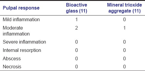
In the BAG group, inflammation was seen in three patients; internal resorption and abscess were not seen at all. In the MTA group, inflammation was seen in one patient and internal resorption and abscess were not seen in any patient. Fisher's exact test showed no significant difference between the two groups (P > 0.05). Dentinal bridge formation was noted in five patients in the BAG group and six patients in the MTA group. No significant difference was observed between the BAG and MTA groups using Chi-square analysis (P = 0.67).
Conclusion:
Based on the results of this study, MTA and BAG can be used for DPC of primary teeth.
Key words: Deciduous tooth, dental pulp capping, mineral trioxide aggregate
Introduction
Dental caries is the most prevalent chronic disease in children. Extensive caries are treated by three pulp treatment techniques: Direct pulp capping (DPC), pulpotomy and pulpectomy depending on the severity of pulp involvement.[1]
DPC is a conservative modality for treatment of traumatically or mechanically exposed pulps. However, the use of DPC is more often limited to primary teeth rather than permanent teeth.[2,3] Calcium hydroxide is the gold standard material for pulp capping of vital pulp tissue. Calcium hydroxide has antibacterial effects and can induce proliferation, healing, and repair of fibroblasts resulting in induction of soft and hard tissue regeneration.[2,4] However, calcium hydroxide interferes with the healing process; the formed dentinal bridge does not provide a proper seal, and the antimicrobial property of calcium hydroxide disappears over time.[5,6,7] DPC with calcium hydroxide in primary teeth has a low success rate.[2] Therefore, other materials have been proposed for DPC of primary teeth. Mineral trioxide aggregate (MTA) is a relatively new material marketed in 1998 under Food and Drug Administration approval.[8] The major components of this material are a tricalcium silicate, tricalcium aluminate, and tricalcium oxide. Sealing ability of this material is very high, it is compatible with tissues and can induce dentinal bridge formation.[9,10,11,12,13,14] Bioactive glass (BAG) is relatively new in dentistry.[15] It is composed of calcium and phosphate in a proportion that can be similar to bone hydroxyapatite. It is biocompatible, can stimulate hard tissue formation and mineralization and has antibacterial properties. It is currently used for bone grafting and as scaffolds.[15,16,17,18,19] Therefore, it appears that BAG and MTA can be used for pulp capping.
The aim of this study was to evaluate dental pulp response of primary teeth after DPC with MTA and BAG.
Materials and Methods
In this double-blind, randomized clinical trial, 22 sound primary canines scheduled for extraction for orthodontic reasons were selected in 11 children aged 78 years (including seven girls and four boys). The sample size was calculated according to a previous study.[20] The inclusion criteria were: Sound primary teeth with root resorption no more than the apical third. The study protocol was approved by the Ethics Committee of Shahed University (40/104951), and it was registered in www.irct.ir (IRCT2015100624397N1). Written informed consent was obtained from the parents. The teeth were randomly divided into two groups: 11 teeth in the MTA group and 11 teeth in the BAG group. The randomization was done using a table of random numbers by someone blinded to the experimental groups. A total of 22 Class V cavities with a diameter of 0.5 mm were prepared in the middle third of the buccal surfaces of the teeth with a carbide bur (D and Z Co., Germany). Cavity preparation was continued until the shadow of the pulp was seen. Each cavity was rinsed with saline and dried with cotton pellets. Dental pulp was exposed with a sterile probe. Hemorrhage was controlled by placing a cotton pellet moistened in sterile saline. Then, MTA (Angelus, Brazil) was placed on the exposure site in 11 teeth, and BAG Biogran (3i Implant Innovations, USA) was placed on the exposure site in the remaining 11 teeth. All the teeth were restored with amalgam.[20] After 2 months, the 22 teeth were extracted and prepared for H and E staining. All the sections were studied by a pathologist blinded to the study design. Histological assessment was carried out according to Fuks et al.[21]
Scoring was done as follows:
None or mild inflammation: 0, moderate inflammation: 1, severe inflammation: 2, necrosis: 3, abscess: 4, resorption: 5.
Dentinal bridge formation was also evaluated in the two groups. Finally, the data were analyzed by Fisher's exact test. In this study, pulp statue of samples was evaluated histopathologically.
Results
The histological tissue changes in the BAG and MTA groups are shown in Table 1. Inflammation was seen in one case in the MTA group and three cases in the BAG group. Fisher's exact test showed no significant differences (P > 0.05). Internal resorption and abscess were not seen in any of the two groups [Table 1].
Table 1.
Tissue changes in bioactive glass, mineral trioxide aggregate groups

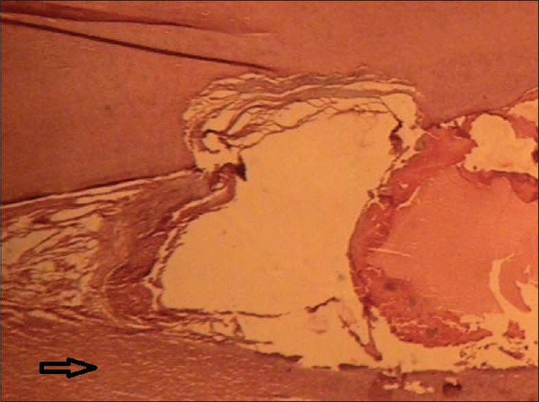
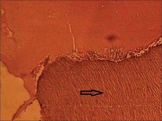
Dentinal bridge formation was seen in five samples in the BAG group [Figure 1]. In the MTA group, the dentinal bridge was found in six samples [Figure 2].
Figure 1.

Dentinal formation in bioactive glass group
Figure 2.

Dentinal formation in mineral trioxide aggregate group
Discussion
The aim of this study was to evaluate pulpal response of primary teeth after pulpotomy with BAG and MTA.
Previous studies have shown that DPC may have poor prognosis in primary teeth since the presence of internal resorption, pulp calcification and necrosis in the primary teeth is likely, and this treatment may cause injury to the surrounding tissues. Fuks et al. reported that undifferentiated mesenchymal cells in the primary pulp differentiate to odontoclasts, leading to internal resorption.[21] However, DPC is a conservative vital pulp therapy eliminating the need for a more invasive treatment modality. Several materials have been used for DPC of primary teeth.
It seems that BAG and MTA are suitable materials for DPC because of their antibacterial properties and biocompatibility.
The results of this study showed that most samples had normal pulp tissue. Salako et al.,[22] also found normal pulp tissue after pulpotomy of dog's teeth; however, the samples in their study and ours were different and differences in the anatomy of human and animal pulp might have affected the results. Similarly, Haghgoo et al. found that human pulp tissue was normal after DPC with BAG and MTA.[20,23]
Based on the results of the current study, internal resorption was not seen in the BAG or MTA groups; BAG and MTA are biocompatible and can induce regeneration of pulpal tissue.
The results of the study by Haghgoo et al. are in accordance with those of the current study.[23]
The results of the current study did not reveal any abscess in any sample of the MTA or BAG groups, which might be attributed to antibacterial properties of these two materials. Menezes et al. and Haghgoo et al. reported similar results as well.[13,20,23]
Based on the results of the current study, dentinal bridge was found in five samples in the BAG group and six samples in the MTA group consistent with the results of studies by Haghgoo et al.[20,23]
The results of a study by Fallahinejad Ghajari et al.,[24] in which clinical and radiographic success rates of DPC with calcium enriched mixture (CEM) cement and MTA were compared, showed no significant differences between treatment outcomes of DPC with CEM cement and MTA. Fallahinejad Ghajari et al.[24] studied success rate of DPC with the two biomaterials clinically and radiographically and differences in the study design and methodology of the two studies might have affected the results.
Limitation
This study evaluated pulpal response of primary teeth histologically. One of the limitations of this study was a selection of sound teeth scheduled for extraction.
Suggestion
In this study, the histological status of dental pulp was evaluated after DPC. It is suggested that dental pulp of primary teeth be evaluated after DPC with BAG and MTA clinically and radiographically.
Conclusion
Based on the results of this study MTA and BAG can be used for direct pulp capping in primary teeth.
Financial support and sponsorship
This study was supported by the Faculty of Dentistry, Shahed University, Tehran, Iran.
Conflicts of interest
There are no conflicts of interest.
References
- 1.Smaïl-Faugeron V, Courson F, Durieux P, Muller-Bolla M, Glenny AM, Fron Chabouis H. Pulp treatment for extensive decay in primary teeth. Cochrane Database Syst Rev. 2014;8:CD003220. doi: 10.1002/14651858.CD003220.pub2. [DOI] [PubMed] [Google Scholar]
- 2.Anna B, Fuks CD. Pulp therapy for the primary dentition. In: Pinkham JR, Cassamassio PS, Field H, editors. Pediatric Dentistry Infancy Through Adolescence. 4th ed. Missouri: Saunder Co.; 2005. p. 383. [Google Scholar]
- 3.McDonald RE, Avery DR, Dean JA. Treatment of deep caries, vital pulp exposure, and pulpless teeth. In: McDonald RE, Avery DR, Dean JA, editors. Dentistry for the Child and Adolescent. 9th ed. Missouri: Mosby Co.; 2011. p. 350. [Google Scholar]
- 4.Schröder U. Effects of calcium hydroxide-containing pulp-capping agents on pulp cell migration, proliferation, and differentiation. J Dent Res. 1985;64:541–8. doi: 10.1177/002203458506400407. [DOI] [PubMed] [Google Scholar]
- 5.Mathwsen RJ, Primosch RE. Fundamental of Pediatric Dentistry. 3rd ed. Chicago: Quintessence Publishing Co.; 1995. p. 259. [Google Scholar]
- 6.Cox CF, Sübay RK, Ostro E, Suzuki S, Suzuki SH. Tunnel defects in dentin bridges: Their formation following direct pulp capping. Oper Dent. 1996;21:4–11. [PubMed] [Google Scholar]
- 7.Cox CF, Bergenholtz G, Heys DR, Syed SA, Fitzgerald M, Heys RJ. Pulp capping of dental pulp mechanically exposed to oral microflora: A 1-2 year observation of wound healing in the monkey. J Oral Pathol. 1985;14:156–68. doi: 10.1111/j.1600-0714.1985.tb00479.x. [DOI] [PubMed] [Google Scholar]
- 8.Nakata TT, Bae KS, Baumgartner JC. Perforation repair comparing mineral trioxide aggregate and amalgam using an anaerobic bacterial leakage model. J Endod. 1998;24:184–6. doi: 10.1016/S0099-2399(98)80180-5. [DOI] [PubMed] [Google Scholar]
- 9.Eidelman E, Holan G, Fuks AB. Mineral trioxide aggregate vs. formocresol in pulpotomized primary molars: A preliminary report. Pediatr Dent. 2001;23:15–8. [PubMed] [Google Scholar]
- 10.Yildiz E, Tosun G. Evaluation of formocresol, calcium hydroxide, ferric sulfate, and MTA primary molar pulpotomies. Eur J Dent. 2014;8:234–40. doi: 10.4103/1305-7456.130616. [DOI] [PMC free article] [PubMed] [Google Scholar]
- 11.Torabinejad M, Hong CU, McDonald F, Pitt Ford TR. Physiological and chemical properties of new end filling material. J Endod. 1995;21:349–53. doi: 10.1016/S0099-2399(06)80967-2. [DOI] [PubMed] [Google Scholar]
- 12.Maroto M, Barbería E, Planells P, García Godoy F. Dentin bridge formation after mineral trioxide aggregate (MTA) pulpotomies in primary teeth. Am J Dent. 2005;18:151–4. [PubMed] [Google Scholar]
- 13.Menezes R, Bramante CM, Letra A, Carvalho VG, Garcia RB. Histologic evaluation of pulpotomies in dog using two types of mineral trioxide aggregate and regular and white Portland cements as wound dressings. Oral Surg Oral Med Oral Pathol Oral Radiol Endod. 2004;98:376–9. doi: 10.1016/S107921040400215X. [DOI] [PubMed] [Google Scholar]
- 14.Mettlach SE, Zealand CM, Botero TM, Boynton JR, Majewski RF, Hu JC. Comparison of mineral trioxide aggregate and diluted formocresol in pulpotomized human primary molars: 42-month follow-up and survival analysis. Pediatr Dent. 2013;35:E87–94. [PMC free article] [PubMed] [Google Scholar]
- 15.Stanley HR, Clark AE, Pameijer CH, Louw NP. Pulp capping with a modified bioglass formula (#A68-modified) Am J Dent. 2001;14:227–32. [PubMed] [Google Scholar]
- 16.Schepers E, de Clercq M, Ducheyne P, Kempeneers R. Bioactive glass particulate material as a filler for bone lesions. J Oral Rehabil. 1991;18:439–52. doi: 10.1111/j.1365-2842.1991.tb01689.x. [DOI] [PubMed] [Google Scholar]
- 17.Shapoff CA, Alexander DC, Clark AE. Clinical use of a bioactive glass particulate in the treatment of human osseous defects. Compend Contin Educ Dent. 1997;18:352–4, 356, 358. [PubMed] [Google Scholar]
- 18.Chacko NL, Abraham S, Rao HN, Sridhar N, Moon N, Barde DH. A Clinical and radiographic evaluation of periodontal regenerative potential of PerioGlas®: A synthetic, resorbable material in treating periodontal infrabony defects. J Int Oral Health. 2014;6:20–6. [PMC free article] [PubMed] [Google Scholar]
- 19.Farooq I, Imran Z, Farooq U, Leghari A, Ali H. Bioactive glass: A material for future. World J Dent. 2012;3:199–201. [Google Scholar]
- 20.Haghgoo R, Jalayer Naderi N. Comparison of calcium hydroxide and bioactive glass after direct pulp capping in primary teeth. J Dent Tehran Univ. 2007;4:155–9. [Google Scholar]
- 21.Fuks AB, Eidelman E, Cleaton-Jones P, Michaeli Y. Pulp response to ferric sulfate, diluted formocresol and IRM in pulpotomized primary baboon teeth. ASDC J Dent Child. 1997;64:254–9. [PubMed] [Google Scholar]
- 22.Salako N, Joseph B, Ritwik P, Salonen J, John P, Junaid TA. Comparison of bioactive glass, mineral trioxide aggregate, ferric sulfate, and formocresol as pulpotomy agents in rat molar. Dent Traumatol. 2003;19:314–20. doi: 10.1046/j.1600-9657.2003.00204.x. [DOI] [PubMed] [Google Scholar]
- 23.Haghgoo R, Jalayer Naderi N. Histopathologic evaluation of pulp after direct pulp capping with calcium hydroxide and mineral trioxide aggregate in primary teeth. J Dent Shahid Beheshti Univ. 2008;26:136–42. [Google Scholar]
- 24.Fallahinejad Ghajari M, Asgharian Jeddi T, Iri S, Asgary S. Direct pulp-capping with calcium enriched mixture in primary molar teeth: A randomized clinical trial. Iran Endod J. 2010;5:27–30. [PMC free article] [PubMed] [Google Scholar]


