Fig. 1.
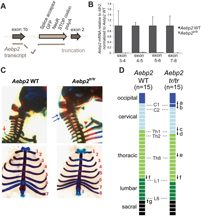
Aebp2 truncation leads to perinatal lethality and anterior homeotic transformations. (A) Insertion of the splice acceptor cassette in front of exon 2 leads to trapping of the transcript and a protein product that contains the first 217 amino acids (aa) of AEBP2, encoded in exon 1b, fused to green fluorescent protein (GFP) and aminoglycoside 3′ phosphotransferase (NeoR). (B) The levels of Aebp2 mRNA transcripts containing exons downstream of the trapping cassette are severely reduced in Aebp2tr/tr mESCs compared with the parental WT Aebp2 mESCs. Error bars indicate s.d. (C) Lateral views of the occipito-cervical region (top panels) and ventral views of the rib cage (bottom) of Aebp2 WT (left) and Aebp2tr/tr (right) foetuses. In Aebp2tr/tr foetuses, the ventral region of the atlas (C1, indicated by a green arrowhead) is fused to that of the axis (C2). Ventral ossification centre of the atlas was laterally expanded and acquired similar features to occipital bone. The dorsal region of the axis was cranio-caudally expanded and partially bifurcated (indicated by blue arrows). The proximal region of the rib associated with the first thoracic vertebra (Th1) was not formed (indicated by a red arrowhead). The prominent dorsal process, which is associated with the Th2 in the Aebp2 WT (indicated by a red arrow), is not formed in the Aebp2tr/tr. Bottom panel also shows association of the 8th rib to the sternum. (D) Schematic summary for homeotic transformations of the axis in the Aebp2tr/tr foetuses.

