Abstract
Ghrelin is an orexigenic gastric peptide hormone secreted when caloric intake is limited. Ghrelin also regulates blood glucose, as emphasized by the hypoglycemia that is induced by caloric restriction in mouse models of deficient ghrelin signaling. Here, we hypothesized that activation of β1-adrenergic receptors (β1ARs) localized to ghrelin cells is required for caloric restriction–associated ghrelin release and the ensuing protective glucoregulatory response. In mice lacking the β1AR specifically in ghrelin-expressing cells, ghrelin secretion was markedly blunted, resulting in profound hypoglycemia and prevalent mortality upon severe caloric restriction. Replacement of ghrelin blocked the effects of caloric restriction in β1AR-deficient mice. We also determined that treating calorically restricted juvenile WT mice with beta blockers led to reduced plasma ghrelin and hypoglycemia, the latter of which is similar to the life-threatening, fasting-induced hypoglycemia observed in infants treated with beta blockers. These findings highlight the critical functions of ghrelin in preventing hypoglycemia and promoting survival during severe caloric restriction and the requirement for ghrelin cell–expressed β1ARs in these processes. Moreover, these results indicate a potential role for ghrelin in mediating beta blocker–associated hypoglycemia in susceptible individuals, such as young children.
Introduction
Ghrelin is a peptide hormone first isolated from the stomach and reported in 1999 (1). Following the unique posttranslational addition of an acyl group by ghrelin O-acyltransferase (GOAT), ghrelin can bind and activate the G protein–coupled growth hormone secretagogue receptor (GHSR), which is the only known ghrelin receptor (1–3). It is via CNS and/or peripheral GHSRs that ghrelin influences growth hormone (GH) secretion, mood, gastrointestinal motility, hippocampal neurogenesis, and many other processes (4–10). Actions of unmodified ghrelin (desacyl-ghrelin), the modes thereof, and the potential significance of the acyl-ghrelin/desacyl-ghrelin ratio are less well described (4). Of all processes influenced by the ghrelin system, eating and BW have received the most attention. Ghrelin is unique among gut hormones in that it is orexigenic, potently stimulating eating (5, 9). Plasma ghrelin rises before meals, after food deprivation, and after weight loss; it falls after eating and is low in most forms of obesity (9, 11). When the action of fasting-induced elevated ghrelin is blocked pharmacologically in mice, rebound overeating is blunted, and when the action of weight loss–associated elevated ghrelin is similarly blocked, as in a rat cancer cachexia model, worsened anorexia and hastened death have been described (12, 13). Exogenous infusions of ghrelin or GHSR agonists similarly increase eating and, in addition, lower energy expenditure and upregulate the expression of lipogenic and fat storage–promoting enzymes (5, 14–17). Ghrelin also shifts food preference toward high-fat diets (HFDs), shifts fuel preference away from metabolic utilization of fat, and engages several food reward behaviors (5, 13, 18, 19). Collectively, these actions are likely contributors to the ability of administered ghrelin to increase BW in normal subjects and maintain BW or delay weight loss in cachectic subjects (5, 15, 17, 20).
The ghrelin system also regulates and is regulated by blood glucose. Suggesting a role for ghrelin in modulating glycemia, ghrelin administration has been shown to increase blood glucose acutely, which may occur via its known actions to lower insulin levels, attenuate insulin sensitivity, stimulate glucagon secretion, induce GH secretion, raise glucocorticoid levels, and/or increase food intake (1, 5, 21–24). Also, both ad libitum–fed Tg mice with hyperghrelinemia due to ghrelin promoter–driven expression of simian virus 40 large T antigen and overnight-fasted rats carrying a gain-of-function GHSR mutation (the GHSR-Q343X isoform, which is associated with a reduced ghrelin-induced GHSR internalization and enhanced sensitivity to administered ghrelin) exhibit higher blood glucose levels than do WT control animals (24, 25). Ex vivo studies with primary cultures of dispersed mouse gastric mucosal cells, approximately 0.3% to 1% of which include ghrelin cells, demonstrate that ghrelin cells can directly sense glucose, enhancing ghrelin release upon exposure to glucose concentrations representative of hypoglycemia and reducing ghrelin release when exposed to high glucose levels (26, 27). Not only does exposure of ghrelin cells to low glucose levels stimulate ghrelin release, but increased sympathoadrenal tone, as occurs during the usual counterregulatory response to hypoglycemia, could also stimulate ghrelin secretion (26, 28). Supportive of this assertion, ghrelin secretion increases when sympathetic nerves are stimulated artificially or when adrenergic agents are infused locally into the gastric submucosa (29, 30). Norepinephrine, epinephrine, and isoproterenol all stimulate ghrelin secretion from ghrelinoma cell lines and from primary cultures of gastric mucosal cells (26, 31–36). Reserpine, which depletes norepinephrine from sympathetic neuron terminals, blocks the overnight fast–induced increase in plasma ghrelin, as does the β1-adrenergic receptor (β1AR) blocker atenolol (34). Importantly, β1AR is the most highly expressed of all nonodorant GPCRs within ghrelin cells (~25-fold higher than the next), the most highly expressed of the adrenergic receptors within ghrelin cells, and the only small-molecule neurotransmitter receptor enriched in ghrelin cells as compared with non-ghrelin gastric mucosal cells (34, 35, 37).
Given this β1AR expression data and stimulation of ghrelin secretion by catecholamines, we designed the current study to test the hypothesis that activation of β1ARs expressed on ghrelin cells is required for the usual ghrelin secretory response, serving as a primary modulator of ghrelin secretion during both short-term and more severe caloric restriction. Furthermore, we tested whether activation of ghrelin cell β1ARs is required to engage the protective glucoregulatory functions of the ghrelin system.
Results
Generation of mice with ghrelin cell–specific deletion of β1ARs.
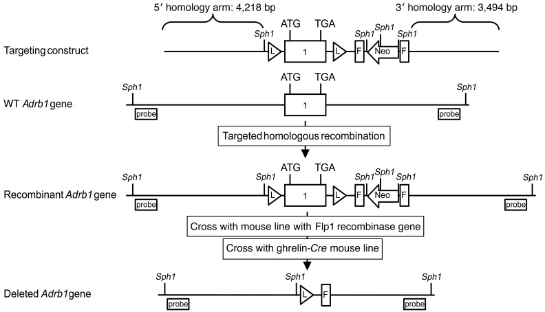
The majority of the mice generated previously using a germline β1AR-KO (Adrb1–/–) approach were reported to die prenatally (38). Here, we selectively deleted β1AR from ghrelin-expressing cells. To do so, a conditional Adrb1-KO (Adrb1fl/fl, referred to herein as β1ARfl/fl) mouse line was first developed by flanking the single exon Adrb1 gene with inserted loxP sites (Figure 1), followed by its genetic cross with our previously reported and validated ghrelin-Cre line that expresses Cre recombinase selectively in ghrelin cells (35, 39). As demonstrated previously, and together with further immunohistochemical and quantitative reverse transcriptase PCR (qPCR) authentication of the ghrelin-Cre line performed here (see Supplemental Methods; Supplemental Figures 1 and 2; supplemental material available online with this article; doi:10.1172/JCI86270DS1), cells with Cre activity include more than 95% of ghrelin cells in the gastric mucosa (from which most circulating ghrelin in adults emanates [refs. 40–44]) and a majority (~69%) of the highly dispersed, far less numerous population of ghrelin cells in the duodenum (35). Other cells with Cre activity include Leydig cells of the testis, epithelial cells of the epididymis, and scarce cells within some pancreatic islets, all of which correspond to known ghrelin cell distribution patterns (44– 49). Germline transmission of the recombinant loxP-flanked Adrb1 gene was validated by PCR analysis of genomic DNA obtained by tail snips (Supplemental Figure 3, A and B). Four experimental groups were generated: β1ARfl/fl ghrelin-CreTg+ mice (referred to herein as GC-β1AR–/– mice) with ghrelin cell–selective deletion of β1AR and 3 control genotypes (WT control: β1ARWT/WT ghrelin-CreTg–, referred to herein as β1ARw/w/G-CreTg–; Cre recombinase control: β1ARWT/WT ghrelin-CreTg+, referred to herein as β1ARw/w/G-CreTg+; and floxed β1AR control: β1ARfl/fl ghrelin-CreTg–, referred to herein as β1ARfl/fl/G-CreTg–). Cre-mediated deletion of the β1AR gene selectively in ghrelin cells from GC-β1AR–/– mice was confirmed by comparing Adrb1 mRNA levels (as determined by qPCR) in FACS-purified gastric ghrelin cells obtained from GC-β1AR–/– mice with Adrb1 mRNA levels in FACS-purified gastric ghrelin cells obtained from β1ARfl/fl/G-CreTg– control mice, after those mice had been placed on a ghrelin humanized Renilla reniformis GFP (ghrelin-hrGFP) reporter background (37). As compared with gastric ghrelin cells from control mice, those from GC-β1AR–/– mice had a 73.5% reduction in Adrb1 mRNA expression levels (Supplemental Figure 3C). Adrb1 expression was unaffected in a set of 7 tissues outside the gastrointestinal tract (Supplemental Table 1).
Figure 1. Generation of conditional β1AR-KO mice and mice with ghrelin cell–selective β1AR deletion.
Schematic diagrams show the homologous recombination targeting strategy to flank the β1AR (Adrb1) coding region with loxP sites, thus creating conditional β1AR-KO mice. Binding sites for Southern blot probes used to detect correctly targeted genomic DNA (after restriction digestion with Sph1) are depicted. Crosses first with Flp1 recombinase mice to remove the frt-Neo-frt cassette that had been included in the targeting construct to find neomycin-resistant ES cell clones and then with ghrelin-Cre mice result in Cre recombinase–mediated removal of the β1AR coding sequence selectively in ghrelin cells.
Ghrelin cell–selective β1AR deletion lowers plasma ghrelin and fasting blood glucose levels.
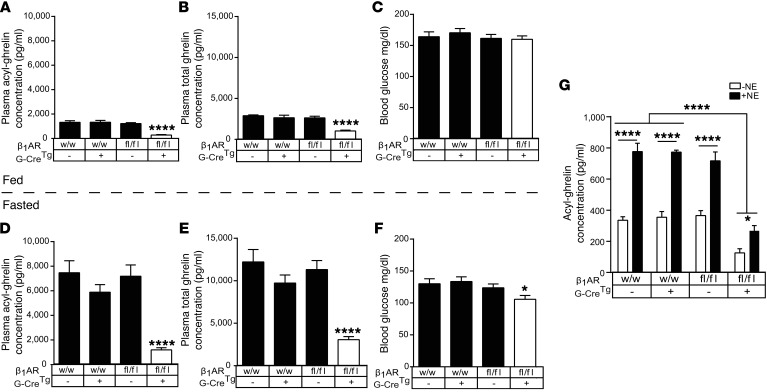
Plasma ghrelin normally increases upon fasting. To study the in vivo impact of β1AR deletion in ghrelin cells on ghrelin secretion and blood glucose levels, we measured plasma ghrelin and blood glucose before and after a 24-hour fast in 8-week-old GC-β1AR–/– mice. These mice showed, on average, 4.5-fold lower ad libitum–fed and 5.8-fold lower fasted plasma acyl-ghrelin levels when compared with levels in the control groups (Figure 2, A and D). Plasma total ghrelin levels in GC-β1AR–/– mice were, on average, 2.2-fold lower in ad libitum–fed and 3.6-fold lower in fasted conditions when compared with levels in the control groups (Figure 2, B and E). These in vivo results were corroborated by ghrelin secretion experiments using ex vivo primary gastric mucosal cell cultures (26): release of both basal and norepinephrine-induced acyl-ghrelin was severely blunted in cells from GC-β1AR–/– mice (Figure 2G). Importantly, while blood glucose levels of ad libitum–fed GC-β1AR–/– mice were comparable to levels in control groups, their fasted blood glucose level was significantly lower, although still within the normal range (Figure 2, C and F), which was similar to the blood glucose response to an acute fast in mice lacking GHSRs (24, 50). The corresponding plasma norepinephrine levels were higher in GC-β1AR–/– mice as compared with levels in the 3 control groups following the 24-hour fast, although no significant changes in plasma epinephrine, insulin, glucagon, GH, or insulin-like growth factor 1 (IGF-1) were noted (Supplemental Figure 4).
Figure 2. Ghrelin cell–selective β1AR deletion reduces plasma ghrelin, resulting in fasting hypoglycemia.
Plasma acyl- and total ghrelin levels in ad libitum–fed (A and B, respectively) and 24-hour–fasted conditions (D and E, respectively) in 8-week-old male mice and their corresponding blood glucose levels (C and F, respectively). n = 19–30. *P < 0.05 and ****P < 0.001, for significant reductions in plasma ghrelin or blood glucose in GC-β1AR–/– mice compared with control groups by 1-way ANOVA, followed by Holm-Sidak’s post-hoc multiple comparisons test. (G) Acyl-ghrelin concentrations in media from primary gastric mucosal cell cultures derived from ad libitum–fed control groups or GC-β1AR–/– mice treated for 6 hours with or without 10 μM norepinephrine (NE). n = 6–9 wells each. *P < 0.05 and ****P < 0.001, for significant increases in ghrelin secretion with norepinephrine treatment compared with vehicle treatment within the same genotype and significant decreases in ghrelin secretion in cultures from GC-β1AR–/– mice compared with control groups, as analyzed by 2-way ANOVA, followed by Holm-Sidak’s post-hoc multiple comparisons test. All values are expressed as the mean ± SEM. w/w, mice with WT β1AR genes; fl/fl, mice homozygous for the loxP-flanked β1AR gene; –, absence of the ghrelin-Cre Tg; +, presence of the ghrelin-Cre Tg.
The plasma ghrelin levels achieved by ghrelin cell–selective β1AR deletion were similar to those achieved by global β1AR blockade or deletion: pharmacological inhibition of β1AR by twice-daily administration of atenolol for 3 days reduced plasma acyl-ghrelin in ad libitum–fed WT mice by 3.2-fold and markedly blunted the usual rise in plasma ghrelin induced by a 24-hour fast (Supplemental Figure 5), as reported previously (34). The accompanying blood glucose values of these fasted 6-week-old mice were unaffected by atenolol. Also, we generated limited numbers of global β1AR-KO mice (Z-β1AR–/–) by crossing the β1ARfl/fl mice with Zp3-Cre mice (51) (see Supplemental Methods for a detailed description). The few surviving Z-β1AR–/– mice, which we confirmed as having global β1AR deletion (Supplemental Figure 6A) and which were phenotypically similar to WT mice in overall BW and appearance, demonstrated lower basal and fasted plasma acyl- and total ghrelin levels (Supplemental Figure 6B).
Ghrelin deficiency in GC-β1AR–/– mice does not greatly impact food intake, BW, or adiposity.
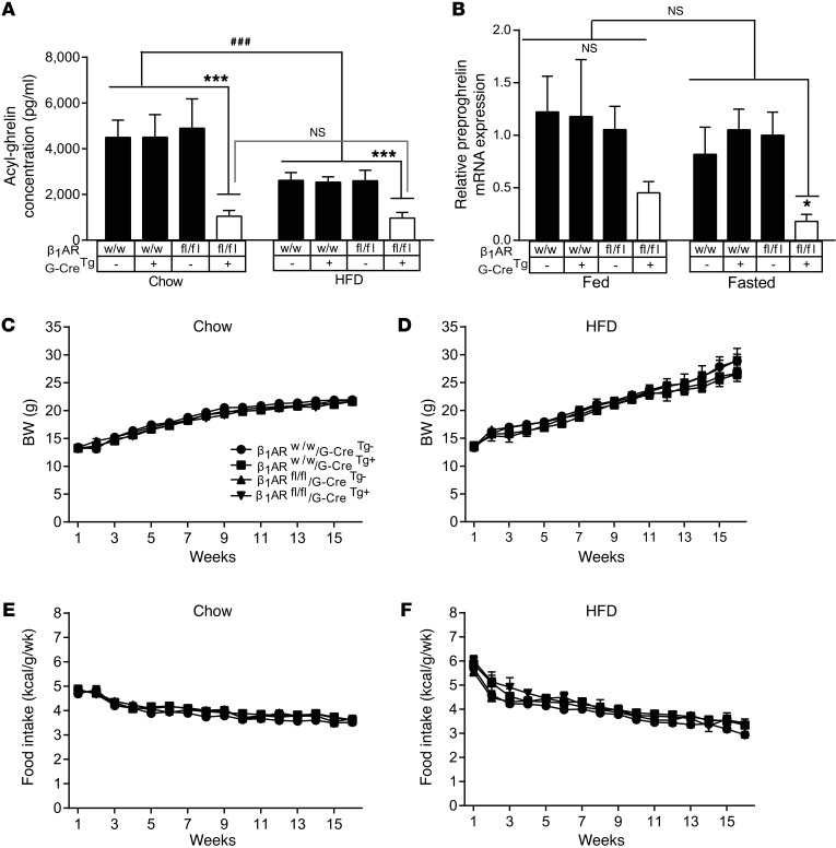
To determine the effect of ghrelin cell β1AR deletion–induced blunting of ghrelin secretion on food intake and BW, we fed female GC-β1AR–/– mice standard chow or an HFD (42% kcal from fat) for 16 weeks. Ad libitum–fed and fasted acyl-ghrelin levels were lower in the female GC-β1AR–/– mice than were levels in control mice at various time points over the 16-week period (Figure 3A; Supplemental Figure 7), similar to our observation in standard chow–fed 8-week-old male mice (Figure 2). While control groups fed an HFD for 16 weeks had reduced fasted plasma ghrelin levels compared with mice fed standard chow, HFD feeding in GC-β1AR–/– mice did not alter plasma ghrelin levels compared with levels in the standard chow–fed GC-β1AR–/– mice (Figure 3A). Chow-fed GC-β1AR–/– mice demonstrated reduced gastric mucosal preproghrelin mRNA expression, although this observation was significant only in the fasted condition (Figure 3B). Preproghrelin mRNA expression levels were not different among the experimental groups fed an HFD (Supplemental Figure 8). No differences in weekly food intake or BW were observed in GC-β1AR–/– mice (Figure 3, C–F). Likewise, body composition and body length were similar at the end of the study (Supplemental Figure 9).
Figure 3. Ghrelin cell–selective β1AR deletion blocks HFD-induced reductions in ghrelin but does not alter BW or ad libitum food intake.
(A) Plasma acyl-ghrelin levels in female mice fasted for 24 hours after 16 weeks of exposure to standard chow or an HFD. n = 11–16. ***P < 0.005, for significant differences in plasma ghrelin in GC-β1AR–/– mice compared with control genotype mice fed the same diet; ###P < 0.005, for significant reductions in plasma acyl-ghrelin in mice fed an HFD compared with mice fed standard chow. No significant changes in plasma acyl-ghrelin in GC-β1AR–/– mice fed chow or an HFD were observed. Data were analyzed by 2-way ANOVA, followed by Holm-Sidak’s post-hoc multiple comparisons test. (B) Quantitative estimation of relative preproghrelin mRNA levels in gastric mucosal cells from ad libitum–fed and 24-hour–fasted mice after 16 weeks of exposure to standard chow. Data are expressed as ΔΔCt values relative to ad libitum–fed β1ARw/w/G-CreTg– control mice. n = 5–7. *P < 0.05, for significant differences in preproghrelin mRNA between fasted GC-β1AR–/– mice and fasted control genotypes. Data were analyzed by 2-way ANOVA, followed by Holm-Sidak’s post-hoc multiple comparisons test. (C and D) BW and (E and F) weekly food intake in mice fed standard chow or an HFD for 16 weeks. n = 11–16. No significant changes were observed among the groups fed the same diet, as analyzed by repeated-measures 2-way ANOVA. Values are expressed as the mean ± SEM.
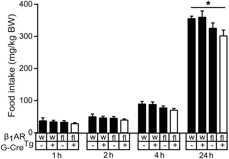
As GHSR antagonist has been shown to reduce food intake in acutely fasted WT mice (13), we also tested whether blunted fasting-induced ghrelin secretion in GC-β1AR–/– mice has a similar effect. Rebound food intake following a 24-hour fast of 8-week-old male mice that had been maintained on a standard chow diet was measured. GC-β1AR–/– mice ingested less standard chow in the 24 hours following fasting than did the β1ARw/w/G-CreTg– WT control group mice (Figure 4).
Figure 4. Mice with ghrelin cell–selective β1AR deletion demonstrate reduced rebound feeding after a 24-hour fast.
Rebound food intake measured in 8-week-old male mice at different time points following a 24-hour fast. n = 19–30. *P < 0.05, for significantly less food consumption by GC-β1AR–/– mice compared with β1ARw/w/G-CreTg– mice, as determined by repeated-measures 2-way ANOVA, followed by Holm-Sidak’s post-hoc multiple comparisons test. All values are expressed as the mean ± SEM. w, mice with WT β1AR genes; fl, mice homozygous for the loxP-flanked β1AR genes; –, absence of the ghrelin-Cre Tg; +, presence of the ghrelin-Cre Tg.
Severe (60%) caloric restriction of GC-β1AR–/– mice induces frank hypoglycemia and increases mortality.
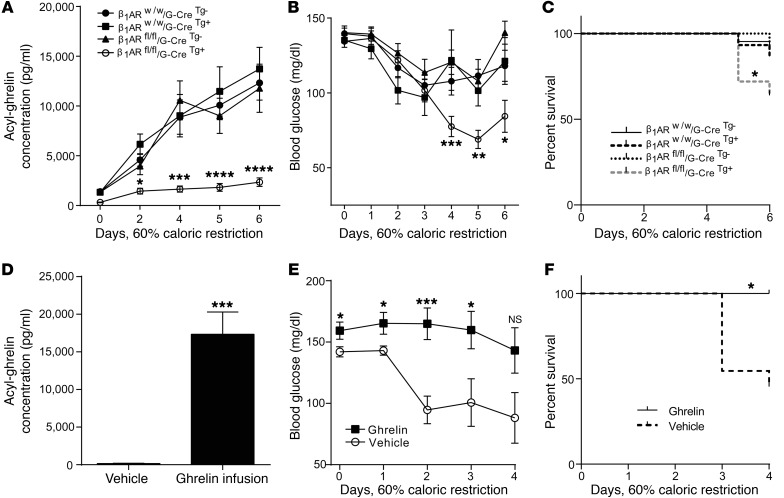
To test the functional significance of ghrelin cell β1ARs during a more severe state of energy insufficiency, we subjected singly housed 8-week-old GC-β1AR–/– mice to 60% caloric restriction (by feeding them 40% of their usual daily calories) for 6 successive days, using a previously reported protocol (39, 52–54). BW declines in GC-β1AR–/– mice were similar to those in control groups (Supplemental Figure 10). Although plasma acyl-ghrelin levels rose in GC-β1AR–/– mice (7.4-fold), this increase was smaller than the 8.8- to 10.3-fold increases observed in the control groups, and the final level achieved was over 80% lower than that in the control groups (Figure 5A). This reduced plasma ghrelin level in GC-β1AR–/– mice was accompanied by frank hypoglycemia (nadir = 45.3 ± 3.7 mg/dl) — while similarly treated control groups retained normoglycemia (nadir = 76.3 ± 5.8 mg/dl) — and increased mortality (36%) compared with a 9%–13% mortality rate in the control groups (Figures 5, B and C). Of note, data presented in Figure 5, B and E were derived from surviving mice and therefore likely underrepresent the actual magnitude in the mean falls in blood glucose levels. No significant differences in corresponding plasma insulin, glucagon, GH, or IGF-1 were detected among the groups of surviving mice (Supplemental Figure 11).
Figure 5. Mice with ghrelin cell–selective β1AR deletion demonstrate a blunted ghrelin response to severe caloric restriction, resulting in hypoglycemia and decreased survival.
Plasma acyl-ghrelin (A), blood glucose (B), and survival (C) in mice given access to a 60% caloric restriction protocol (40% of their usual daily calories for 6 consecutive days). Reduced plasma ghrelin in GC-β1AR–/– mice was accompanied by lower blood glucose and reduced survival. n = 13–22 (A–C). Continuous s.c. acyl-ghrelin delivery via Alzet osmotic minipumps to GC-β1AR–/– mice during the severe caloric restriction restored plasma ghrelin levels (D), prevented hypoglycemia (E), and promoted survival (F). n = 8–11 (D–F). Data in A, B, D, and E were analyzed by 2-way ANOVA, followed by Holm-Sidak’s post-hoc multiple comparisons test. *P < 0.05, **P < 0.01, ***P < 0.05, and ****P < 0.001, for significant differences in plasma acyl-ghrelin and blood glucose levels in GC-β1AR–/– mice as compared with levels in control group mice (A and B) and significant differences between acyl-ghrelin and vehicle-infused GC-β1AR–/– mice (D and E). Survival curves (C and F) were calculated by the Kaplan-Meier method, with comparisons among groups using the Mantel-Cox log-rank test. *P < 0.05, for significant differences in survival among the groups (C and F).
To confirm the role of reduced ghrelin secretion in the marked hypoglycemia and increased mortality experienced by GC-β1AR–/– mice, a separate cohort of GC-β1AR–/– mice was administered 4 mg/kg/day acyl-ghrelin s.c. via an osmotic minipump during the severe caloric restriction period. Ghrelin infusion, which increased plasma ghrelin to a level slightly higher than that attained physiologically in control mice during severe caloric restriction (Figures 5, A and D), not only increased blood glucose in ad libitum–fed GC-β1AR–/– mice (day 0 in Figure 5E) but also prevented the fall in blood glucose in GC-β1AR–/– mice upon severe caloric restriction and resulted in 100% survival (Figure 5, E and F).
Administration of atenolol to juvenile mice induces hypoglycemia accompanied by reduced plasma ghrelin levels.
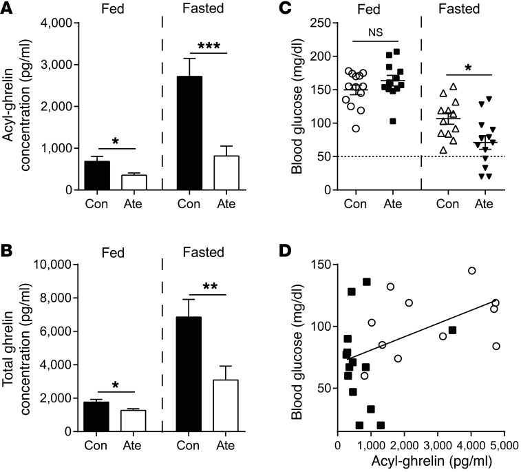
Beta blockers are widely prescribed drugs to treat cardiac failure and hypertension. While hypoglycemia unawareness is a concern in adult diabetic subjects treated with beta blockers (55), life-threatening hypoglycemic episodes are not an infrequent occurrence in fasted infants and toddlers treated with beta blockers, e.g., for infantile hemangioma and arrhythmias (56–59). To test whether beta blocker–induced hypoglycemia in young patients could be related to reductions in plasma ghrelin levels, we designed a preclinical model in which 3-week-old WT mice were administered atenolol or vehicle twice daily for 3 days. Plasma acyl- and total ghrelin levels measured in fed conditions or after a 24-hour fast were both significantly reduced on the third day with atenolol administration (Figure 6, A and B). Atenolol treatment significantly lowered blood glucose (in many cases to below 50 mg/dl) when measured after fasting, but not in the fed state (Figure 6C). Plasma acyl-ghrelin levels positively correlated with blood glucose levels in these fasted 3-week-old mice (Figure 6D). The correlation was seemingly influenced more by the vehicle-treated mice than by the atenolol-treated mice (which, as a group, had lower ghrelin levels), suggesting a threshold concentration of ghrelin above which its protective glucoregulatory functions are active. Plasma insulin levels did not differ with atenolol treatment in the fasted 3-week-old mice, but plasma IGF-1 was significantly lower with atenolol treatment (Supplemental Figure 12). The reduction in blood glucose levels with atenolol treatment was accompanied by induction of the hepatic gluconeogenic genes encoding peroxisome proliferative activated receptor γ, coactivator 1 α (PGC1α [Ppargc1a]), phosphoenolpyruvate carboxykinase 1 (PEPCK [Pck1]), and glucose-6-phosphatase (G6P [G6pc]) (Supplemental Figure 13). Other hepatic genes encoding for the gluconeogenic enzymes hepatic nuclear factor 4, α (HNF4α [Hnf4a]) and pyruvate carboxylase (PCX [Pcx]) and the glycogenolytic enzyme liver glycogen phosphorylase (PYGL [Pygl]) did not change with atenolol treatment. Although plasma IGF-1 was reduced by atenolol, mRNA levels of Igf1 and insulin-like growth factor–binding protein 1 (Igfbp1) were unchanged (Supplemental Figures 12 and 13). Unlike in fasted 3-week-old mice, neither fed 3-week-old mice (Figure 6C) nor fed or fasted 6-week-old mice (Supplemental Figure 5) had atenolol-induced reductions in plasma ghrelin levels associated with hypoglycemia.
Figure 6. Beta blocker administration to 3-week-old WT mice induces hypoglycemia associated with reduced fasting plasma ghrelin.
Plasma acyl-ghrelin (A), total ghrelin (B), and corresponding blood glucose (C) levels in ad libitum–fed or 24-hour–fasted 3-week-old male C57BL/6N pups administered atenolol (Ate) (10 mg/kg) or vehicle (Con) twice daily for 3 days. n = 9–13. *P < 0.05, **P < 0.01, and ***P < 0.005, for each condition by unpaired Student’s t test. (D) Scatter plot of fasted blood glucose versus corresponding plasma acyl-ghrelin levels in mice treated with atenolol or vehicle. White circles indicate mice treated with vehicle, and black squares indicate mice treated with atenolol; the line indicates the linear regression fit for the data points (r2 = 0.2). P = 0.03, for significant Pearson’s correlation coefficient between blood glucose and acyl-ghrelin levels.
Discussion
Until now, advances in our understanding of β1AR biology, including as it regards ghrelin secretion, have been limited mainly to pharmacological approaches because of the poor survival rates of germline β1AR-KO mice (38). The new conditional β1AR-KO mouse line reported here has enabled us to test the functional significance of ghrelin cell β1ARs, including their effects on ghrelin secretion, blood glucose regulation, survival response to a starvation-like state, food intake, and BW. Using GC-β1AR–/– mice, which reproduce and survive normally, we now provide genetic evidence of required ghrelin cell β1AR expression to regulate basal ghrelin release and to maintain preproghrelin gene transcription and stimulate ghrelin secretion in response to caloric restriction. Furthermore, we reveal the required roles for ghrelin cell β1ARs in preventing falls in blood glucose levels during caloric restriction (including preventing the development of marked hypoglycemia in both severely calorie-restricted adult mice and acutely fasted young mice), in supporting survival in the setting of a starvation-like state, and in encouraging rebound food intake following a short-term fast.
There are several noteworthy discussion points and implications of these results. Hypoglycemia was the most conspicuous phenotype resulting from the abrogated ghrelin secretion in GC-β1AR–/– mice, developing within just a few days of exposure to severe caloric restriction and probably contributing to the pronounced mortality. This protocol, which rapidly and markedly depletes fat stores and emulates a starvation state (28), has the same effect in mice that lack ghrelin (54), GOAT (52, 60), or GHSR (53) and in mice with targeted ghrelin cell degradation (39). However, the reproducibility of this effect and the dependency of ghrelin secretion in this starvation model on ghrelin cell β1AR expression were not expected, especially since low glucose can itself stimulate ghrelin secretion (26). Notably, although this effect of low glucose is observed in cultured gastric ghrelin cells (26), the same phenomenon when occurring in vivo seems unable to mount a ghrelin secretory response of sufficient magnitude to prevent further blood glucose falls and preserve life. We conclude that β1AR-driven ghrelin secretion induced by caloric restriction is protective, particularly in starvation-like states during which it prevents marked hypoglycemia and sustains life.
Given our results with severely calorie-restricted GC-β1AR–/– mice and overnight-fasted young WT mice administered atenolol, we also propose that unintended blockade of ghrelin cell β1ARs in beta blocker–treated individuals and the ensuing reduction in plasma ghrelin might, in a permissive setting, predispose them to severe hypoglycemia and death (Figure 7). As mentioned above, life-threatening hypoglycemia is a well-recognized adverse effect of beta blocker therapy in young children (57, 58, 61). Several cases of hypoglycemia in newborns and toddlers with hemangioma treated with the nonselective beta blocker propranolol have been reported and most often occurred when there was poor oral intake such as during an overnight fast or concomitant infection (56). These cases have prompted the establishment of a standardized consensus–derived set of best practices for propranolol-treated children with infantile hemangioma, including recommendations for frequent meals and avoidance of prolonged fasts, periprocedural support with glucose-containing parenteral fluids, discontinuation of propranolol during illness (especially in the setting of restricted oral intake), and listing hypoglycemia as a side effect of beta blocker therapy (56, 57). Severe hypoglycemic episodes have also been reported in young children taking beta blockers for prevention of arrhythmias (57). The correlation of plasma ghrelin levels with blood glucose in fasted 3-week-old mice administered atenolol or vehicle (Figure 6D) supports the assertion that reduced plasma ghrelin underlies the above-described, sometimes profound cases of beta blocker–induced hypoglycemia reported in children. Why hypoglycemia does not occur with reportable frequency in adults on beta blocker therapy is unclear, although neither did it occur here in fasted 6-week-old mice administered atenolol. This discrepant, age-dependent susceptibility to hypoglycemia related to a blunted ghrelin response deserves further study, but may be associated with lower fat or glycogen reserves or differential glucose utilization rates in children versus adults (56) or perhaps a greater dependency on ghrelin for blood glucose regulation in the young. Other groups besides the young that are predicted to be susceptible to hypoglycemia and possibly death as a result of beta blocker–induced or other causes of reduced ghrelin secretion include cachectic individuals and individuals with anorexia nervosa, in whom high levels of ghrelin have been described and normally may serve a protective function (7, 12, 62, 63). Consideration should also be given to the possible involvement of an insufficient ghrelin response in the more common condition of transitional neonatal hypoglycemia in normal newborns (64) or in cases of exercise-induced hypoglycemia in beta blocker–treated individuals (65).
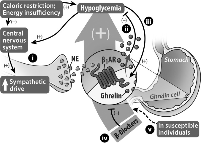
Figure 7. Proposed models of caloric restriction–induced ghrelin secretion to restore glucose homeostasis and of beta blocker–induced hypoglycemia.
(i) Caloric restriction acting through the CNS enhances sympathetic drive onto ghrelin cells that are distributed within the gastric mucosa. Norepinephrine released from the sympathetic nerve terminals activates β1ARs located on the ghrelin cells to induce ghrelin synthesis and secretion. (ii) In turn, the released acyl-ghrelin engages several processes, including GH secretion, that tend to limit falls in blood glucose. Reduced blood glucose can also stimulate a sympathetic response and, furthermore, (iii) can directly stimulate ghrelin cells to release ghrelin, although this direct stimulus only accounts for a fraction of the usual secretory response of ghrelin cells to caloric restriction. When body fat is depleted as a consequence of severe caloric restriction, the ghrelin cell β1AR–mediated release of ghrelin functions as a critical defense against marked falls in blood glucose, thereby promoting survival. (iv) Administration of beta blockers results in unintended blockade of ghrelin cell β1ARs, causing blunted ghrelin secretion. (v) When lack of normal ghrelin secretion caused by β1AR blockade is combined with food restriction, susceptible individuals, such as young children and probably also adults with cachexia or anorexia nervosa, become predisposed to developing life-threatening hypoglycemia.
As mentioned in the Introduction, ghrelin has at its disposal several potential downstream effector hormones and tissues through which it may act to sustain blood glucose levels in fasted states. One of its key glucoregulatory mediators is GH, for which ghrelin serves as a potent secretagogue during caloric restriction (1, 52). Whereas plasma GH progressively rises in WT mice subjected to a week of 60% caloric restriction, peaking just prior to the addition of food, this rise is blunted in similarly treated GOAT–/– and ghrelin–/– mice, which exhibit an approximately 2-fold reduction in plasma GH accompanying hypoglycemia when measured on the final day of caloric restriction (52, 54, 60). The lower GH and blood glucose levels are both preventable by continuous ghrelin infusion or by continuous GH infusion during the caloric restriction (52). The previous studies with GOAT–/– and ghrelin–/– mice revealed a reduced generation of substrates for gluconeogenesis during 60% caloric restriction, which in the case of GOAT–/– mice was associated with reduced hepatic autophagy (54, 60). Those previous studies also suggested that ghrelin’s protective effects on blood glucose and survival during 60% caloric restriction are directly attributable to its GH secretagogue activity and in turn (via a non–IGF-1–signaling mechanism) to stimulation of hepatic autophagy, together with other as-yet undetermined processes, to supply substrates for gluconeogenesis (54, 60). In the absence of ghrelin, fat-depleted mice with limited access to food, as achieved using the 60% caloric restriction protocol, are unable to maintain sufficient rates of gluconeogenesis, even though there is an attempt via upregulation of gluconeogenic enzyme gene expression (e.g., PEPCK and G6Pase) (52). While the severely calorie-restricted GC-β1AR–/– mice in the current study did not demonstrate reduced GH levels, as had been detected previously in GOAT–/– and ghrelin–/– mice, unlike the previous studies, in which the blood samples for GH estimation were collected from all mice that underwent the severe caloric restriction protocol (including those that were in a moribund state due to hypoglycemia) (52), here, samples were not taken from moribund animals and instead only included those from mice that survived the entire 6 days of the protocol. This also explains the reduced sample size of plasma GH values from GC-β1AR–/– mice (Supplemental Figure 4E). Had samples been obtained from all GC-β1AR–/– mice in the severe caloric restriction study, including those that did not survive (just prior to their demise), we believe that an impaired GH response as a result of ghrelin secretion deficiency would become evident, similar to what was observed in the previous studies.
A blunted GH pathway response as a result of lowered ghrelin levels, though, was detected here in the fasted 3-week-old C57BL/6N mice treated with atenolol. In particular, following the 24-hour fast, lowered plasma IGF-1 accompanied the lowered blood glucose and lowered ghrelin levels in mice treated with atenolol as compared with levels in controls. Thus, despite likely differences in the amounts of stored glucose, stored fat, and potential circulating gluconeogenic substrates in 24-hour fasted 3-week-old mice versus adult mice exposed to a week-long, fat-depleting 60% caloric restriction protocol, in both scenarios, hypoglycemia results from an experimentally induced insufficient ghrelin response and a resulting insufficient GH response (albeit seemingly normally mediated by IGF-1 in the former scenario versus hepatic phosphorylated-STAT5 in the latter scenario) (60). Also of note, while gene expression of the hepatic gluconeogenic enzymes PGC1α, PEPCK, and G6P was induced here in the fasted, atenolol-treated 3-week-old mice, it is apparent that this likely compensatory response is nonetheless inadequate to prevent hypoglycemia from developing. Similarly, gluconeogenesis previously had been determined to be insufficient in 60% calorie-restricted GOAT–/– mice, despite upregulation of PEPCK and G6P, presumably due to an insufficient availability of gluconeogenic substrates (52).
Ghrelin also has the capacity to influence blood glucose by stimulating food intake, which was experimentally restricted in the current study, and by raising circulating glucocorticoids and/or reducing insulin sensitivity (23, 50, 66), which were not specifically examined here. Furthermore, ghrelin can modify the secretion of several pancreatic islet hormones that regulate blood glucose including the reduction of insulin secretion and stimulation of glucagon secretion (via either direct actions on pancreatic β cells [21, 23, 67] and α cells [24] or indirect actions on hypothalamic AgRP neurons [53] or somatostatin-secreting pancreatic δ cells [68]). However, insulin and glucagon levels in GC-β1AR–/– mice subjected to the 60% caloric restriction protocol were like those in similarly treated control groups. These results, along with similar observations in 60% calorie-restricted GOAT–/– mice (52), suggest that ghrelin modulation of insulin or glucagon levels may not be a significant contributor to ghrelin’s actions to sustain blood glucose in states of severe caloric restriction. Rather, these effects on insulin and glucagon probably play a more prominent role in ghrelin’s glucoregulatory efficacy in other scenarios. For instance, previously, we have demonstrated exaggerated drops in plasma glucagon associated with exaggerated drops in blood glucose (albeit not frank hypoglycemia) in overnight-fasted GHSR-deficient mice as compared with levels in WT mice (24, 53). The unaltered insulin and glucagon levels observed here also suggest that the scarce expression of ghrelin-Cre within pancreatic islets did not alter the overall usual sympathetic influence on islet hormone release during hypoglycemia (69, 70).
It is also noteworthy that signals from the autonomic nervous system so prominently impact ghrelin secretion, especially given the inherent nutrient-sensing, enteroendocrine nature of most ghrelin cells (71, 72). While recognition by ghrelin cells of dietary metabolites — and, in particular, their sensing of low glucose levels — could drive the modest increase in plasma ghrelin levels observed in GC-β1AR–/– mice upon a 24-hour fast or severe caloric restriction, the levels achieved are far lower than those in mice with intact ghrelin cell β1AR signaling. Thus, it could be that the major impact of nutrients such as glucose on ghrelin secretion is inhibitory and, instead, that signals from the brain — such as those carried by the sympathetic nerves — serve as the main stimulatory regulators of ghrelin release. The entrainment of preprandial rises in plasma ghrelin levels to set meal schedules (73) also suggests the importance of higher-order inputs to ghrelin cells from the brain in stimulating ghrelin secretion. Another related observation is the significant elevation of plasma norepinephrine levels in fasted GC-β1AR–/– mice. This could be due to a loss of usual ghrelin inhibition on sympathetic drive, as parenteral and intracerebroventricular administration of ghrelin is known to reduce plasma norepinephrine levels and inhibit sympathetic activity (74–76), or to a compensatory increase in sympathoadrenal drive in an attempt to boost ghrelin levels or in response to the low fasted blood glucose levels (77). Parenthetically, the specific role of catecholaminergic input from the sympathetic nervous system in β1AR-driven ghrelin secretion, as opposed to catecholamines produced by the adrenal glands, is supported by studies showing failed induction of ghrelin secretion by parenteral administration of epinephrine to achieve levels mirroring those seen in severe stress (29).
Another important topic of discussion relates to the physiological role of secreted ghrelin in eating and BW. There is a significant dichotomy in the results obtained using ghrelin gain-of-function models as compared with and among ghrelin system loss-of-function studies. As such, while administered ghrelin stimulates food intake, increases BW, and promotes adiposity (5, 14, 78), and rats carrying the above-described gain-of-function GHSR-Q343X isoform exhibit increased adiposity when older and improved BW maintenance during chronic caloric restriction (25), many studies with recombinant mice lacking a functional ghrelin system show no or only modest feeding and/or BW phenotypes (4, 39, 52, 79–83), while others suggest that intact ghrelin signaling is required for normal eating and BW responses, especially to hedonically rewarding diets (13, 79, 82). Here, deletion of ghrelin cell β1AR–dependent ghrelin secretion blunted rebound food intake following a 24-hour fast, suggesting a modest role for β1AR-mediated increases in endogenous ghrelin secretion in the usual feeding response to a short-term fast. However, this deletion did not impact ad libitum feeding responses, BW regulation, or body composition. Of related interest, plasma ghrelin levels are usually lower in diet-induced obesity, which is often touted as being a compensatory response to prevent exaggerated BW gain (11). Here, mice lacking ghrelin cell β1ARs failed to further reduce plasma ghrelin levels in response to chronic HFD feeding, suggesting that obesity-related reductions in plasma ghrelin levels are mediated by reduced sympathetic drive or, as demonstrated previously using cultured gastric mucosal cells from diet-induced obese mice (27), are mediated by a reduced responsiveness of ghrelin cells to sympathetic stimulation. Regardless, in the current study, the HFD-fed GC-β1AR–/– mice did not diverge in BW from that of control animals, despite divergent ghrelin secretion responses to HFD feeding.
As final discussion points, the present study also demonstrated a differential magnitude of acyl-ghrelin versus total ghrelin reductions due to ghrelin cell–selective β1AR deletion, which we speculate could result from differential effects on acyl-ghrelin versus desacyl-ghrelin release or alterations to GOAT activity and/or other enzymes responsible for ghrelin biosynthesis, maturation, or degradation (2, 3). Additionally, it should be noted that, besides being a powerful tool to investigate the role of β1AR signaling in ghrelin cells, the β1ARfl/fl line reported here should prove valuable in future studies of other important β1AR-mediated processes, such as cardiac function, smooth muscle tone, the hypoglycemia counterregulatory response, and adipose tissue physiology. Also, given our findings in calorie-restricted GC-β1AR–/– mice, we would predict that other conditions associated with enhanced sympathetic drive, such as psychosocial stress and cold exposure — both of which have been shown to be associated with increased plasma ghrelin levels — may involve robust β1AR-dependent ghrelin secretion responses (50, 84–87) on which the full physiological responses to those conditions may depend. Furthermore, the occurrence of hypoglycemia in calorie-restricted GC-β1AR–/– mice and the prevention thereof by ghrelin administration suggest that closer blood glucose monitoring might be prudent in cachectic individuals if beta blocker therapy becomes indicated, just as has already been recommended for children with infantile hemangioma treated with propranolol, for whom we now propose that reduced ghrelin secretion contributes to the beta blocker–associated hypoglycemia. Finally, our current results can be added to the accumulating evidence suggesting that the ghrelin system has only subtle effects on eating and BW when food is readily available and plentiful, but that it nonetheless remains essential for the maintenance of life-sustainable glycemic levels when energy stores approach those of starvation states.
Methods
Detailed experimental procedures for standard animal techniques, qPCR, and cell culture are described in the Supplemental Methods.
Animal procedures.
Mice were housed under standard laboratory conditions and were provided ad libitum access to standard chow (Harlan Teklad 2016, with an energy density of 3 kcal/g, of which 12% of kcal are derived from fat) and water unless otherwise specified.
Generation of β1ARfl/fl mice.
A mouse line containing a recombinant β1AR gene (Adrb1) flanked by loxP sites was generated by BAC recombineering techniques in EL250 and EL350 cells (Figure 1) using previously described methods (88, 89). Briefly, a 111,024-bp BAC clone (BMQ-52i16) containing the entire Adrb1 gene was obtained from Source Bioscience and transformed into EL350 cells. To generate the targeting construct, a 4,210-bp fragment containing the complete Adrb1 coding sequence and homology arms was cloned into a commercially available pGEM vector (Promega), which had been altered to contain the thymidine kinase gene (used for negative selection of ES cell clones). A previously described loxP-neomycin resistance gene (Neo)-loxP cassette from pL452 was inserted 607 bp upstream of the Adrb1 start codon using homologous recombination, followed by removal of Neo-loxP by arabinose induction of Cre recombinase (90). Next, another loxP-frt-Neo-frt cassette from pL451 was inserted 517 bp downstream of the Adrb1 stop codon. These loxP insertion sites were chosen on the basis of a comparison of the 5′- and 3′-UTR regions of the Adrb1 gene in an array of mammalian species and were within regions with the least homology near the start and stop codons. The resulting targeting construct was prepared and used for electroporation into 129/SvEvTac (SM-1 cells) by the UT Southwestern Medical Center Transgenic Technology Core Facility. Correctly targeted ES cell clones were identified by Southern blot and PCR analyses. Three embryonic stem (ES) cell clones were selected for blastocyst injection, and germline transmission was established for 2 clones. The resulting pups were backcrossed with C57BL/6N mice for 3 generations. To remove the Neo-Frt sequence, the lines were then crossed with “Flp1 recombinase” mice [B6.Cg-Tg(ACTFLPe)9205Dym/J; The Jackson Laboratory] that had been backcrossed for more than 10 generations onto the C57BL/6N background in our colony. One of the lines was chosen for subsequent breeding to generate experimental mice. Furthermore, to demonstrate germline transmission of the loxP-flanked Adrb1 allele, heterozygous β1ARWT/fl mice were crossed with each other, yielding the predicted numbers of mice with 2 copies of the loxP-flanked Adrb1 allele, mice with 2 copies of the WT Adrb1 allele, and heterozygotes, as determined using a PCR strategy on genomic DNA of resulting pups (Supplemental Figure 3).
Generation of mice with ghrelin cell–selective β1AR deletion.
Ghrelin-Cre mice (35, 39) on a pure C57BL/6N genetic background were crossed with heterozygous β1ARWT/fl mice to generate breeders harboring 1 copy of the loxP-flanked Adrb1 allele and 1 copy of the ghrelin-Cre Tg (G-CreTg+). These mice were bred with heterozygous Adrb1WT/fl mice to generate the 4 study groups, which included mice containing 2 loxP-flanked Adrb1 alleles or 2 WT Adrb1 alleles, all with or without 1 copy of the ghrelin-Cre Tg. The primers used in the study to validate the genotypes of all the genetically engineered mice are listed in Supplemental Table 2.
Long-term feeding studies.
Female mice were fed either standard chow or an HFD (Harlan Teklad TD88137, with an energy density of 4.5 kcal/g, of which 42% of kcal are derived from fat) for 16 weeks. Fed plasma acyl-ghrelin levels were sampled at 4 and 12 weeks of the study, and 24-hour–fasted acyl-ghrelin levels were sampled at 8 and 16 weeks of the study. After 16 weeks, body composition analyses were performed using an EchoMRI-100 (Echo Medical Systems), body (nose-to-anus) length was measured under anesthesia using a ruler, and then half the cohort was sacrificed in an ad libitum–fed state, while the other half was sacrificed after a 24-hour fast. Gastric mucosal cells were harvested from excised stomachs (see Supplemental Methods) and were then treated with RNA-STAT 60 (Tel-Test Inc.) for preproghrelin mRNA quantification.
Severe (60%) caloric restriction protocol.
Eight-week-old male mice were provided access to 40% of their usual daily calories in the form of their usual diet, as described previously, for 6 days (52, 53). All mice were individually housed during the study and acclimatized for 1 week before starting the caloric restriction protocol. During the acclimatization period, daily food intake was measured for 5 days to determine the mean usual daily caloric intake for each mouse. At the start of the experiment, the percentage of body fat mass of the mice as determined using the EchoMRI-100 system was between 8% and 10%. Mice subjected to this protocol typically experience a drop in body fat percentage to less than 2% after only 2 days (52, 53). We entered 25 GC-β1AR–/– mice, 22 β1ARw/w/G-CreTg– mice, 15 β1ARw/w/G-CreTg+ mice, and 21 β1ARfl/fl/G-CreTg– mice into this study, although noted that 9, 2, 2, and 2 mice, respectively, had died by the sixth day of 60% caloric restriction (Figure 5C), leading us to discontinue the protocol on the sixth day instead of waiting 7 days or more, as had previously been published (39, 52–54, 60).
Determination of blood glucose and ghrelin levels.
Blood samples were collected by a quick superficial temporal vein (submandibular) bleed into EDTA-coated microtubes containing a protease inhibitor, p-hydroxymercuribenzoic acid (final concentration 1 mM), kept on ice. Blood glucose concentrations were measured using the hand-held AlphaTRAK 2 (Abbott Animal Health) blood glucose monitoring system. The samples were immediately centrifuged at 4°C at 1,500 g for 15 minutes, and HCl was added to the supernatant to achieve a final concentration of 0.1N (for stabilization of acyl-ghrelin). Processed samples were stored at –80°C in small aliquots until analysis of acyl-ghrelin or total ghrelin levels. Ghrelin concentrations in the plasma and cell culture media were determined by using ELISA kits from Millipore-Merck (catalog EZRGRA-90K for acyl-ghrelin and catalog EZRGRT-91K for total ghrelin). The endpoint calorimetric assays were performed using a PowerWave XS Microplate spectrophotometer and KC4 Junior software (BioTek Instruments).
Isolation of primary gastric mucosal cells.
Primary gastric mucosal cells were isolated by a combined enzymatic and mechanical dispersion method as described previously (26, 34, 35) and in more detail in the Supplemental Methods.
Atenolol administration.
Atenolol (10 mg/kg BW i.p.) or its vehicle (2 mM HCl) was administered over 3 days to 3- or 6-week-old male WT C57BL/6N mice as described previously (34). Briefly, this occurred at 7 am and 7 pm on days 1 and 2 and at 7 am on day 3. For the 6-week-old mice, blood was collected at 8:30 am on day 2 (ad libitum–fed condition), after which the same mice were fasted for 24 hours and blood again collected at 8:30 am on day 3 (24-hour–fasted condition). For 3-week-old mice, the same dosing regimen was followed, but blood collections for the ad libitum–fed and 24-hour–fasted conditions were performed in separate cohorts of mice on day 3, one cohort of which was not fasted and the other cohort of which was fasted, respectively.
Statistics.
All data are expressed as the mean ± SEM. Two-tailed statistical analysis and graph preparations were performed using GraphPad Prism 6.0 (GraphPad Software). A Student’s t test, 1-way ANOVA, 2-way ANOVA, or repeated-measures 2-way ANOVA, followed by the appropriate post-hoc comparison test, was used to test for significant differences among test groups. Survival curves were calculated by the Kaplan-Meier estimate method and compared using the Mantel-Cox log-rank test. The strength of the linear relationship between 2 sets of variables was compared by Pearson’s correlation coefficient. Outliers were detected by Grubb’s test. P values of less than 0.05 were considered statistically significant.
Study approval.
All animal procedures and use of mice were approved by the IACUC of UT Southwestern Medical Center.
Author contributions
BKM, SOL, and JMZ conceptualized the experiments; BKM, SOL, PV, and CH performed the experiments and analyzed data; BKM, SOL, and JMZ wrote the manuscript; BKM and JMZ secured funding; and JMZ supervised the research activity.
Supplementary Material
Acknowledgments
This work was supported by the NIH (R01 DK103884); an International Research Alliance with the Novo Nordisk Foundation Center for Basic Metabolic Research at the University of Copenhagen; the Diana and Richard C. Strauss Professorship in Biomedical Research; the Mr. and Mrs. Bruce G. Brookshire Professorship in Medicine (to JMZ); and the Hilda and Preston Davis Foundation Postdoctoral Fellowship Program in Eating Disorders Research (to BKM). We thank the members of the Molecular Pathology Core facility, the Flow Cytometry Core facility, the Transgenic Core facility and the Animal Resource Center at UT Southwestern Medical Center for their exceptional service and support. Catecholamine assays were performed by the Vanderbilt University Medical Center (VUMC) Hormone Assay and Analytical Services Core (supported by NIH grants DK059637 and DK020593).
Footnotes
Conflict of interest: The authors have declared that no conflict of interest exists.
Reference information:J Clin Invest. 2016;126(9):3467–3478. doi:10.1172/JCI86270.
Contributor Information
Sherri Osborne-Lawrence, Email: sosborne_lawrence@yahoo.com.
Prasanna Vijayaraghavan, Email: Prasanna.Vijayaraghavan@UTSouthwestern.edu.
Chelsea Hepler, Email: Chelsea.Hepler@UTSouthwestern.edu.
References
- 1.Kojima M, Hosoda H, Date Y, Nakazato M, Matsuo H, Kangawa K. Ghrelin is a growth-hormone-releasing acylated peptide from stomach. Nature. 1999;402(6762):656–660. doi: 10.1038/45230. [DOI] [PubMed] [Google Scholar]
- 2.Yang J, Brown MS, Liang G, Grishin NV, Goldstein JL. Identification of the acyltransferase that octanoylates ghrelin, an appetite-stimulating peptide hormone. Cell. 2008;132(3):387–396. doi: 10.1016/j.cell.2008.01.017. [DOI] [PubMed] [Google Scholar]
- 3.Gutierrez JA, et al. Ghrelin octanoylation mediated by an orphan lipid transferase. Proc Natl Acad Sci U S A. 2008;105(17):6320–6325. doi: 10.1073/pnas.0800708105. [DOI] [PMC free article] [PubMed] [Google Scholar]
- 4.Müller TD, et al. Ghrelin. Mol Metab. 2015;4(6):437–460. doi: 10.1016/j.molmet.2015.03.005. [DOI] [PMC free article] [PubMed] [Google Scholar]
- 5.Tschöp M, Smiley DL, Heiman ML. Ghrelin induces adiposity in rodents. Nature. 2000;407(6806):908–913. doi: 10.1038/35038090. [DOI] [PubMed] [Google Scholar]
- 6.Nogueiras R, Tschöp MH, Zigman JM. Central nervous system regulation of energy metabolism: ghrelin versus leptin. Ann N Y Acad Sci. 2008;1126:14–19. doi: 10.1196/annals.1433.054. [DOI] [PMC free article] [PubMed] [Google Scholar]
- 7.Lutter M, et al. The orexigenic hormone ghrelin defends against depressive symptoms of chronic stress. Nat Neurosci. 2008;11(7):752–753. doi: 10.1038/nn.2139. [DOI] [PMC free article] [PubMed] [Google Scholar]
- 8.Cruz CR, Smith RG. The growth hormone secretagogue receptor. Vitam Horm. 2008;77:47–88. doi: 10.1016/S0083-6729(06)77004-2. [DOI] [PubMed] [Google Scholar]
- 9.Cummings DE. Ghrelin and the short- and long-term regulation of appetite and body weight. Physiol Behav. 2006;89(1):71–84. doi: 10.1016/j.physbeh.2006.05.022. [DOI] [PubMed] [Google Scholar]
- 10.Walker AK, et al. The P7C3 class of neuroprotective compounds exerts antidepressant efficacy in mice by increasing hippocampal neurogenesis. Mol Psychiatry. 2015;20(4):500–508. doi: 10.1038/mp.2014.34. [DOI] [PMC free article] [PubMed] [Google Scholar]
- 11.Cummings DE, et al. Plasma ghrelin levels after diet-induced weight loss or gastric bypass surgery. N Engl J Med. 2002;346(21):1623–1630. doi: 10.1056/NEJMoa012908. [DOI] [PubMed] [Google Scholar]
- 12.Fujitsuka N, et al. Potentiation of ghrelin signaling attenuates cancer anorexia-cachexia and prolongs survival. Transl Psychiatry. 2011;1: doi: 10.1038/tp.2011.25. [DOI] [PMC free article] [PubMed] [Google Scholar]
- 13.Perello M, et al. Ghrelin increases the rewarding value of high-fat diet in an orexin-dependent manner. Biol Psychiatry. 2010;67(9):880–886. doi: 10.1016/j.biopsych.2009.10.030. [DOI] [PMC free article] [PubMed] [Google Scholar]
- 14.Nakazato M, et al. A role for ghrelin in the central regulation of feeding. Nature. 2001;409(6817):194–198. doi: 10.1038/35051587. [DOI] [PubMed] [Google Scholar]
- 15.Wren AM, et al. Ghrelin causes hyperphagia and obesity in rats. Diabetes. 2001;50(11):2540–2547. doi: 10.2337/diabetes.50.11.2540. [DOI] [PubMed] [Google Scholar]
- 16.Asakawa A, et al. Ghrelin is an appetite-stimulatory signal from stomach with structural resemblance to motilin. Gastroenterology. 2001;120(2):337–345. doi: 10.1053/gast.2001.22158. [DOI] [PubMed] [Google Scholar]
- 17.Strassburg S, et al. Long-term effects of ghrelin and ghrelin receptor agonists on energy balance in rats. Am J Physiol Endocrinol Metab. 2008;295(1):E78–E84. doi: 10.1152/ajpendo.00040.2008. [DOI] [PMC free article] [PubMed] [Google Scholar]
- 18.Shimbara T, et al. Central administration of ghrelin preferentially enhances fat ingestion. Neurosci Lett. 2004;369(1):75–79. doi: 10.1016/j.neulet.2004.07.060. [DOI] [PubMed] [Google Scholar]
- 19.Skibicka KP, Hansson C, Egecioglu E, Dickson SL. Role of ghrelin in food reward: impact of ghrelin on sucrose self-administration and mesolimbic dopamine and acetylcholine receptor gene expression. Addict Biol. 2012;17(1):95–107. doi: 10.1111/j.1369-1600.2010.00294.x. [DOI] [PMC free article] [PubMed] [Google Scholar]
- 20.DeBoer MD, et al. Ghrelin treatment causes increased food intake and retention of lean body mass in a rat model of cancer cachexia. Endocrinology. 2007;148(6):3004–3012. doi: 10.1210/en.2007-0016. [DOI] [PubMed] [Google Scholar]
- 21.Dezaki K, et al. Endogenous ghrelin in pancreatic islets restricts insulin release by attenuating Ca2+ signaling in beta-cells: implication in the glycemic control in rodents. Diabetes. 2004;53(12):3142–3151. doi: 10.2337/diabetes.53.12.3142. [DOI] [PubMed] [Google Scholar]
- 22.Dezaki K, Kakei M, Yada T. Ghrelin uses Galphai2 and activates voltage-dependent K+ channels to attenuate glucose-induced Ca2+ signaling and insulin release in islet beta-cells: novel signal transduction of ghrelin. Diabetes. 2007;56(9):2319–2327. doi: 10.2337/db07-0345. [DOI] [PubMed] [Google Scholar]
- 23.Tong J, et al. Ghrelin suppresses glucose-stimulated insulin secretion and deteriorates glucose tolerance in healthy humans. Diabetes. 2010;59(9):2145–2151. doi: 10.2337/db10-0504. [DOI] [PMC free article] [PubMed] [Google Scholar]
- 24.Chuang JC, et al. Ghrelin directly stimulates glucagon secretion from pancreatic alpha-cells. Mol Endocrinol. 2011;25(9):1600–1611. doi: 10.1210/me.2011-1001. [DOI] [PMC free article] [PubMed] [Google Scholar]
- 25.Chebani Y, et al. Enhanced responsiveness of Ghsr Q343X rats to ghrelin results in enhanced adiposity without increased appetite. Sci Signal. 2016;9(424): doi: 10.1126/scisignal.aae0374. [DOI] [PubMed] [Google Scholar]
- 26.Sakata I, et al. Glucose-mediated control of ghrelin release from primary cultures of gastric mucosal cells. Am J Physiol Endocrinol Metab. 2012;302(10):E1300–E1310. doi: 10.1152/ajpendo.00041.2012. [DOI] [PMC free article] [PubMed] [Google Scholar]
- 27.Uchida A, Zechner JF, Mani BK, Park WM, Aguirre V, Zigman JM. Altered ghrelin secretion in mice in response to diet-induced obesity and Roux-en-Y gastric bypass. Mol Metab. 2014;3(7):717–730. doi: 10.1016/j.molmet.2014.07.009. [DOI] [PMC free article] [PubMed] [Google Scholar]
- 28.Goldstein JL, Zhao TJ, Li RL, Sherbet DP, Liang G, Brown MS. Surviving starvation: essential role of the ghrelin-growth hormone axis. Cold Spring Harb Symp Quant Biol. 2011;76:121–127. doi: 10.1101/sqb.2011.76.010447. [DOI] [PubMed] [Google Scholar]
- 29.Mundinger TO, Cummings DE, Taborsky GJ. Direct stimulation of ghrelin secretion by sympathetic nerves. Endocrinology. 2006;147(6):2893–2901. doi: 10.1210/en.2005-1182. [DOI] [PubMed] [Google Scholar]
- 30.de la Cour CD, Norlén P, Håkanson R. Secretion of ghrelin from rat stomach ghrelin cells in response to local microinfusion of candidate messenger compounds: a microdialysis study. Regul Pept. 2007;143(1-3):118–126. doi: 10.1016/j.regpep.2007.05.001. [DOI] [PubMed] [Google Scholar]
- 31.Gagnon J, Anini Y. Insulin and norepinephrine regulate ghrelin secretion from a rat primary stomach cell culture. Endocrinology. 2012;153(8):3646–3656. doi: 10.1210/en.2012-1040. [DOI] [PubMed] [Google Scholar]
- 32.Iwakura H, et al. Oxytocin and dopamine stimulate ghrelin secretion by the ghrelin-producing cell line, MGN3-1 in vitro. Endocrinology. 2011;152(7):2619–2625. doi: 10.1210/en.2010-1455. [DOI] [PubMed] [Google Scholar]
- 33.Mani BK, et al. Role of calcium and EPAC in norepinephrine-induced ghrelin secretion. Endocrinology. 2014;155(1):98–107. doi: 10.1210/en.2013-1691. [DOI] [PMC free article] [PubMed] [Google Scholar]
- 34.Zhao TJ, et al. Ghrelin secretion stimulated by {beta}1-adrenergic receptors in cultured ghrelinoma cells and in fasted mice. Proc Natl Acad Sci U S A. 2010;107(36):15868–15873. doi: 10.1073/pnas.1011116107. [DOI] [PMC free article] [PubMed] [Google Scholar]
- 35.Engelstoft MS, et al. Seven transmembrane G protein-coupled receptor repertoire of gastric ghrelin cells. Mol Metab. 2013;2(4):376–392. doi: 10.1016/j.molmet.2013.08.006. [DOI] [PMC free article] [PubMed] [Google Scholar]
- 36.Koyama H, et al. Comprehensive Profiling of GPCR Expression in Ghrelin-Producing Cells. Endocrinology. 2016;157(2):692–704. doi: 10.1210/en.2015-1784. [DOI] [PubMed] [Google Scholar]
- 37.Sakata I, et al. Characterization of a novel ghrelin cell reporter mouse. Regul Pept. 2009;155(1-3):91–98. doi: 10.1016/j.regpep.2009.04.001. [DOI] [PMC free article] [PubMed] [Google Scholar]
- 38.Rohrer DK, et al. Targeted disruption of the mouse beta1-adrenergic receptor gene: developmental and cardiovascular effects. Proc Natl Acad Sci U S A. 1996;93(14):7375–7380. doi: 10.1073/pnas.93.14.7375. [DOI] [PMC free article] [PubMed] [Google Scholar]
- 39.McFarlane MR, Brown MS, Goldstein JL, Zhao TJ. Induced ablation of ghrelin cells in adult mice does not decrease food intake, body weight, or response to high-fat diet. Cell Metab. 2014;20(1):54–60. doi: 10.1016/j.cmet.2014.04.007. [DOI] [PMC free article] [PubMed] [Google Scholar]
- 40.Dornonville de la Cour C, et al. A-like cells in the rat stomach contain ghrelin and do not operate under gastrin control. Regul Pept. 2001;99(2-3):141–150. doi: 10.1016/S0167-0115(01)00243-9. [DOI] [PubMed] [Google Scholar]
- 41.Rindi G, et al. Characterisation of gastric ghrelin cells in man and other mammals: studies in adult and fetal tissues. Histochem Cell Biol. 2002;117(6):511–519. doi: 10.1007/s00418-002-0415-1. [DOI] [PubMed] [Google Scholar]
- 42.Yabuki A, et al. Characterization and species differences in gastric ghrelin cells from mice, rats and hamsters. J Anat. 2004;205(3):239–246. doi: 10.1111/j.0021-8782.2004.00331.x. [DOI] [PMC free article] [PubMed] [Google Scholar]
- 43.Date Y, et al. Ghrelin, a novel growth hormone-releasing acylated peptide, is synthesized in a distinct endocrine cell type in the gastrointestinal tracts of rats and humans. Endocrinology. 2000;141(11):4255–4261. doi: 10.1210/endo.141.11.7757. [DOI] [PubMed] [Google Scholar]
- 44.Wierup N, Sundler F, Heller RS. The islet ghrelin cell. J Mol Endocrinol. 2014;52(1):R35–R49. doi: 10.1530/JME-13-0122. [DOI] [PubMed] [Google Scholar]
- 45.Wierup N, Sundler F. Ultrastructure of islet ghrelin cells in the human fetus. Cell Tissue Res. 2005;319(3):423–428. doi: 10.1007/s00441-004-1044-x. [DOI] [PubMed] [Google Scholar]
- 46.Heller RS, et al. Genetic determinants of pancreatic epsilon-cell development. Dev Biol. 2005;286(1):217–224. doi: 10.1016/j.ydbio.2005.06.041. [DOI] [PubMed] [Google Scholar]
- 47.Prado CL, Pugh-Bernard AE, Elghazi L, Sosa-Pineda B, Sussel L. Ghrelin cells replace insulin-producing beta cells in two mouse models of pancreas development. Proc Natl Acad Sci U S A. 2004;101(9):2924–2929. doi: 10.1073/pnas.0308604100. [DOI] [PMC free article] [PubMed] [Google Scholar]
- 48.Arnes L, Hill JT, Gross S, Magnuson MA, Sussel L. Ghrelin expression in the mouse pancreas defines a unique multipotent progenitor population. PLoS One. 2012;7(12): doi: 10.1371/journal.pone.0052026. [DOI] [PMC free article] [PubMed] [Google Scholar]
- 49.Ilhan T, Erdost H. Effects of capsaicin on testis ghrelin expression in mice. Biotech Histochem. 2013;88(1):10–18. doi: 10.3109/10520295.2012.724083. [DOI] [PubMed] [Google Scholar]
- 50.Chuang JC, et al. Ghrelin mediates stress-induced food-reward behavior in mice. J Clin Invest. 2011;121(7):2684–2692. doi: 10.1172/JCI57660. [DOI] [PMC free article] [PubMed] [Google Scholar]
- 51.Lewandoski M, Wassarman KM, Martin GR. Zp3-cre, a transgenic mouse line for the activation or inactivation of loxP-flanked target genes specifically in the female germ line. Curr Biol. 1997;7(2):148–151. doi: 10.1016/S0960-9822(06)00059-5. [DOI] [PubMed] [Google Scholar]
- 52.Zhao TJ, et al. Ghrelin O-acyltransferase (GOAT) is essential for growth hormone-mediated survival of calorie-restricted mice. Proc Natl Acad Sci U S A. 2010;107(16):7467–7472. doi: 10.1073/pnas.1002271107. [DOI] [PMC free article] [PubMed] [Google Scholar]
- 53.Wang Q, et al. Arcuate AgRP neurons mediate orexigenic and glucoregulatory actions of ghrelin. Mol Metab. 2014;3(1):64–72. doi: 10.1016/j.molmet.2013.10.001. [DOI] [PMC free article] [PubMed] [Google Scholar]
- 54.Li RL, Sherbet DP, Elsbernd BL, Goldstein JL, Brown MS, Zhao TJ. Profound hypoglycemia in starved, ghrelin-deficient mice is caused by decreased gluconeogenesis and reversed by lactate or fatty acids. J Biol Chem. 2012;287(22):17942–17950. doi: 10.1074/jbc.M112.358051. [DOI] [PMC free article] [PubMed] [Google Scholar]
- 55.Sanon VP, et al. Hypoglycemia from a cardiologist’s perspective. Clin Cardiol. 2014;37(8):499–504. doi: 10.1002/clc.22288. [DOI] [PMC free article] [PubMed] [Google Scholar]
- 56.Drolet BA, et al. Initiation and use of propranolol for infantile hemangioma: report of a consensus conference. Pediatrics. 2013;131(1):128–140. doi: 10.1542/peds.2012-1691. [DOI] [PMC free article] [PubMed] [Google Scholar]
- 57.Hussain T, Greenhalgh K, McLeod KA. Hypoglycaemic syncope in children secondary to beta-blockers. Arch Dis Child. 2009;94(12):968–969. doi: 10.1136/adc.2008.145052. [DOI] [PubMed] [Google Scholar]
- 58.Holland KE, Frieden IJ, Frommelt PC, Mancini AJ, Wyatt D, Drolet BA. Hypoglycemia in children taking propranolol for the treatment of infantile hemangioma. Arch Dermatol. 2010;146(7):775–778. doi: 10.1001/archdermatol.2010.158. [DOI] [PubMed] [Google Scholar]
- 59.Poterucha JT, Bos JM, Cannon BC, Ackerman MJ. Frequency and severity of hypoglycemia in children with beta-blocker-treated long QT syndrome. Heart Rhythm. 2015;12(8):1815–1819. doi: 10.1016/j.hrthm.2015.04.034. [DOI] [PubMed] [Google Scholar]
- 60.Zhang Y, Fang F, Goldstein JL, Brown MS, Zhao TJ. Reduced autophagy in livers of fasted, fat-depleted, ghrelin-deficient mice: reversal by growth hormone. Proc Natl Acad Sci U S A. 2015;112(4):1226–1231. doi: 10.1073/pnas.1423643112. [DOI] [PMC free article] [PubMed] [Google Scholar]
- 61.Horev A, Haim A, Zvulunov A. Propranolol induced hypoglycemia. Pediatr Endocrinol Rev. 2015;12(3):308–310. [PubMed] [Google Scholar]
- 62.Otto B, et al. Weight gain decreases elevated plasma ghrelin concentrations of patients with anorexia nervosa. Eur J Endocrinol. 2001;145(5):669–673. [PubMed] [Google Scholar]
- 63.Garcia JM, et al. Active ghrelin levels and active to total ghrelin ratio in cancer-induced cachexia. J Clin Endocrinol Metab. 2005;90(5):2920–2926. doi: 10.1210/jc.2004-1788. [DOI] [PubMed] [Google Scholar]
- 64.Stanley CA, et al. Re-evaluating “transitional neonatal hypoglycemia”: mechanism and implications for management. J Pediatr. 2015;166(6):1520–5.e1. doi: 10.1016/j.jpeds.2015.02.045. [DOI] [PMC free article] [PubMed] [Google Scholar]
- 65.Zaloga GP, Dons RF. Exercise-induced hypoglycemia following propranolol in a patient after gastric fundoplication surgery. Dig Dis Sci. 1984;29(12):1164–1166. doi: 10.1007/BF01317094. [DOI] [PubMed] [Google Scholar]
- 66.Sun Y, Asnicar M, Saha PK, Chan L, Smith RG. Ablation of ghrelin improves the diabetic but not obese phenotype of ob/ob mice. Cell Metab. 2006;3(5):379–386. doi: 10.1016/j.cmet.2006.04.004. [DOI] [PubMed] [Google Scholar]
- 67.Kurashina T, et al. The β-cell GHSR and downstream cAMP/TRPM2 signaling account for insulinostatic and glycemic effects of ghrelin. Sci Rep. 2015;5: doi: 10.1038/srep14041. [DOI] [PMC free article] [PubMed] [Google Scholar]
- 68.DiGruccio MR, et al. Comprehensive alpha, beta and delta cell transcriptomes reveal that ghrelin selectively activates delta cells and promotes somatostatin release from pancreatic islets. Mol Metab. 2016;5(7):449–548. doi: 10.1016/j.molmet.2016.04.007. [DOI] [PMC free article] [PubMed] [Google Scholar]
- 69.Thorens B. Neural regulation of pancreatic islet cell mass and function. Diabetes Obes Metab. 2014;16 Suppl 1:87–95. doi: 10.1111/dom.12346. [DOI] [PubMed] [Google Scholar]
- 70.Ahrén B. Autonomic regulation of islet hormone secretion--implications for health and disease. Diabetologia. 2000;43(4):393–410. doi: 10.1007/s001250051322. [DOI] [PubMed] [Google Scholar]
- 71.Furness JB, Rivera LR, Cho HJ, Bravo DM, Callaghan B. The gut as a sensory organ. Nat Rev Gastroenterol Hepatol. 2013;10(12):729–740. doi: 10.1038/nrgastro.2013.180. [DOI] [PubMed] [Google Scholar]
- 72.Dong CX, Brubaker PL. Ghrelin, the proglucagon-derived peptides and peptide YY in nutrient homeostasis. Nat Rev Gastroenterol Hepatol. 2012;9(12):705–715. doi: 10.1038/nrgastro.2012.185. [DOI] [PubMed] [Google Scholar]
- 73.Frecka JM, Mattes RD. Possible entrainment of ghrelin to habitual meal patterns in humans. Am J Physiol Gastrointest Liver Physiol. 2008;294(3):G699–G707. doi: 10.1152/ajpgi.00448.2007. [DOI] [PubMed] [Google Scholar]
- 74.Nagaya N, et al. Effects of ghrelin administration on left ventricular function, exercise capacity, and muscle wasting in patients with chronic heart failure. Circulation. 2004;110(24):3674–3679. doi: 10.1161/01.CIR.0000149746.62908.BB. [DOI] [PubMed] [Google Scholar]
- 75.Mano-Otagiri A, Ohata H, Iwasaki-Sekino A, Nemoto T, Shibasaki T. Ghrelin suppresses noradrenaline release in the brown adipose tissue of rats. J Endocrinol. 2009;201(3):341–349. doi: 10.1677/JOE-08-0374. [DOI] [PubMed] [Google Scholar]
- 76.Matsumura K, Tsuchihashi T, Fujii K, Abe I, Iida M. Central ghrelin modulates sympathetic activity in conscious rabbits. Hypertension. 2002;40(5):694–699. doi: 10.1161/01.HYP.0000035395.51441.10. [DOI] [PubMed] [Google Scholar]
- 77.Hoffman RP. Sympathetic mechanisms of hypoglycemic counterregulation. Curr Diabetes Rev. 2007;3(3):185–193. doi: 10.2174/157339907781368995. [DOI] [PubMed] [Google Scholar]
- 78.Kamegai J, Tamura H, Shimizu T, Ishii S, Sugihara H, Wakabayashi I. Chronic central infusion of ghrelin increases hypothalamic neuropeptide Y and Agouti-related protein mRNA levels and body weight in rats. Diabetes. 2001;50(11):2438–2443. doi: 10.2337/diabetes.50.11.2438. [DOI] [PubMed] [Google Scholar]
- 79.Zigman JM, et al. Mice lacking ghrelin receptors resist the development of diet-induced obesity. J Clin Invest. 2005;115(12):3564–3572. doi: 10.1172/JCI26002. [DOI] [PMC free article] [PubMed] [Google Scholar]
- 80.Sun Y, Ahmed S, Smith RG. Deletion of ghrelin impairs neither growth nor appetite. Mol Cell Biol. 2003;23(22):7973–7981. doi: 10.1128/MCB.23.22.7973-7981.2003. [DOI] [PMC free article] [PubMed] [Google Scholar]
- 81.Sun Y, Butte NF, Garcia JM, Smith RG. Characterization of adult ghrelin and ghrelin receptor knockout mice under positive and negative energy balance. Endocrinology. 2008;149(2):843–850. doi: 10.1210/en.2007-0271. [DOI] [PMC free article] [PubMed] [Google Scholar]
- 82.Wortley KE, et al. Absence of ghrelin protects against early-onset obesity. J Clin Invest. 2005;115(12):3573–3578. doi: 10.1172/JCI26003. [DOI] [PMC free article] [PubMed] [Google Scholar]
- 83.Wortley KE, et al. Genetic deletion of ghrelin does not decrease food intake but influences metabolic fuel preference. Proc Natl Acad Sci U S A. 2004;101(21):8227–8232. doi: 10.1073/pnas.0402763101. [DOI] [PMC free article] [PubMed] [Google Scholar]
- 84.Kristenssson E, et al. Acute psychological stress raises plasma ghrelin in the rat. Regul Pept. 2006;134(2-3):114–117. doi: 10.1016/j.regpep.2006.02.003. [DOI] [PubMed] [Google Scholar]
- 85.Sgoifo A, et al. Social stress, autonomic neural activation, and cardiac activity in rats. Neurosci Biobehav Rev. 1999;23(7):915–923. doi: 10.1016/S0149-7634(99)00025-1. [DOI] [PubMed] [Google Scholar]
- 86.Celi FS, et al. Minimal changes in environmental temperature result in a significant increase in energy expenditure and changes in the hormonal homeostasis in healthy adults. Eur J Endocrinol. 2010;163(6):863–872. doi: 10.1530/EJE-10-0627. [DOI] [PMC free article] [PubMed] [Google Scholar]
- 87.Stengel A, Goebel M, Luckey A, Yuan PQ, Wang L, Taché Y. Cold ambient temperature reverses abdominal surgery-induced delayed gastric emptying and decreased plasma ghrelin levels in rats. Peptides. 2010;31(12):2229–2235. doi: 10.1016/j.peptides.2010.08.026. [DOI] [PMC free article] [PubMed] [Google Scholar]
- 88.Muyrers JP, Zhang Y, Stewart AF. Techniques: Recombinogenic engineering--new options for cloning and manipulating DNA. Trends Biochem Sci. 2001;26(5):325–331. doi: 10.1016/S0968-0004(00)01757-6. [DOI] [PubMed] [Google Scholar]
- 89.Lee EC, et al. A highly efficient Escherichia coli-based chromosome engineering system adapted for recombinogenic targeting and subcloning of BAC DNA. Genomics. 2001;73(1):56–65. doi: 10.1006/geno.2000.6451. [DOI] [PubMed] [Google Scholar]
- 90.Liu P, Jenkins NA, Copeland NG. A highly efficient recombineering-based method for generating conditional knockout mutations. Genome Res. 2003;13(3):476–484. doi: 10.1101/gr.749203. [DOI] [PMC free article] [PubMed] [Google Scholar]
Associated Data
This section collects any data citations, data availability statements, or supplementary materials included in this article.


