. 2016 Aug 15;5:e18312. doi: 10.7554/eLife.18312

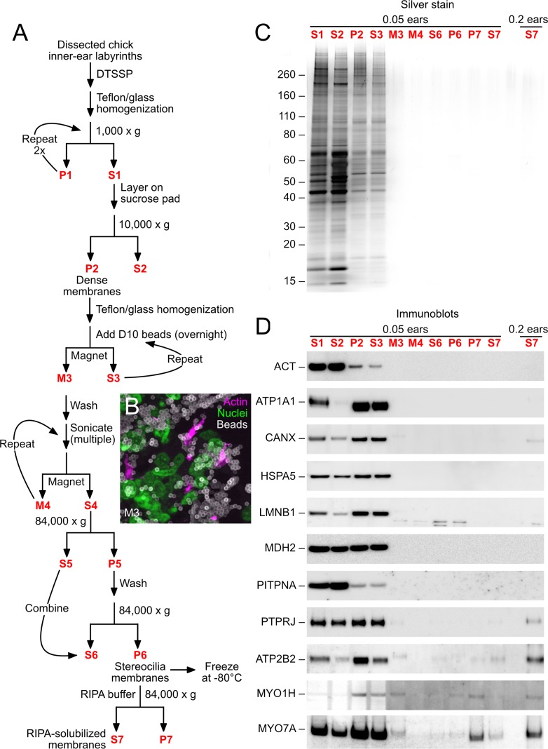
Figure 1. D10 chick stereocilia-membrane enrichment procedure.

(A) Flow chart for stereocilia membrane enrichment. Bold red lettering highlights the principal steps of the procedure. (B) Imaging of M3 fraction of D10 beads; samples were labeled with phalloidin (magenta, for actin), DAPI (green, for nuclei), and anti-mouse IgG (white, for D10 antibody on beads). Aggregation of beads and binding of nuclei (and other contaminants) limits the enrichment gained with this step. (C) SDS-PAGE analysis of enrichment fractions; gel stained with silver. Note that fraction S7 was loaded in a second lane with 4x more material. (D) Immunoblot analysis of purification fractions. For actin (ACT), lanes 1–10 had 0.01 ear-equivalents loaded and lane 11 had 0.2 ears. For all other immunoblots, lanes 1–10 had 0.05 ear-equivalents loaded and lane 11 had 0.2 ears. Other than actin, all proteins are referred to by their official gene symbols.

DOI: http://dx.doi.org/10.7554/eLife.18312.003

Figure 1.

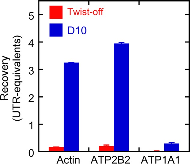
Figure 1—figure supplement 1. Protein recovery with D10 and twist-off methods.

Figure 1—figure supplement 1.

Samples (0.3 and 1.0 ear-equivalents) of hair bundles purified by twist-off and stereocilia membranes purified by the D10 purification were subjected to immunoblotting with antibodies against actin, ATP2B2, and ATP1A1. Aliquots of whole utricle (0.05–1.2 utricles) were run on the same gel for calibration. Fiji was used to measure the immunoblot intensity for each protein in each lane, and the calibration curve was used to determine the amount of each protein recovered in twist-offs or D10 preps relative to the amount in whole utricle. Linear regression was used to determine the recovery (in utricle-equivalents) per ear of starting material. Because seven sensory epithelia were sampled, actin and ATP2B2 were present in the D10 prep at levels considerably higher per ear than was seen in the whole utricles. ATP1A1 was not detected in twist-offs, and while substantial amounts were detected in the D10 prep, they were relatively low compared to the amount present in a whole utricle. Mean ± standard error for the regression fit are plotted.