

Abstract
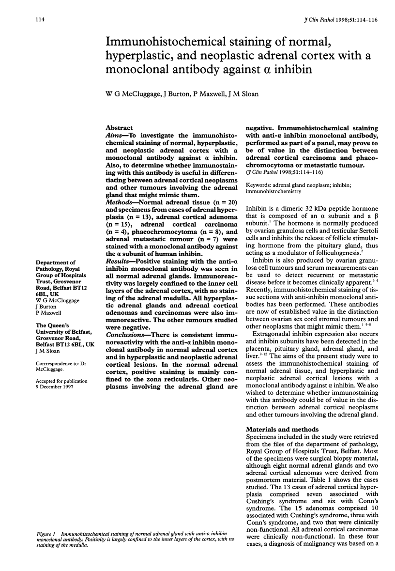
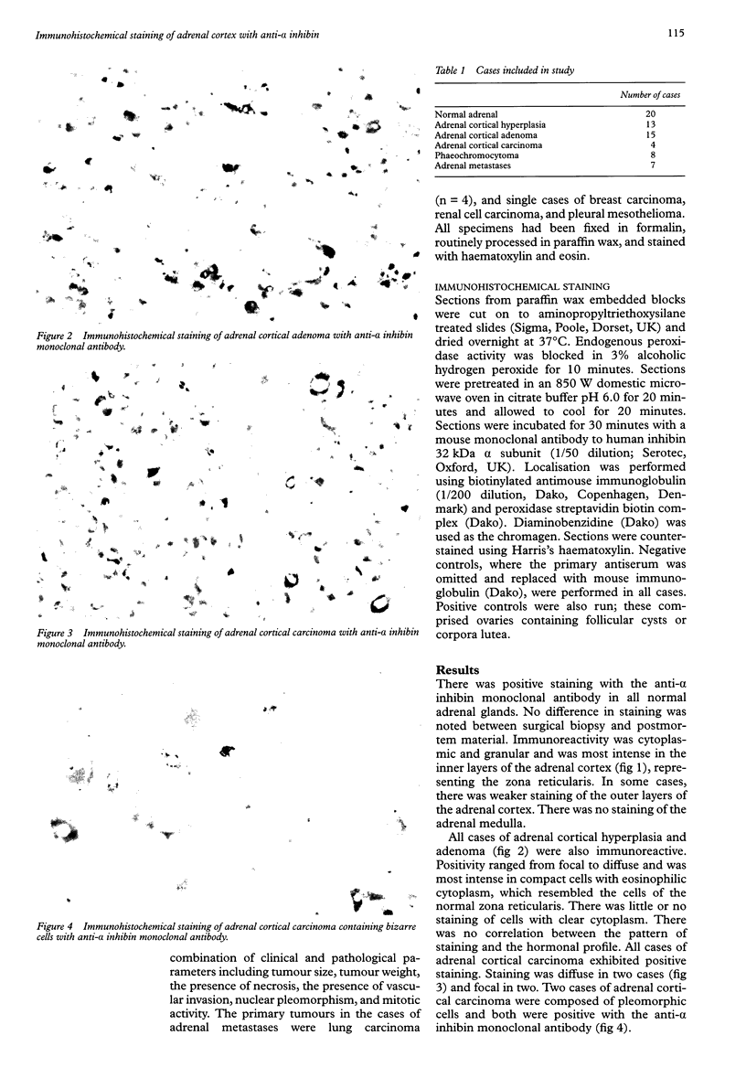
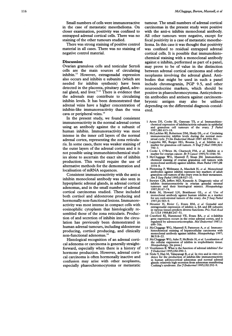
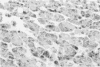
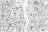
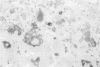
AIMS: To investigate the immunohisto-chemical staining of normal, hyperplastic, and neoplastic adrenal cortex with a monoclonal antibody against alpha inhibin. Also, to determine whether immunostaining with this antibody is useful in differentiating between adrenal cortical neoplasms and other tumours involving the adrenal gland that might mimic them. METHODS: Normal adrenal tissue (n = 20) and specimens from cases of adrenal hyperplasia (n = 13), adrenal cortical adenoma (n = 15), adrenal cortical carcinoma (n = 4), phaeochromocytoma (n = 8), and adrenal metastatic tumour (n = 7) were stained with a monoclonal antibody against the alpha subunit of human inhibin. RESULTS: Positive staining with the anti-alpha inhibin monoclonal antibody was seen in all normal adrenal glands. Immunoreactivity was largely confined to the inner cell layers of the adrenal cortex, with no staining of the adrenal medulla. All hyperplastic adrenal glands and adrenal cortical adenomas and carcinomas were also immunoreactive. The other tumours studied were negative. CONCLUSIONS: There is consistent immunoreactivity with the anti-alpha inhibin monoclonal antibody in normal adrenal cortex and in hyperplastic and neoplastic adrenal cortical lesions. In the normal adrenal cortex, positive staining is mainly confined to the zona reticularis. Other neoplasms involving the adrenal gland are negative. Immunohistochemical staining with anti-alpha inhibin monoclonal antibody, performed as part of a panel, may prove to be of value in the distinction between adrenal cortical carcinoma and phaeochromocytoma or metastatic tumour.
Full text
PDF
Images in this article
Selected References
These references are in PubMed. This may not be the complete list of references from this article.
- Arora D. S., Cooke I. E., Ganesan T. S., Ramsdale J., Manek S., Charnock F. M., Groome N. P., Wells M. Immunohistochemical expression of inhibin/activin subunits in epithelial and granulosa cell tumours of the ovary. J Pathol. 1997 Apr;181(4):413–418. doi: 10.1002/(SICI)1096-9896(199704)181:4<413::AID-PATH789>3.0.CO;2-U. [DOI] [PubMed] [Google Scholar]
- Cooke I., O'Brien M., Charnock F. M., Groome N., Ganesan T. S. Inhibin as a marker for ovarian cancer. Br J Cancer. 1995 May;71(5):1046–1050. doi: 10.1038/bjc.1995.201. [DOI] [PMC free article] [PubMed] [Google Scholar]
- Crawford R. J., Hammond V. E., Evans B. A., Coghlan J. P., Haralambidis J., Hudson B., Penschow J. D., Richards R. I., Tregear G. W. Alpha-inhibin gene expression occurs in the ovine adrenal cortex, and is regulated by adrenocorticotropin. Mol Endocrinol. 1987 Oct;1(10):699–706. doi: 10.1210/mend-1-10-699. [DOI] [PubMed] [Google Scholar]
- Flemming P., Wellmann A., Maschek H., Lang H., Georgii A. Monoclonal antibodies against inhibin represent key markers of adult granulosa cell tumors of the ovary even in their metastases. A report of three cases with late metastasis, being previously misinterpreted as hemangiopericytoma. Am J Surg Pathol. 1995 Aug;19(8):927–933. doi: 10.1097/00000478-199508000-00008. [DOI] [PubMed] [Google Scholar]
- Lappöhn R. E., Burger H. G., Bouma J., Bangah M., Krans M., de Bruijn H. W. Inhibin as a marker for granulosa-cell tumors. N Engl J Med. 1989 Sep 21;321(12):790–793. doi: 10.1056/NEJM198909213211204. [DOI] [PubMed] [Google Scholar]
- McCluggage W. G., Maxwell P., Patterson A., Sloan J. M. Immunohistochemical staining of hepatocellular carcinoma with monoclonal antibody against inhibin. Histopathology. 1997 Jun;30(6):518–522. doi: 10.1046/j.1365-2559.1997.5580774.x. [DOI] [PubMed] [Google Scholar]
- McCluggage W. G., Maxwell P., Sloan J. M. Immunohistochemical staining of ovarian granulosa cell tumors with monoclonal antibody against inhibin. Hum Pathol. 1997 Sep;28(9):1034–1038. doi: 10.1016/s0046-8177(97)90056-3. [DOI] [PubMed] [Google Scholar]
- McLachlan R. I., Robertson D. M., Healy D. L., Burger H. G., de Kretser D. M. Circulating immunoreactive inhibin levels during the normal human menstrual cycle. J Clin Endocrinol Metab. 1987 Nov;65(5):954–961. doi: 10.1210/jcem-65-5-954. [DOI] [PubMed] [Google Scholar]
- Meunier H., Rivier C., Evans R. M., Vale W. Gonadal and extragonadal expression of inhibin alpha, beta A, and beta B subunits in various tissues predicts diverse functions. Proc Natl Acad Sci U S A. 1988 Jan;85(1):247–251. doi: 10.1073/pnas.85.1.247. [DOI] [PMC free article] [PubMed] [Google Scholar]
- Nishi Y., Haji M., Takayanagi R., Yanase T., Ikuyama S., Nawata H. In vivo and in vitro evidence for the production of inhibin-like immunoreactivity in human adrenocortical adenomas and normal adrenal glands: relatively high secretion from adenomas manifesting Cushing's syndrome. Eur J Endocrinol. 1995 Mar;132(3):292–299. doi: 10.1530/eje.0.1320292. [DOI] [PubMed] [Google Scholar]
- Rishi M., Howard L. N., Bratthauer G. L., Tavassoli F. A. Use of monoclonal antibody against human inhibin as a marker for sex cord-stromal tumors of the ovary. Am J Surg Pathol. 1997 May;21(5):583–589. doi: 10.1097/00000478-199705000-00012. [DOI] [PubMed] [Google Scholar]
- Stewart C. J., Jeffers M. D., Kennedy A. Diagnostic value of inhibin immunoreactivity in ovarian gonadal stromal tumours and their histological mimics. Histopathology. 1997 Jul;31(1):67–74. doi: 10.1046/j.1365-2559.1997.5780819.x. [DOI] [PubMed] [Google Scholar]
- Voutilainen R. What is the function of adrenal inhibins? Eur J Endocrinol. 1995 Mar;132(3):290–291. doi: 10.1530/eje.0.1320290. [DOI] [PubMed] [Google Scholar]