Abstract
The normal value of alpha angle is controversial. The aim of this study was to compare the alpha angle in asymptomatic volunteers versus patients who had undergone surgery for symptomatic cam-type femoroacetabular impingement (FAI) and determine a diagnostic cut-off value for symptomatic cam impingement. This is a diagnostic test study. Cases were defined as those patients who had undergone surgery for symptomatic cam or mixed type FAI. Controls were defined as asymptomatic volunteers, with no history of hip pain who had undergone a computed tomography (CT) scan of the abdomen and pelvis for a non-joint or bone-related reason. In both groups, the alpha angle was measured in an oblique axial CT reconstruction of the femoral neck. A logistic regression model was first estimated and a receiver operating characteristics (ROC) curve was then calculated. The diagnostic cut-off value selected was the one that maximizes sensitivity and specificity. Data were analysed from 38 consecutive cases of cam or mixed FAI and 101 controls. The average alpha angle was 67°(±12°) among cases and 48°°(±5°) among controls. An odds ratio of 1.28 [1.18–1.39] was obtained. A ROC curve of 0.96 [0.93–0.99] was calculated, and using an alpha angle of 57° as the diagnostic cut-off value, provided a sensitivity of 92% and a specificity of 95%. If a patient complains of hip pain and an alpha angle of 57° is found in CT, strongly suggest that cam impingement is causing the pain.
INTRODUCTION
Femoroacetabular impingement (FAI) is an anatomic and functional condition that results in a mechanical conflict in the hips, producing pathological contact between the acetabulum and the femoral head–neck junction. This can lead to labral–chondral injury, pain and limited range of motion. Some authors have suggested a relationship between FAI deformity and the subsequent development of osteoarthritis [1, 2].
Three types of FAI have been described: pincer, cam and mixed. Pincer FAI is characterized by focal or general over-coverage of the femoral head by the acetabulum, while cam-type FAI is characterized by the presence of an aspherical portion of the femoral head–neck junction. The mixed type is diagnosed when both types of impingements are found [3, 4].
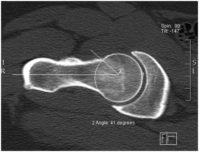
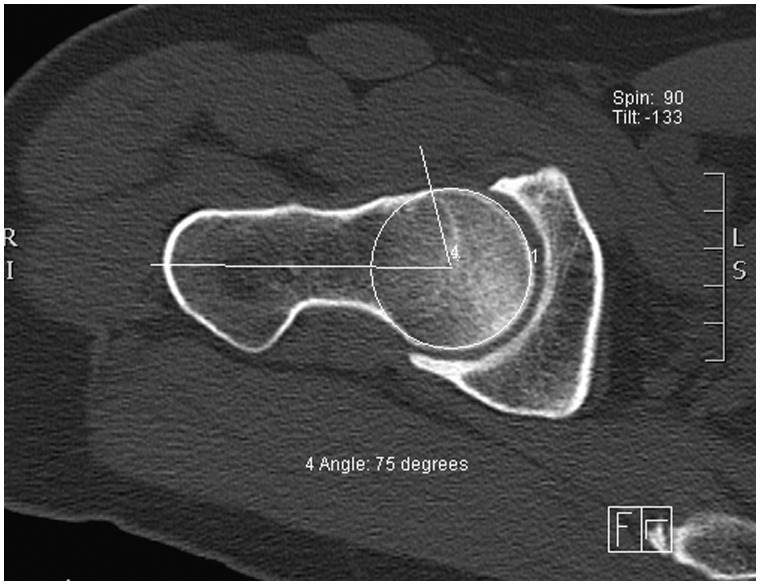
The alpha angle is a radiological measure that has been proposed for the diagnosis and evaluation of surgical treatment in cam type FAI (Figs. 1 and 2) [5]. The normal value of the angle is controversial; even more so is the value to be considered pathological. First described in 2002, Notzli et al. [6] suggested that the pathological value was greater than 50°. In this study, the alpha angle was measured using magnetic resonance imaging (MRI). Tannast et al. [7], in a review published in 2007, proposed the same cut-off value, but with computed tomography (CT).
Fig. 1.
Shows the measurement of an angle alpha in an asymptomatic individual. It was measured in an oblique axial CT reconstruction of the femoral neck, at the anterolateral region.
Fig. 2.
Shows the measurement of an angle alpha in symptomatic patients who underwent surgery. It was measured in an oblique axial CT reconstruction of the femoral neck, at the anterolateral region.
In 2005, Beaulé et al. [8] proposed a cut-off value of 50°. The study compares alpha angle, measured in CT, between symptomatic and asymptomatic individuals. Five years later, the same author published an article, in which, the average alpha angle, measured in MRI, was 50.15° in asymptomatic population [9]. Allen et al. in 2009, using radiographs, and Kang et al. in 2010 using CT, reported that angle values of more than 55.5° and 55°, respectively, have to be considered pathological; only asymptomatic volunteers were included in both studies [5, 10].
In an earlier performed study by the current group, we found that a cut-off value of 50° using CT would characterize 28% of the asymptomatic population as pathological [11]. Lastly Pollard et al. [12], in 2010 using radiographs, established that the average normal value of the alpha angle is 47.5°, with a 95% confidence interval of 46–49°. The initial studies that set pathological cut-off values between 50° and 55° for the alpha angle have been invalidated by later studies performed on healthy populations, in which greater values for the alpha angle have been found in asymptomatic volunteers. In addition, those initials studies lacked the appropriate statistical methodology to determine cut-off values for diagnostic tests [13, 14]. The aim of this study was to compare measurements of the alpha angle in asymptomatic volunteers and patients who had undergone surgery for symptomatic cam-type FAI and determine a diagnostic cut-off value using a regression model and ROC curve.
MATERIALS AND METHODS
A case–control type diagnostic test study was designed and approved by our institution’s ethics review board. All participants provided informed consent. Cases were defined as patients who had undergone arthroscopy surgery for symptomatic FAI in our institution between 2011 and 2012. Controls were defined as patients with no history of hip pain who underwent a CT scan for a non-joint or bone-related reason.
Patients that met inclusion criteria for cases were those who presented hip pain referred to the inguinal region, a positive flexion–adduction–internal rotation (FADIR) test on physical examination and a positive lidocaine test, and subsequently underwent surgical treatment via hip arthroscopy. For the lidocaine test, a radiologist, who specializes in the musculoskeletal area, injected 3 cc of 5% lidocaine directly into the hip under an ultrasound guide. A FADIR test was performed before and after the injection, and was considered positive when the patient subjectively experienced a 50% pain decrease in a visual analogue scale. In addition, inclusion in the study required the presence of an intraoperative cam type FAI pathomorphology and osteochondroplasty procedure performed. Exclusion criteria were previous hip surgeries, history of hip dysplasia and patients in whom surgical procedures were performed only in the acetabulum during arthroscopy. A retrospective review of clinical records was conducted using the above inclusion and exclusion criteria to identify cases for this study.
The control population consisted of individuals who consulted in our institution for a non-joint or bone-related reason, and who required an abdominal and pelvic CT for diagnosis. Prior to enrolment, volunteers completed a questionnaire, which included asking for current or past history of hip-related pain or hip surgery. Any positive answer led to the volunteer being excluded from the study. Recruitment of controls was conducted prospectively during 2012.
All CT scans were performed in our institution. CT images were obtained using a SOMATOM Sensation 64 Siemens equipment. The acquisition protocol used a 1.5-mm section thickness with a 0.3-mm interval reconstruction. Information was reprocessed to multi-planar reconstructions of 3 mm. In both cohorts, the alpha angle was measured in an oblique axial CT reconstruction of the femoral neck, in the anterolateral region at 1:30. In this location, the neck was divided into thirds, the measurement was taken in the middle third. Only the affected hip was measured in cases, while controls underwent measurement of both hips, with the average of the left and right hip recorded. Measurements were performed in both groups by the chief musculoskeletal radiology (J.D.) of our institution. No analysis of interobserver and intraobserver agreement was made. Figures 1 and 2 show two examples of the femoral alpha angle measurement performed in the study.
STATISTICAL ANALYSIS
Descriptive analysis was first conducted, and average and standard deviations of the alpha angle of both groups were reported. In the second step, a logistic regression model was estimated, in which the presence of FAI (case/control) was used as the dependent variable and the alpha angle measurement as the independent variable. A Hosmer–Lemeshow goodness-of-fit test was done to test logistic regression assumptions (this is considered appropriate if P > 0.15). The receiver operating characteristics (ROC) curve was calculated to determine the discrimination capacity. The area under the curve was interpreted according to Hosmer and Lemeshow recommendations (Table I) [14]. Finally, probabilities associated with each cut-off value of the alpha angle were calculated, with the value that maximized sensitivity and specificity selected as the diagnostic cut-off value [13, 15]. A significance level of 0.05 was established and 95% confidence interval was reported. All analyses were performed using Stata v11.2 (StataCorp LP, College Station, TX, USA).
Table I.
Shows discrimination ability of ROC curve according to the value of area under the curve based on Hosmer and Lemeshow [14]
| Area under ROC curve | Discrimination |
|---|---|
| 0.50–0.60 | Luck |
| 0.61–0.70 | Low |
| 0.71–0.80 | Acceptable |
| 0.81–0.90 | Very good |
| 0.91–1 | Excellent |
RESULTS
Thirty-eight patients (82.6%) that underwent surgery for FAI between 2011 and 2012 at our institution were included; all of them underwent hip arthroscopy. Eight patients were excluded due to the presence of pincer-type only FAI without any evidence of cam-type pathomorphology. A total of 101 asymptomatic volunteers were recruited. The average age was 36.1 years (±11.8°) among cases and 36.8 years (±14.4°) among controls, gender and age were summarize in Table II. The average Wiberg angle was 38° (±7.2°) among controls and 39° (±4.1°) among cases. A non-parametric median test was performed showing no difference (P = 0.30), and a logistic regression was estimated showing a non-significant odds ratio 0.98 [0.92–1.04].
Table II.
Shows descriptive analysis of age and gender by groups
| Case | Controls | P (test) | |
|---|---|---|---|
| Male | 21/38 (55.26%) | 41/101 (40.59%) | – |
| Female | 17/38 (44.74%) | 60/101 (59.41%) | 0.13 (Fisher exact) |
| Age (years) | 36.12 (±11.82) | 36.82 (±14.43) | 0.95 (Wilcoxon unpaired) |
| Male age (years) | 30.20 (±11.78) | 36.50 (±13.18) | 0.10 (Wilcoxon unpaired) |
| Female (years) | 40.79 (±09.78) | 37.29 (±16.24) | 0.17 (Wilcoxon unpaired) |
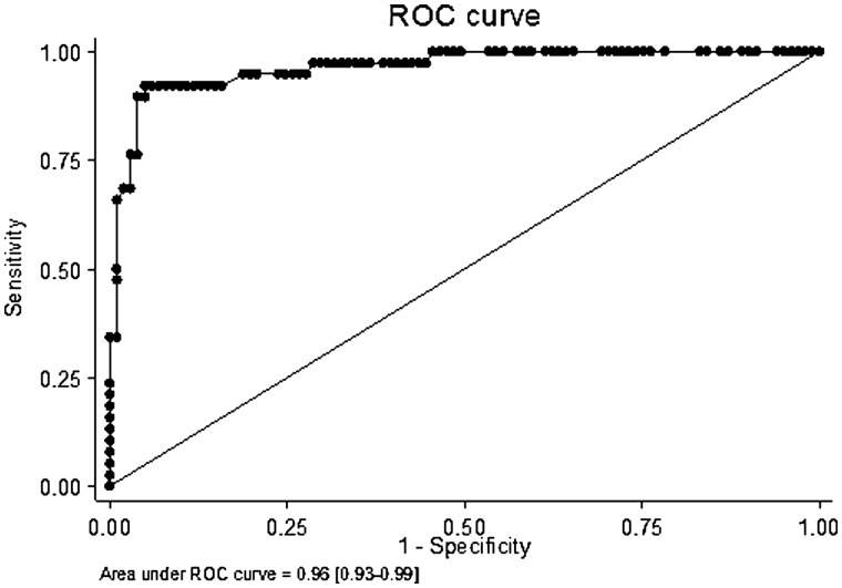
The average alpha angle was 66.8° (±12.2°) among cases and 47.8° (±5.3°) among controls. A logistic regression model was estimated and an odds ratio of 1.28 [1.18–1.39] was obtained, meaning that, as the angle alpha increases, the risk of being symptomatic increases, this was statistically significant. The area under the ROC curve was 0.96 [0.93–0.99] (Fig. 3), which means an excellent discrimination level (Table I). An alpha angle of 57° maximized sensitivity (92%) and specificity (95%), and correctly classified 94% of patients in this study. Table III shows the sensitivity and specificity estimated by the regression model at 50, 57, 60 and 65 degrees of alpha angle.
Fig. 3.
Shows ROC curve obtained after the estimation of the logistic regression model. The area under the curve was 0.96, which is excellent based on Hosmer and Lemeshow [14].
Table III.
Shows the sensitivity and specificity estimated by the logistic regression model at different cut-off values of femoral alpha angle
| Cut off value | Sensivity (%) | Specificity (%) |
|---|---|---|
| >50° | 97 | 74 |
| >57° | 92 | 95 |
| >60° | 75 | 95 |
| >65° | 48 | 96 |
DISCUSSION
The alpha angle has been frequently cited as a measure of cam-type FAI. However, there are controversies in the contemporary literature regarding the ability of this angle to discriminate between asymptomatic and symptomatic individuals, and even more controversy regarding the cut-off value that defines cam-type FAI. In a study published in 2002, Notzli et al. [6] included 39 patients with hip symptoms, positive physical exam and positive MRI for cam-type FAI and compared those patients with 40 controls. However, this study only showed that both groups had different average values for the alpha angle, with the authors suggesting that an angle >50° should be used as the cut-off value. In 2005, Beaulé et al. [8] published a retrospective study that included 30 patients who had undergone surgery for cam-type FAI and 12 healthy individuals. The alpha angle was measured using CT and the selected cut-off value was 50.5° because that gave 100% specificity to the sample. It should be noted that there was only one control every three cases, and the methodology for choosing the cut-off value was based on a descriptive analysis without an estimated model such as logistic regression.
In a retrospective review of 2803 anteroposterior radiographs, Gosvig et al. [16] suggested a pathological value of 83° for men and 57° for women. These values were set using two standard deviations above the mean. However, the medical reasons to request the radiographs were not mentioned in the article; as the radiographies were retrospectively reviewed, no clinical elements were used for inclusion or exclusion criteria and some patients who were included had osteoarthritis. In addition, the use of the standard deviation is not the most accurate method to determine a diagnostic cut-off value.
Allen et al. [10], in a study that included 113 individuals, found that an alpha angle of 60° has an odds ratio of 2.59 of having hip pain. Nonetheless, the odds ratio of this study was calculated respecting the contralateral hip of the same individual; in others words, the contralateral asymptomatic hip was used as the control group. Theoretically, this assumption is wrong for morphological analysis; it cannot be assumed that the left and right hips provide independent observations, taking into account the fact that both have the same genetic basis, makes findings difficult to interpret.
Sutter et al. [17] performed a study that included 53 asymptomatic individuals and 53 patients with symptomatic cam-type FAI. They measured the alpha angle in radially reformatted MRI images, assessing the contour of the head–neck junction. They found that a diagnostic cut-off value of 60° in the anterolateral region had a sensitivity of 72–76% and specificity of 73–80%. This finding is similar to that reported in this study, stating that the cut-off value is greater than the 50°–55° suggested by earlier studies. However, measurement of the alpha angle in radians, as performed by Sutter et al., is mostly used for investigative purpose and not in clinical practice. In addition, Sutter et al. used a control/case ratio of only 1; in contrast, we used three times more controls than cases to determinate the cut off value. Finally, to discriminate patients with pincer-type FAI from those with cam or mixed type FAI, Sutter et al. used an alpha angle of <55° as an exclusion criteria for patients (cases) with symptomatic FAI. In this study, the exclusion was made on the basis of the arthroscopic finding of a bump. This must be taken into account as a potential source of bias, as Sutter et al. used an outcome of the study as a criteria for inclusion or exclusion. We think that the most important finding of Sutton et al. study is that the anterolateral region is the area with better discrimination (the same region where we perform our single measurement), nevertheless the cut-off value can be overestimated as they exclude patients based on the alpha angle measurement.
A strength of this study is that the recruitment of healthy individuals (controls) was prospective, measuring the alpha angle with CT scanning with no unnecessary radiation. Furthermore, we recruited a case/control ratio of 3:1, which, we believe, gives the study greater reliability.
As the proportion of female/male was different between groups we conducted a Fisher exact test to determine if this difference was significant. A P value of 0.13 was obtained, meaning that it was not statically significant (Table II). Secondly, in an earlier performed study by the current group, alpha angle measurement in healthy people was not associated with gender, weight, height or age [18].
Another important strength is that the cases presented symptoms, physical examination and an intraarticular lidocaine test consistent with cam-type FAI. Finally, the use of a logistic regression model is a valid methodology to obtain diagnostic cut-off points for a pathology [13, 14]. Another strength of the study is that no difference in Wiberg angle was found between both cohorts, so pincer has little influence in the hip pain of the case groups.
The lack of analysis of inter or intraobserver agreement is an important issue in this study. The same author (J.D.), who is the chief of musculoskeletal radiology at our institution, performed all measurements for both cohorts. Taking this into consideration, we can say that the measurements were performed by a highly trained observer, and if any measurement error exists, it is a systematic error. Moreover, beyond the radiology report of the measurement, he did not participate in the clinic’s decision whether the patients should or should not need hip arthroscopy.
A weakness of this study is that the cases were recruited retrospectively. It must be noted that in our institution, patients who undergo hip arthroscopy have a special sheet in their clinic record that states symptom location, physical examination findings, CT measurements and arthroscopy findings. However, as patients were included retrospectively, we cannot assess how the angle measured influenced the indications for the procedure and the indication for osteochondroplasty. Another weakness is that control volunteers were recruited without a physical examination of the hip.
We are aware of alpha angle measurement limitations. As FAI is a dynamic condition, asymptomatic individuals could have a high alpha angle, but no pain, only because the patient does not place CAM pathomorphology in risk with an activity that use extreme hip range of motion [19, 20]. On the other hand, the measurement on CT scan was only a single measurement and multiple measurements along the femoral head–neck junction may add further diagnostic ability [7].
CONCLUSION
The alpha angle value measured in an oblique CT reconstruction of the femoral neck has a high discriminating capacity for the diagnosis of symptomatic cam-type FAI. If a patient complains of hip pain and an alpha angle of 57° is found in CT, strongly suggest that cam impingement is causing the pain.
ACKNOWLEDGEMENTS
We acknowledge the contribution of the research, orthopaedics and radiology department of the Hospital Clinico Universidad de Chile.
CONFLICT OF INTEREST STATEMENT
None declared.
REFERENCES
- 1.Ganz R, Leunig M, Leunig-Ganz K, et al. The etiology of osteoarthritis of the hip: an integrated mechanical concept. Clin Orthop Relat Res 2008; 466: 264–72. [DOI] [PMC free article] [PubMed] [Google Scholar]
- 2.Beall DP, Sweet CF, Martin HD, et al. Imaging findings of femoroacetabular impingement syndrome. Skeletal Radiol 2005; 34: 691–701. [DOI] [PubMed] [Google Scholar]
- 3.Navarro N, Orellana C, Moreno M, et al. Atrapamiento femoroacetabular. Seminarios De La Fundación Española De Reumatología 2012; 13: 15–22. [Google Scholar]
- 4.Audenaert EA, Peeters I, Vigneron L, et al. Hip morphological characteristics and range of internal rotation in femoroacetabular impingement. Am J Sports Med 2012; 40: 1329–36. [DOI] [PubMed] [Google Scholar]
- 5.Kang AC, Gooding AJ, Coates MH, et al. Computed tomography assessment of hip joints in asymptomatic individuals in relation to femoroacetabular impingement. Am J Sports Med 2010; 38: 1160–5. [DOI] [PubMed] [Google Scholar]
- 6.Notzli HP, Wyss TF, Stoecklin CH, et al. The contour of the femoral head-neck junction as a predictor for the risk of anterior impingement. J Bone Joint Surg Br 2002; 84: 556–60. [DOI] [PubMed] [Google Scholar]
- 7.Tannast M, Siebenrock KA, Anderson SE. Femoroacetabular impingement: radiographic diagnosis—what the radiologist should know. Am J Radiol 2007; 188: 1540–52. [DOI] [PubMed] [Google Scholar]
- 8.Beaulé P, Zaragoza E, Motamedi K, et al. Three-dimensional computed tomography of the hip in the assessment of femoroacetabular impingement. J Orthop Res 2005; 23: 1286–92. [DOI] [PubMed] [Google Scholar]
- 9.Hack K, Di Primio G, Rakhra K, et al. Prevalence of cam-type femoroacetabular impingement morphology in asymptomatic volunteers. J Bone Joint Surg 2010; 92: 2436–44. [DOI] [PubMed] [Google Scholar]
- 10.Allen D, Beaulé PE, Ramadan O, et al. Prevalence of associated deformities and hip pain in patients with cam-type femoroacetabular impingement. J Bone Joint Surg Br 2009; 91: 589–94. [DOI] [PubMed] [Google Scholar]
- 11.Barrientos C, Diaz J, Barahona M, et al. Caracterización morfológica de la cadera mediante tomografía axial computada en población chilena: implicancias en el pinzamiento femoroacetabular. Rev Chilena Ortop Y Traum 2012; 53: 70–7. [Google Scholar]
- 12.Pollard TC, Villar RN, Norton MR, et al. Femoroacetabular impingement and classification of the cam deformity: the reference interval in normal hips. Acta Orthop 2010; 81: 134–41. [DOI] [PMC free article] [PubMed] [Google Scholar]
- 13.Kleinbaum DG, Kupper LL, Muller KE. Applied Regression Analysis and Other Multivariate Analysis Methods. Boston: PWS-Kent Publishing Company; 1988. [Google Scholar]
- 14.Hosmer Jr DW, Lemshow S. Applied Logistic Regression. New York: John Wiley & Sons, 2004. [Google Scholar]
- 15.Burgueño MJ, García-Bastos JL, González-Buitrago JM. Las curvas ROC en la evaluación de las pruebas diagnósticas. Med Clin 1995; 104: 661–70. [PubMed] [Google Scholar]
- 16.Gosvig KK, Jacobsen S, Palm H, et al. A new radiological index for assessing asphericity of the femoral head in cam impingement. J Bone Joint Surg Br 2007; 89: 1309–16. [DOI] [PubMed] [Google Scholar]
- 17.Sutter R, Dietrich TJ, Zingg PO. et al. How useful is the alpha angle for discriminating between symptomatic patients with cam-type femoroacetabular impingement and asymptomatic volunteers? Radiology 2012; 264: 514–21. [DOI] [PubMed] [Google Scholar]
- 18.Barahona M, Barrientos C, Cavada G. Análisis multivariado de la morfología de la cadera en imágenes obtenidas por tomografía computada en población asintomática Editorial Académica española. 1 edición septiembre 2015. p. 69. ISBN-13: 978-3-659-09798-0, ISBN-10: 3659097985, EAN: 9783659097980.
- 19.Ito K, Minka-II MA, Leunig M, et al. Femoroacetabular impingement and the cam-effect. A MRI-based quantitative anatomical study of the femoral head-neck offset. J Bone Joint Surg 2001; 83: 171.. [DOI] [PubMed] [Google Scholar]
- 20.Kapron AL. Radiographic prevalence of femoroacetabular impingement in collegiate football players. J Bone Joint Surg 2011; 93: e111. [DOI] [PubMed] [Google Scholar]


