

Abstract
A pseudotail is a very rare, dermal appendage arising from the lumbosacral region with an association with spinal dysraphism. We report a case of a pseudotail in a healthy newborn female with sonographic imaging of a tethered cord and dermal sinus tract with MRI and surgical correlation.
Keywords: Pseudotail, Spine ultrasound, Tethered cord, Dermal sinus
Riassunto
La pseudo-coda è un'appendice dermica molto rara, della regione lombo-sacrale, associata a disrafismo spinale. Segnaliamo il caso di una pseudo-coda in una bambina appena appena nata sana, con immagini ecografiche della colonna e della formazione e della colonna con il tramite cutaneo con MRI e la correlazione chirurgica.
Introduction
A pseudotail is a very rare, fingerlike dermal appendage of the lumbosacral region that has a high association with underlying spinal dysraphism [1, 2]. There is a paucity of information on sonographic imaging of these lesions in the radiographic literature with no reported cases of a pseudotail associated with a dermal sinus tract. We present a case of a pseudotail in a healthy newborn female with excellent sonographic imaging of a tethered cord and dermal sinus tract with MRI and surgical correlation.
Case report
A healthy, one-day old female born at 38 weeks gestation presented with a soft, skin covered, dermal appendage approximately 1 cm in diameter and 6 cm in length (Fig. 1) located above the gluteal cleft, just right of midline, near the lumbosacral junction. She voided normally. There was no evident weakness, numbness, or neurologic limitation.
Fig. 1.

Pre-operative image of the pseudotail at 4 months of age
Ultrasound examination of the spine revealed a low lying, tethered cord to the level of L4 (Fig. 2a). A dermal sinus tract was also seen extending from the thecal sac at the level of S1 to the dermal appendage (Fig. 2b) which was also seen at MRI (Fig. 3). Splaying of the posterior elements below the level of S1 was consistent with mild, occult dysraphism (image not shown).
Fig. 2.

a Neonatal spine ultrasound revealing a tethered cord (arrow) at the level of L4. b Dermal sinus tract (arrow) extending from the thecal sac at the level of S1 to the pseudotail
Fig. 3.

MRI revealing low lying cord and a dermal sinus tract (arrow)
When the patient was 2 months old, the pseudotail was surgically removed. At surgery, the dermal sinus tract was seen extending to the level of the conus (Fig. 4). The sinus tract was resected and the filum terminale was sectioned. The histopathologic findings were consistent with a fat containing, fibroepithelial polyp with no bone, cartilage, or skeletal muscle. The patient recovered well after surgery with no limitations.
Fig. 4.
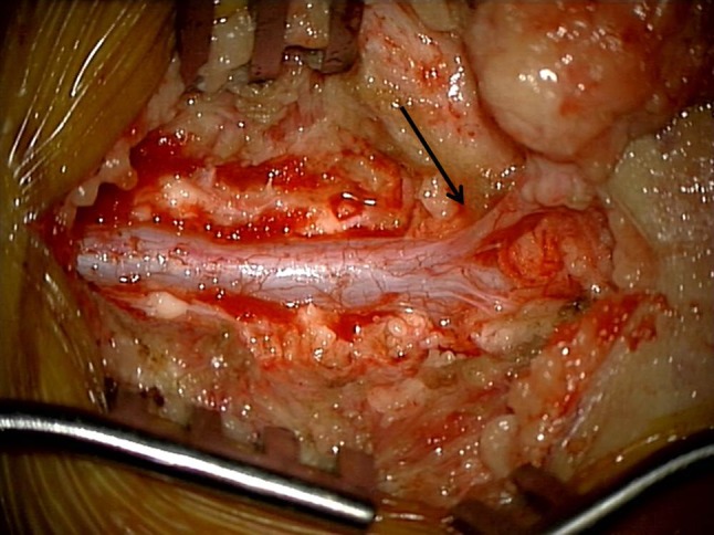
Intraoperative photo confirming the tethered cord with a dermal sinus tract (arrow) extending to the pseudotail
Discussion
A true, or vestigial, tail is described as a skin covered embryonic remnant at the caudal spine that contains vertebrae, muscle, and neurovascular tissue. Any other dermal appendage of the caudal spine by default has been referred to as a “pseudotail” and variably reported as a lumbosacral protrusion of tissue to include prolongation of coccygeal vertebrae, lipoma, teratoma, or glioma [3]. Classically, as in our case, a pseudotail is in the form of a soft, fat containing, skin covered mobile appendage.
Management of any true tail or pseudotail involves a thorough neurologic examination and imaging of the spine with ultrasound and/or MRI for evaluation of possible dysraphism and tethered cord. This is relevant since an incidence of spinal dysraphism and tethered cord have a reported association with human tails at 49 and 20 %, respectively [4].
Spinal ultrasound provides good visualization of congenital anomalies in the newborn owing to the incomplete ossification of the vertebral elements. In our case ultrasound was able to visualize the low lying tethered cord, dermal sinus tract and occult dysraphism. The spinal ultrasound correlated well with what was shown on MRI and found intraoperatively.
Conclusion
Spinal ultrasound imaging in a newborn with a pseudotail can provide excellent resolution of associated abnormalities such as a dermal sinus tract causing a tethered cord and thus aid in management and prognosis.
Compliance with ethical standards
Funding
No funding was required or received for this case report.
Conflict of interest
The authors Clark and Davidson declare no conflict of interest.
Ethical approval
As a single case report, this article does not contain any studies with animal or human participants performed by any of the authors. This case has been approved for submission per the author’s Department of Clinical Investigation.
Informed consent
Informed consent was obtained from the parents of the infant prior to all surgical consultations.
Footnotes
The views expressed in this manuscript are those of the authors and do not reflect the official policy of the Department of the Army, Department of Defense, or the US Government.
Contributor Information
Paul Clark, Phone: 571-409-0055, Email: paul.clark9.mil@mail.mil.
Laurence Davidson, Email: laurence.davidson.mil@mail.mil.
References
- 1.Albright AL, Pollack IF, Adelson PD (2007) Principles and Practice of Pediatric Neurosurgery, 2nd ed. Thieme publishers, pp 378–79
- 2.Singh DK, Kumar B, Sinha VD, et al. The human tail: rare lesion with occult spinal dysraphism—a case report. J Pediatr Surg. 2008;43:E41–E43. doi: 10.1016/j.jpedsurg.2008.04.030. [DOI] [PubMed] [Google Scholar]
- 3.Dao AH, Netsky MG. Human tails and pseudotails. Hum Pathol. 1984;15:449–453. doi: 10.1016/S0046-8177(84)80079-9. [DOI] [PubMed] [Google Scholar]
- 4.Lu FL, Wang PJ, Teng RJ, et al. The human tail. Pediatr Neurol. 1998;19(3):230–233. doi: 10.1016/S0887-8994(98)00046-0. [DOI] [PubMed] [Google Scholar]