

Abstract
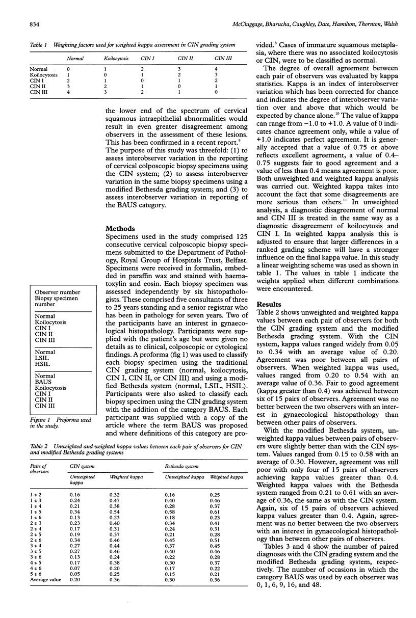
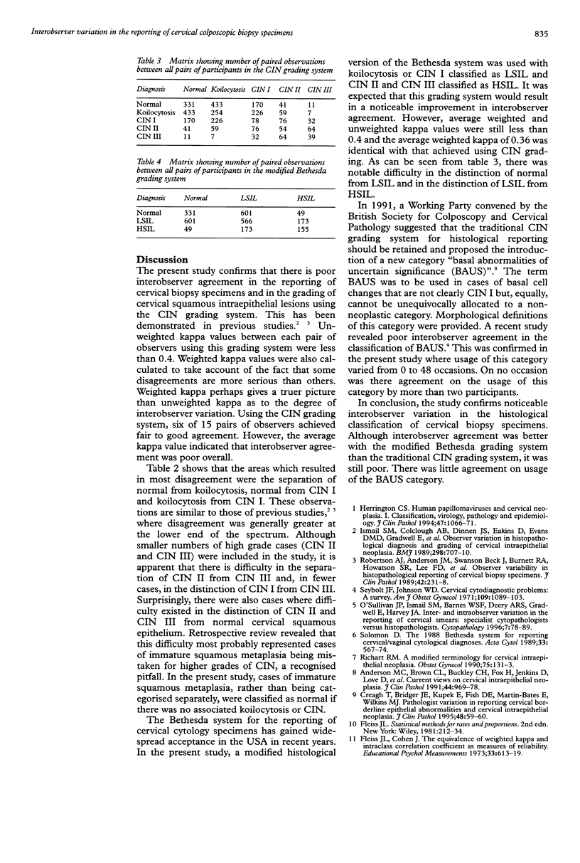
AIMS: To assess interobserver variation in reporting cervical colposcopic biopsy specimens and to determine whether a modified Bethesda grading system results in better interobserver agreement than the traditional cervical intraepithelial neoplasia (CIN) grading system. METHODS: One hundred and twenty five consecutive cervical colposcopic biopsy specimens were assessed independently by six histopathologists. Specimens were classified using the traditional CIN grading system as normal, koilocytosis, CIN I, CIN II, or CIN III. The specimens were also classified using a modified Bethesda grading system as either normal, low grade squamous intraepithelial lesion (LSIL) or high grade squamous intraepithelial lesion (HSIL). Participants were also asked to categorise biopsy specimens by the CIN system with the addition of the recently proposed category "basal abnormalities of uncertain significance (BAUS)". The degree of agreement between participants was assessed by kappa statistics. RESULTS: Using the CIN system, interobserver agreement was generally poor: unweighted and weighted kappa values between individual pairs of observers ranging from 0.05 to 0.34 (average 0.20) and from 0.20 to 0.54 (average 0.36), respectively. With the modified Bethesda system, interobserver agreement was better but still poor: unweighted and weighted kappa values ranging from 0.15 to 0.58 (average 0.30) and from 0.21 to 0.61 (average 0.36), respectively. There was little or no agreement between observers in the diagnosis of BAUS. CONCLUSIONS: Interobserver agreement in the reporting of cervical colposcopic biopsy specimens using the CIN grading system is poor. Agreement, while still poor, is better when a modified Bethesda grading system is used. There is little or no consensus in the diagnosis of BAUS.
Full text
PDF
Selected References
These references are in PubMed. This may not be the complete list of references from this article.
- Anderson M. C., Brown C. L., Buckley C. H., Fox H., Jenkins D., Lowe D. G., Manners B. T., Melcher D. H., Robertson A. J., Wells M. Current views on cervical intraepithelial neoplasia. J Clin Pathol. 1991 Dec;44(12):969–978. doi: 10.1136/jcp.44.12.969. [DOI] [PMC free article] [PubMed] [Google Scholar]
- Creagh T., Bridger J. E., Kupek E., Fish D. E., Martin-Bates E., Wilkins M. J. Pathologist variation in reporting cervical borderline epithelial abnormalities and cervical intraepithelial neoplasia. J Clin Pathol. 1995 Jan;48(1):59–60. doi: 10.1136/jcp.48.1.59. [DOI] [PMC free article] [PubMed] [Google Scholar]
- Herrington C. S. Human papillomaviruses and cervical neoplasia. I. Classification, virology, pathology, and epidemiology. J Clin Pathol. 1994 Dec;47(12):1066–1072. doi: 10.1136/jcp.47.12.1066. [DOI] [PMC free article] [PubMed] [Google Scholar]
- Ismail S. M., Colclough A. B., Dinnen J. S., Eakins D., Evans D. M., Gradwell E., O'Sullivan J. P., Summerell J. M., Newcombe R. G. Observer variation in histopathological diagnosis and grading of cervical intraepithelial neoplasia. BMJ. 1989 Mar 18;298(6675):707–710. doi: 10.1136/bmj.298.6675.707. [DOI] [PMC free article] [PubMed] [Google Scholar]
- O'Sullivan J. P., Ismail S. M., Barnes W. S., Deery A. R., Gradwell E., Harvey J. A., Husain O. A., Kocjan G., McKee G., Olafsdottir R. Inter- and intra-observer variation in the reporting of cervical smears: specialist cytopathologists versus histopathologists. Cytopathology. 1996 Apr;7(2):78–89. doi: 10.1046/j.1365-2303.1996.38682386.x. [DOI] [PubMed] [Google Scholar]
- Richart R. M. A modified terminology for cervical intraepithelial neoplasia. Obstet Gynecol. 1990 Jan;75(1):131–133. [PubMed] [Google Scholar]
- Robertson A. J., Anderson J. M., Beck J. S., Burnett R. A., Howatson S. R., Lee F. D., Lessells A. M., McLaren K. M., Moss S. M., Simpson J. G. Observer variability in histopathological reporting of cervical biopsy specimens. J Clin Pathol. 1989 Mar;42(3):231–238. doi: 10.1136/jcp.42.3.231. [DOI] [PMC free article] [PubMed] [Google Scholar]
- The 1988 Bethesda System for reporting cervical/vaginal cytologic diagnoses. Developed and approved at the National Cancer Institute Workshop, Bethesda, Maryland, U.S.A., December 12-13, 1988. Acta Cytol. 1989 Sep-Oct;33(5):567–574. [PubMed] [Google Scholar]