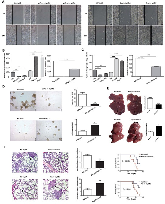
Figure 2. Prp19 enhances invasive potentials of HCC cells both in vitro and in vivo.

A. The migratory capacity of stable Huh7 cells mis-expressing Prp19 was analysed by wound-healing assay. B. Stable Huh7 and Hep3B cells mis-expressing Prp19 was subjected to transwell assay. C. The invasive capacity of Huh7 and Hep3B cells was analysed using transwell filter chambers coated with Matrigel. D. Representative images of colonies formed by stable Huh7 cells mis-expressing Prp19 in soft agar. E. Representative images of tumor in the liver of nude mice given orthotopic implantation of xenograft generated by stable Huh7 cells mis-expressing Prp19. Black arrows indicated metastatic lesions in liver. F. Representative images of hematoxylin & eosin staining of lung tissue sections derived from nude mice injected with stable Huh7 cells mis-expressing Prp19 in lateral tail vein. Black arrows indicated pulmonary metastasis (Original magnification ×100). The cumulative survival of nude mice in each group (n=7) was analysed by GraghPad Prism5. Migrated and invaded cells were plotted as the average number of cells per field of view from three different experiments. Error bar represents stand error of mean. *P < 0.05, **P < 0.01, ***P < 0.001. NC, negative control; NV, null vector.
