Abstract
High-throughput sequencing and GC-MS (gas chromatography-mass spectrometry) were jointly used to reveal the bacterial succession and metabolite changes during flax (Linum usitatissimum L.) retting. The inoculation of Bacillus cereus HDYM-02 decreased bacterial richness and diversity. This inoculum led to the replacement of Enterobacteriaceae by Bacillaceae. The level of aerobic Pseudomonadaceae (mainly Azotobacter) and anaerobic Clostridiaceae_1 gradually increased and decreased, respectively. Following the addition of B. cereus HDYM-02, the dominant groups were all degumming enzyme producers or have been proven to be involved in microbial retting throughout the entire retting period. These results could be verified by the metabolite changes, either degumming enzymes or their catalytic products galacturonic acid and reducing sugars. The GC-MS data showed a clear separation between flax retting with and without B. cereus HDYM-02, particularly within the first 72 h. These findings reveal the important bacterial groups that are involved in fiber retting and will facilitate improvements in the retting process.
Flax (Linum usitatissimum L.) is a fiber-bearing plant and has been widely grown and exploited for thousands of years. Flax fiber is one of the oldest natural bast fibers and has been used in the textile, material and medical fields because of its desirable properties1,2,3. Products made of flax fiber, as well as fibers from other plants such as hemp, kenaf, ramie and jute, can be substituted for fiberglass/epoxy materials and are thus environmentally friendly and helpful to relieve the petroleum crisis. The separation of bast fibers from plant straws can be achieved using to mechanical or chemical processes, which usually result in poor-quality fibers4,5. Retting is widely applied to extract bast fibers from either straws or decorticated fibers using enzymes produced by microbes to degrade the gummy substances linking bast fibers together such as pectin, lignin, hemicellulose and wax6. Retting with commercial enzymes is likely cost-prohibitive, although high-quality fibers are produced7. Therefore, microbial retting offers the best chance for the large-scale production of high-quality fibers.
Several degumming enzyme producers, particularly pectinolytic bacterial strains, including Bacillus, Clostridium and Pseudomonas, have been isolated and re-inoculated into retting ecosystem, resulting in shorter retting time and better fiber qualities8,9,10,11,12. Recently, several researchers have identified the bacterial community in the retting ecosystem using modern molecular techniques, such as 16S gene library13 and denatured gradient gel electrophoresis (DGGE)12,14,15, and have shown that the common dominant groups present during retting are Clostridiaceae, Pseudomonadaceae and Bacillaceae. Visi et al. used next-generation semiconductor sequencing to conclude that the community structure during kenaf retting the was first driven by the switch to anaerobic conditions and subsequently by possible competition for nitrogen16. Retting is a substantially complicated ecosystem that involves interaction between biotic and abiotic factors, i.e., microbes and metabolites. Investigations of the relationship between bacterial succession and metabolite changes are the best way to understand and improve the retting process. However, little is known about the specific microbes involved in degumming, the process for degrading gummy substances and the dynamic amendment of inoculated strains to retting solution.
In this research, B. cereus HDYM-02 was used as an inoculum to estimate its effects on the transition of both the bacterial community and metabolites. B. cereus HDYM-02, isolated from flax retting solution, was a degumming enzyme producer17, which conferred its biocompatibility in practical applications of microbial retting of flax straws18,19,20. For the first time, high-throughput sequencing and gas chromatography-mass spectrometry (GC-MS) were jointly used to reveal the relationship between bacterial succession and the metabolite changes during flax retting. The results derived from this study discovered dominant and unreported bacterial groups that were involved in retting and offered a profile of the metabolite transitions. These findings suggested the possibilities for facilitating improvements to the retting process, with shorter times and better fiber qualities.
Materials and Methods
Microorganism and culture
Bacterial strain B. cereus HDYM-02 was isolated from a flax retting solution21 and stored in 30% glycerol at −80 °C. Fresh media was inoculated with the −80 °C freezer stocks, and the cultures were grown overnight in 250 ml of Luria-Bertani medium in 500-ml Erlenmeyer flasks at 37 °C with shaking. Cells were collected by centrifugation, suspended and adjusted to 108 cells/ml in tap water as the microbial inoculum for flax retting.
Flax retting
Fibrous flax seeds were purchased from Heilongjiang Academy of Agriculture Sciences, China, and had been homogenized by the seller such that the seeds were even and similar in shape and size. The seeds were planted in black soil of an experimental field at East University of Heilongjiang, China, N 45°66 and E 126°36, where the active accumulated temperature was in the range of 2700 to 3400 °C. Straws were harvested 105 days post-planting during May to mid-August and then dried in the field. The middle part of flax straws was chosen for retting and cut to a length of approximately 600 mm, with a diameter of approximately 1.3 mm to achieve uniformity across all experiments and ensure that the straws would fit within the retting tank. The retting experiments were performed in 30 L aluminum tanks filled with flax straws and tap water at a ratio of 1:15 at 35 °C for 120 h. The microbial retting tank was denoted as BA and was inoculated with 5 × 10⁸ B. cereus HDYM-02 cells per gram of flax straw. The control tank without the microbial inoculum underwent natural retting and was denoted as CK. In this study, each retting experiment was performed in six independent replicates.
Protein and enzyme assays
The total extracellular protein content was determined based on the Lowry procedure using bovine serum albumin (BSA) as the standard22. The pectinase activity was determined using the method described by Sampriya et al.23. Ten microliters of properly diluted crude enzyme were added to 490 μl of a 0.1% pectin solution and incubated at 65 °C for 5 min. Then, 1.5 ml of dinitrosalicylic acid was added to the reaction mixture, which was heated up in boiling water for 15 min. The absorbance was measured at 540 nm. One unit of enzyme activity was defined as the amount of enzyme that released 1 μg of galacturonic acid per minute under certain conditions. The mannanase activity was determined using the method reported by Akino et al. with slight modifications24. The reaction mixture, which consisted of 0.1 ml of properly diluted crude enzyme and 0.9 ml of reducing sugar-free konjaku flour (0.5%, w/v), was incubated at 55 °C for 30 min. Then, 3 ml of dinitrosalicylic acid was added to the reaction mixture, which was heated up in boiling water for 5 min. The absorbance was measured at 550 nm. One unit of activity was defined as the amount of enzyme that produced 1 μmol of mannose per minute.
DNA extraction, PCR amplification and high-throughput sequencing
Three out of six randomly selected retting solution samples were collected at 24, 48, 72, 96 and 120 h, respectively. The community DNA was extracted with the TIANamp Bacteria DNA Kit (DP302) (TIANGEN, China) according to the manufacture’s protocols. The DNA extracts were stored at −20 °C prior to PCR amplification. The V4 and V5 hypervariable regions of the 16S rRNAs (Escherichia coli positions 515–907) were selected for PCR. The primers were 515F (5′-GTGCCAGCMGCCGCGG-3′) and 907R (5′-CCGTCAATTCMTTTRAGTTT-3′), where the barcode is an eight-base sequence unique to each sample25. The PCR reactions were performed in triplicate 20-μL mixtures containing 4 μL of 5× FastPfu Buffer, 2 μL of 2.5 mM dNTPs, 0.8 μL of each primer (5 μM), 0.4 μL of FastPfu polymerase, and 10 ng of template DNA. The PCR procedure consisted of an initial 2 min denaturation at 95 °C; 25 cycles of denaturing at 94 °C for 30 s, annealing at 55 °C for 30 s, and extension at 72 °C for 30 s; and a final extension at 72 °C for 5 min.
The amplicons were extracted from 2% agarose gels and purified using the AxyPrep DNA Gel Extraction Kit (Axygen Biosciences, Union City, CA, U.S.) according to the manufacturer’s instructions and quantified using QuantiFluor™ -ST (Promega, U.S.). The purified amplicons were pooled in equimolar amounts and paired-end sequenced (2 × 250) on an Illumina MiSeq platform according to standard protocols. The raw reads were deposited into the NCBI Sequence Read Archive (SRA) database (Accession Number: SRP068887).
Sequence analysis and phylogenetic classification
The raw fastq files were demultiplexed and quality-filtered using QIIME (version 1.17)26. The operational Units (OTUs) were clustered with a 97% similarity cutoff using UPARSE (version 7.1 http://drive5.com/uparse/)27 and chimeric sequences were identified and removed using UCHIME28. The phylogenetic affiliation of each 16S rRNA gene sequence was analyzed by RDP Classifier (http://rdp.cme.msu.edu/)29 against the silva (SSU115)16S rRNA database using a confidence threshold of 70%30. The clusters were constructed at a 3% dissimilarity cutoff and served as OTUs for generating predictive rarefaction models and for determining the ACE (abundance-based coverage estimators) and the Chao 1 richness and Shannon-Weaver diversity indices. The interrelationships between the bacterial communities from both the BA and CK samples were visualized using PCA (principle component analysis).
Metabolite analysis
All supernatants of the retting solution samples were lyophilized and dissolved in 200 μl of pyridinamine (150 mg/ml) and 200 μl of N-methyl-N-(trimethylsilyl)trifluoroacetamide (containing 1% trimethylchlorosilane), and incubated at 70 °C for 1 h. Then, the samples were mixed with 300 μl of dichloromethane and filtered with 0.22-μm filtration membranes. The resulting solutions were transferred into tubes and their GC-MS spectra were obtained using a 7890A/5975C GC-MS spectrometer (Agilent, USA). Individual metabolites from the GC-MS spectra were identified and quantified using the Agilent OpenLAB CDS Chemstation. The metabolites were identified by searching the NIST11.5 database and converted to AIA form. The AIA data were processed, including data extraction, peak matching, Rt adjustment, visualization and normalization, using R 3.1.3 (Smooth Sidewalk, released on 2015-03-09) for the multivariate statistical analysis. The PCA model was established and verified with SIMCA-P 11.5. The reducing sugar content was determined by the DNS method31. Galacturonic acid was assayed using the method of Dietz and Rouse32.
Statistical analysis
Three samples out of the six replicates were used for high-throughput sequencing. The data represent the mean values of three independent replicates ±SD (standard deviation) at each time point. All six samples were used for GC-MS and the enzyme and protein assays. The data represent the mean values of the six independent replicates ±SD at each time point. Differences between groups were compared using ANOVA and Tukey’s test. Differences were considered significant when the p value was less than 0.05. The data were statistically analyzed using JMP 9.0.2 (SAS Institute Inc., USA).
Results
Overall bacterial phylogeny and community diversity
In total, we obtained 614,136 quality sequences from six retting solution samples (three BA and three CK) at five time points (24, 48, 72, 96 and 120 h), and an average of 16,558-28,409 sequences were obtained per sample (mean = 20,471). The read lengths ranged from 301 to 400 with an average of 396. The sequence information and calculated bacterial diversity index were listed in Table 1. The mean values of ACE, Chao1 and Shannon-Weaver values in the BA samples were significantly lower than those in the CK samples, which indicated a decrease in both bacterial richness and diversity with the of addition of B. cereus HDYM-02. The results coincided with the slight decrease in the average sums of reads and OTUs in the BA samples (101, 588 reads, 346 OTUs) compared with the CK samples (103, 124 reads, 349 OTUs).
Table 1. Summary of the sequencing data sets and statistical analysis of retting solution samples.
| Sample | No. of reads | Average read length (bp) | OTUs | ACE | Chao1 | Shannon-Weaver |
|---|---|---|---|---|---|---|
| BA-24 h | 25034 ± 998 | 396.28 | 62 ± 3.51 | 69 ± 4.16 | 66 ± 4.00 | 1.81 ± 0.05 |
| BA-48 h | 19507 ± 674 | 395.93 | 63 ± 4.04 | 66 ± 2.64 | 65 ± 3.21 | 2.24 ± 0.08 |
| BA-72 h | 17604 ± 1557 | 395.95 | 73 ± 3.06 | 82 ± 4.58 | 77 ± 4.58 | 2.24 ± 0.05 |
| BA-96 h | 20750 ± 1820 | 396.07 | 77 ± 4.51 | 83 ± 4.58 | 83 ± 4.58 | 2.47 ± 0.07 |
| BA-120 h | 18693 ± 538 | 396.04 | 71 ± 3.78 | 77 ± 3.46 | 75 ± 4.00 | 2.04 ± 0.06 |
| sum value of BA | 101588 ± 5587a | 346 ± 12.58a | 377 ± 18.23a | 366 ± 20.03a | 10.8 ± 0.09a | |
| CK-24 h | 16558 ± 991 | 395.90 | 60 ± 3.00 | 64 ± 3.51 | 81 ± 3.06 | 2.20 ± 0.09 |
| CK-48 h | 28409 ± 1004 | 395.89 | 74 ± 5.67 | 79 ± 2.08 | 83 ± 3.51 | 2.03 ± 0.06 |
| CK-72 h | 23826 ± 939 | 395.95 | 66 ± 3.06 | 73 ± 3.06 | 71 ± 4.58 | 2.23 ± 0.06 |
| CK-96 h | 16584 ± 934 | 396.00 | 73 ± 5.03 | 116 ± 3.51 | 90 ± 3.60 | 2.38 ± 0.08 |
| CK-120 h | 17747 ± 714 | 396.03 | 76 ± 3.06 | 80 ± 3.00 | 80 ± 3.60 | 2.61 ± 0.03 |
| sum value of CK | 103124 ± 4583a | 349 ± 8.08a | 412 ± 4.36b | 405 ± 5.29b | 11.46 ± 0.09b |
Different letters indicate significant variances between BA and CK samples.
Bacterial community structure
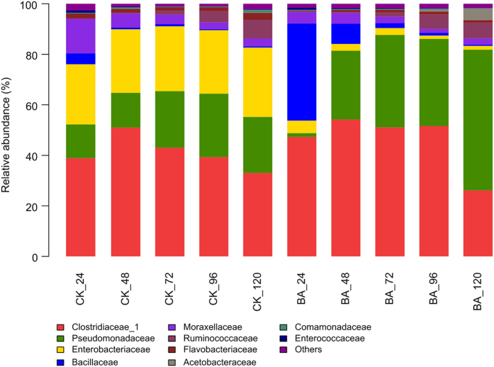
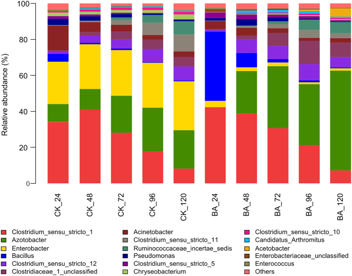
The five phyla identified in both the BA and CK samples were Actinobacteria, Cyanobacteria, Bacteroidetes, Firmicutes and Proteobacteria. The latter three phyla were predominant and comprised the following eight classes: Flavobacteriia, Sphingobacteriia, Bacilli, Clostridia, Negativicutes, Alphaproteobacteria, Betaproteobacteria and Gammaproteobacteria. These eight classes were subdivided into fourteen orders: Flavobacteriales, Sphingobacteriales, Bacillales, Lactobacillales, Clostridiales, Selenomonadales, Caulobacterales, Rhizobiales, Rhodospirillales, Sphingomonadales, Burkholderiales, Enterobacteriales, Pseudomonadales and Xanthomonadales. The bacterial communities at the family and genus levels were shown in Figs 1 and 2, respectively. The common, predominant bacteria present in all of the retting solution samples were represented by the families Clostridiaceae and Pseudomonadaceae. Enterobacteriaceae comprised a relatively large proportion of the community throughout naturally retting process, i.e., CK samples, which was replaced by Bacillacea within the BA_24 sample due to the addition of B. cereus HDYM-02. The bacterial compositions were similar between the BA and CK samples. Nevertheless, the abundance of several families was distinguished by the addition of B. cereus HDYM-02. The most remarkable difference was the increased Bacillaceae (average abundance in BA: 10.0% and CK 1.4%) and decreased Enterobacteriaceae (average abundance in BA: 2.6% and CK 25.4%) abundances. Moreover, the average abundance of Pseudomonadaceae and Clostridiaceae was enhanced in the BA samples (31.1% and 46.1%, respectively) as compared to the CK samples (19.4% and 41.0%, respectively).
Figure 1. Bacterial community structures at family level.
The abundance is presented in terms of a percentage of bacterial sequences in retting solution samples.
Figure 2. Bacterial community structures at genus level.
The abundance is presented in terms of a percentage of bacterial sequences in retting solution samples.
As shown in Fig. 3, PCA analysis revealed a significant separation of the bacterial communities between the BA and CK samples. The cumulative percentage variance of the species explained by PC1 and PC2 were 59.88% and 24.24%, respectively. Following the addition of B. cereus HDYM-02, the BA_24 h sample obviously separated from the other nine samples. Enterobacteriaceae, Clostridiaceae and Pseudomonadaceae represented the largest three contributions to the PCA (Table 2). These three families were also predominant and notably fluctuated in the retting solution samples (Fig. 2).
Figure 3. PCA analysis of retting solution samples based on the composition of bacterial communities.
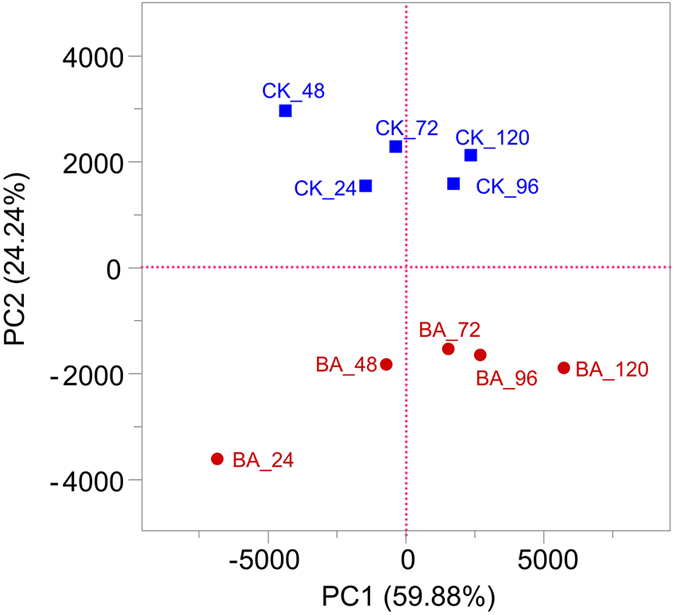
Table 2. Loading value of PCA of retting solution samples based on the composition of bacterial communities.
| No. of OTU | Represented family | Represented genus | Loading value | |
|---|---|---|---|---|
| PC1 | 63 ± 3.01 | Clostridiaceae_1 | Clostridium_sensu_stricto_1 | −0.73 |
| 59 ± 2.82 | Bacillaceae | Bacillus | −0.34 | |
| 88 ± 3.86 | Pseudomonadaceae | Azotobacter | 0.52 | |
| PC2 | 38 ± 1.21 | Enterobacteriaceae | Enterobacter | 0.84 |
| 59 ± 1.95 | Bacillaceae | Bacillus | −0.43 |
Absolute loading values greater than 0.3 were used to indicate significance among OTUs.
Changes in metabolites
A total of 460 organic metabolites were identified based on the GC-MS spectra from all of the retting solution samples, including hydrocarbons and their derivatives, acids, esters, sterols, aldehydes, and aromatics. A PCA was performed using all metabolites identified in GC-MS spectra to examine the effects of B. cereus HDYM-02 on the variations of metabolites during the entire retting period, a (Fig. 4). Two principal components explained 78.17% of the total variance. The PCA plots showed differences between the BA and CK samples. Generally, the spots shifts indicated that continuous metabolic changes occurred during the retting process. Spots representing the BA samples were more scattered than those representing CK samples, suggesting faster succession and a different composition of metabolites. Retting processed very rapidly between 24 h to 72 h, particularly for the BA samples, which was in accordance with the galacturonic acid profile shown in Fig. 5. Thereafter, the spots moved slowly until 120 h, indicating that the metabolites in the retting solution samples changed slowly during this period. Nineteen out of 460 metabolites were considered as significantly changed during retting the period based on the VIP (variable importance in the projection) and P values (Supplemental Table 1).
Figure 4. PCA based on GC-MS spectra of metabolites obtained from the retting solution samples.
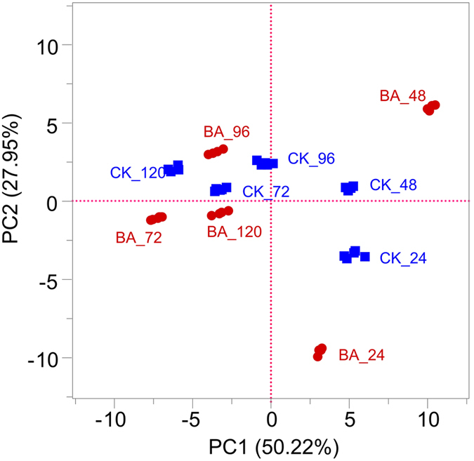
Figure 5. Changes in galacturonic acid and reducing sugar of retting solution samples.
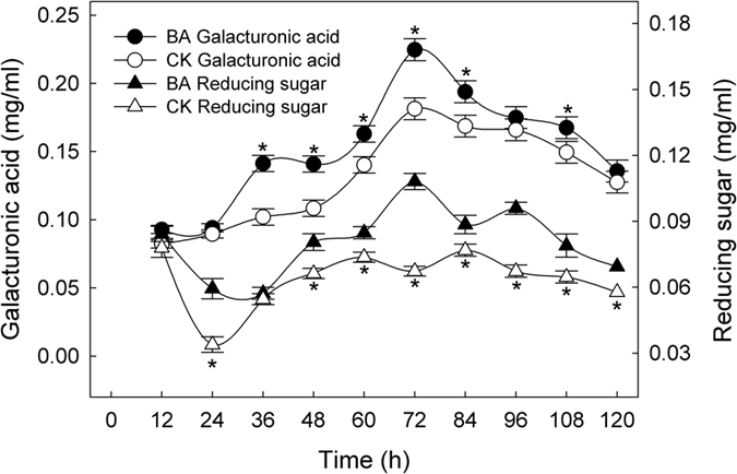
*Indicate significant differences between BA and CK samples at each time point.
Galacturonic acid and reducing sugars, residues of gummy substances present in flax straw, were selected as indicators of the extent of degumming during retting process (Fig. 5). Generally, the dynamic changes in these residues showed two coincident features: fluctuations in the wave pattern, i.e., the rise-fall-rise-fall cycle, and higher concentrations in the BA samples than in the CK samples. In the BA and CK samples, the peak values of galacturonic acid were 0.22 mg/ml and 0.18 mg/ml at 72 h, respectively, whereas the peak values of the reducing sugars were 0.11 mg/ml at 72 h and 0.08 mg/ml at 84 h, respectively.
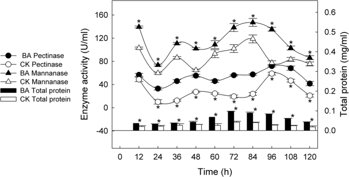
Changes in degumming enzymes and proteins
The dynamic changes in the degumming enzymes, including pectinase and mannanase, and proteins were illustrated in Fig. 6. A similar wave pattern as that of galacturonic acid and the reducing sugars shown in Fig. 5 was observed. The BA samples consistently displayed higher enzyme activity than the CK samples. The peak values for pectinase were 71.86 U/ml in the BA_96 h sample and 58.92 U/ml in the CK_96 h sample, whereas the peak values for mannanase were 147.21 U/ml in the BA_84 h sample and 118.75 U/ml in the CK_84 h sample. Degumming enzymes were the main extracellular proteins in the retting solutions. Consequently, the protein concentration was generally higher in the BA samples than in the CK samples. The highest values appeared at 72 h and were 0.10 mg/ml and 0.04 mg/ml, respectively.
Figure 6. Changes in degumming enzymes and extracellular total protein of retting solution samples.
*Indicate significant differences between BA and CK samples at each time point.
Discussion
Bacterial succession
A small number of studies have used modern molecular techniques based on the 16S rRNA to explore the bacterial communities in retting solutions of bast fiber plants, such as clone library in jute13 and DGGE in jute15 and flax12,14. Recently, one high-throughput sequencing platform, semiconductor sequencing, has been applied in investigations of the bacterial retting community of kenaf under different retting conditions at the order level16. In this study, high-throughput sequencing was first used to reveal the bacterial succession from phylum to species during the entire retting period for flax straw. Two basic retting experiments were initially conducted. CK was performed to mimic and reveal a natural retting process, including the contributions of the inherent microbes present in flax straws and tap water. BA was performed to estimate the effects of the bacterial strain B. cereus HDYM-02 on bacterial succession, structure and transition.
As shown in Fig. 2, although the bacterial composition at the family level was very similar between the CK and BA samples, the abundances among the bacterial genera were clearly changed following the addition of B. cereus HDYM-02, as expected. Enterobacteriaceae represented a large proportion of the community in the CK samples throughout the entire retting period, suggesting that it was naturally associated with the flax straw. In the BA samples, Bacillaceae almost totally replaced Enterobacteriaceae within 24 h, which almost completely disappeared throughout the whole retting period. The competition for oxygen and nutrients resulted in the replacement of Enterobacteriaceae by Bacillaceae, in accordance with the replacement of the inherent Entrerobacteriales by Bacillales after inoculating Bacillus and Paenibacillus strains during kenaf retting16. In this study, the competition among aerobes following inoculation with B. cereus HDYM-02 also led to the decrease of Chryseobacterium, Flavobacteriaceae, and Acinetobacter, Moraxellaceae (Fig. 3), two families that were also found in flax retting solutions using DGGE12.
The other dominant family in this research was Pseudomonadaceae (Fig. 1), which was subdivided into Azotobacter and Pseudomonas (Fig. 2). Pseudomonas is the dominant genus involved in the microbial retting of flax12,14 and kenaf16. Nevertheless, in this study, the average abundance of Pseudomonas was less than 2.0% across all of the retting samples. Azotobacter was unexpectedly found to be the major component of Pseudomonadaceae. Additionally, the average abundance of Azotobacter was significantly enhanced by the addition of B. cereus HDYM-02, i.e., 17.5% in the CK and 28.0% in the BA samples. Azotobacter widely exists in soil as a nitrogen fixer under aerobic conditions, but has never been identified in flax retting solutions. Surprisingly, in this study, Azotobacter rather than Pseudomonas, gradually became the dominant group, particularly in the BA samples (55.4%, BA 120 h). It maybe speculated that Azotobacter thrived to cope with the nutrient depletion by fixing nitrogen. Notably, Azotobacter has been reported as consortium component applied in microbial retting for coir fiber33, suggesting its retting capacity. An in-depth investigation of this group with respect to its succession and function during flax retting must generate further interest.
The family Clostridiaceae_1 was found to be another major microbial group in both the CK and BA samples during the entire retting period (Fig. 1), indicating their naturally association with the flax straw, similar to kenaf16. Short-length sequencing always loses accuracy as it moves lower in the taxonomic arrangement (i.e., more sequences start to be unclassified), which resulted in the assignment of several Clostridium sensu stricto rather than specific species (Fig. 2). As retting proceeded, several reports showed a later colonization of strictly anaerobic Clostridiaceae, such as C. acetobutylicm and C. felsineum, due to the environmental transition from aerobic to anaerobic conditions11. Visi et al. concluded that at the fourth day of kenaf retting, Clostridium was established and began fixing nitrogen as oxygen levels decreased and nutrient were depleted. Moreover, the heavy inoculation of Bacillus could accelerate the establishment of these bacteria16. Similar results were observed in this study, i.e., an apparent increase in Clostridiaceae abundance occurred from 24 h to 48 h in both the CK (38.9% to 51.0%) and BA (47.3% to 54.2%) samples. However, as retting proceeded, the levels of the anaerobic Clostridium gradually declined. Alternatively, the levels of the aerobic Azotobacter increased and acted as a nitrogen fixer, similar to Clostridium, indicating that the retting system functioned well. This discrepancy might result from the different experimental conditions. The retting experiments were performed in tanks without covers, unlike those performed in airtight containers16.
Changes in metabolites
As shown in Fig. 4, the PCA score plots of all metabolites from the GC-MS-based metabolic analysis showed clear differences in retting extents between the samples retted with and without B. cereus HDYM-02. Generally, the points representing the BA samples were more scattered than those representing the CK samples, indicating that more apparent transitions in the metabolites resulted from the addition of B. cereus HDYM-02. The distances among the points representing BA samples were much more remote, which meant that flax retting with B. cereus HDYM-02 proceeded faster, particularly during the first 72 h. As retting proceeded from 72 to 120 h, the points gradually clustered, i.e., the differences in metabolites between the BA and CK samples were reduced.
Six categories of lignin and wax residues, including 4,6-dimethyl-dodecane, tetradecanoic acid, phthalic acid, n-hexadecanoic acid, 4-(2-propenyl)-phenol, and octadecanoic acid, originating from the surface and inside of the bast fiber6 showed the largest abundances at 72 h, which were significantly larger in the BA samples than in the CK samples (Supplemental Table 1). These available results revealed that the lignins and waxes inside the flax straws were degraded more thoroughly following the addition of B. cereus HDYM-02, suggesting a more effective retting within 72 h, particularly in the BA samples. In addition to fungi, bacteria are also important decomposers of lignins34 and waxes35. Several Bacilli have been proven to be lignin-degraders36,37, including B. cereus38. In this study, the other dominant retting species Azotobacter (Fig. 2), also has once been reported to be able to grow within lignin as carbon source39. Moreover, Azotobacter are capable of degrading many molecules with complicated structures, such as phenols40,41, carbohydrates42 and organic acids43. It could be speculated that Azotobacter played an important role in the retting process by degrading lignins and waxes. The data obtained from the GC-MS spectra should be further explored to identify some metabolites as biomarkers of certain phases of the retting period.
Pectin and hemicellulose are the main gummy substances present in the straws of fiber plants, which can be degraded by the degumming enzymes produced by microbes such as pectinolytic bacteria10,44, and mannanase- and xylanase-producing bacteria45,46. Afterwards, the presence of galacturonic acid and reducing sugars, which are residues of gummy substances, can indicate the degumming extent47,48,49. Both galacturonic acid and reducing sugars were maintained at higher concentrations in the BA samples than in the CK samples throughout the entire retting period (Fig. 5), which, consequently, were consistent with the degumming enzymes and total protein concentrations (Fig. 6).
This result suggested that there was a more thorough removal of gummy substances within flax straws in the BA samples, which could be explained by the bacterial succession (Figs 1 and 2). When retting proceeded to 24 h, the discrepancy was clear because Bacillaceae, mainly Bacillus, almost completely replaced Enterobacteriaceae in the BA samples. The B. cereus HDYM-02 inoculum together with many other Bacilli, have been proven to produce degumming enzymes, such as pectinase, pectate lyase, mannanase and xylanase50,51,52,53. In contrast, members of Enterobacteriaceae rarely produce degumming enzymes. As retting proceeded from 48 h to the end of the process, the most distinct feature in the BA samples compared with the CK samples was the increase in Azotobacter. To date, no studies have directly reported this genus as a degumming enzyme producer. However, evidence shows that Azotobacter can promote the pectinolytic quantities during the composting of agro-industrial refuse54. In addition, Azotobacter has been reported as consortium component applied to microbial retting for coir fiber33, suggesting its retting capacity. Given its abundant proportion and apparent importance during retting, Azotobacter warrants further investigation.
In addition, another notable group was Paenibacillaceae, whose members showed a slightly higher abundance in the BA samples, including Paenibacillus, Brevibacillus and Saccharibacillus. Paenibacillus has been a promising species in the field of microbial retting16 because numerous isolates have been found to produce varying types of pectinases55. Moreover, Paenibacillus can produce antibiotics, which may lead to the changes in the bacterial composition during the retting process56. Pectate lyase activity has been detected in Brevibacillus and Saccharibacillus, suggesting that these bacteria can degrade pectin57. Therefore, it could be speculated that the Paenibacillaceae present in the BA samples had a greater contribution to the degradation of gummy substances during the retting period to some extent.
In short, this study showed that flax retting with B. cereus HDYM-02 as an inoculum significantly changed bacterial succession and efficiently accelerated the retting process compared to natural retting. Following the addition of B. cereus HDYM-02, Bacillaceae almost totally replaced Enterobacteriaceae. Pseudomonadaceae, mainly Azotobacter, continuously increased from 48 h to 120 h and became one of the dominant groups. Clostridiaceae_1 was slightly enhanced, represented approximately half of the proportion of this family, and gradually decreased until 120 h. Following the addition of B. cereus HDYM-02, aerobic or anaerobic pectionolytic bacteria dominated throughout the retting period, resulting in clearly differences in metabolite transitions, particularly galacturonic acid and reducing sugars. In addition to the data on the degumming enzymes and total proteins, the inoculation of B. cereus HDYM-02 induced a more thorough degradation of gummy substances. The combination of high-throughput sequencing and the GC-MS technique was effective for monitoring the microbial community and metabolite production, which might offer a better and further understanding of the process and function of retting ecosystems. This study could be helpful to facilitate improvements in the retting process, with shorter times and better fiber qualities.
Additional Information
How to cite this article: Zhao, D. et al. Bacterial succession and metabolite changes during flax (Linum usitatissimum L.) retting with Bacillus cereus HDYM-02. Sci. Rep. 6, 31812; doi: 10.1038/srep31812 (2016).
Supplementary Material
Acknowledgments
This work was supported by National Nature Science Foundation of China (31270534), Distinguished Young Scholars of Heilongjiang University (JCL201305), National Nature Science Youth Foundation of China (31300355), and Post Doctorate Foundation of Heilongjiang Province (LBH-Z15214).
Footnotes
Author Contributions D.Z., P.L., C.P. and R.D. performed experiments. D.Z. and P.L. prepared Figures D.Z. contributed to write the manuscript text. D.Z., W.P. and J.G. designed the study. All authors reviewed the manuscript.
References
- Siengchin S. & Wongmanee S. Mechanical and impact properties of PLA/2 × 2 twill and 4 × 4 hopsack weave flax textile composites produced by the interval hot Pressing technique. Mechanics of Composite Materials 50, 387–394 (2014). [Google Scholar]
- Wang H. G., Xian G. J. & Li H. Grafting of nano-TiO2 onto flax fibers and the enhancement of the mechanical properties of the flax fiber and flax fiber/epoxy composite. Composites Part A: Applied Science and Manufacturing 76, 172–180 (2015). [Google Scholar]
- Kulma A. et al. New flax producing bioplastic fibers for medical purposes. Industrial Crops and Products 68, 80–89 (2015). [Google Scholar]
- Parikh D., Chen Y. & Sun L. Reducing automotive interior noise with natural fiber nonwoven floor covering systems. Textile Research Journal 76, 813–820 (2006). [Google Scholar]
- Ramaswamy G. N., Ruff C. G. & Boyd C. R. Effect of bacterial and chemical retting on kenaf fiber quality. Textile Research Journal 64, 305–308 (1994). [Google Scholar]
- Akin D. E. In Industrial Applications of Natural Fibres (ed. Mussig Jorg) Ch. 4, 87–108 (A John Wiley and Sons, Ltd., 2010). [Google Scholar]
- Akin D. E. et al. Optimization for enzyme-retting of flax with pectate lyase. Industrial Crops and Products 25, 136–146 (2007). [Google Scholar]
- Tamburini E., León A. G., Perito B. & Mastromei G. Characterization of bacterial pectinolytic strains involved in the water retting process. Environ. Microbiol. 5, 730–736 (2003). [DOI] [PubMed] [Google Scholar]
- Di Candilo M. et al. Effects of selected pectinolytic bacterial strains on water‐retting of hemp and fibre properties. J. Appl. Microbiol. 108, 194–203 (2010). [DOI] [PubMed] [Google Scholar]
- Das B. et al. Effect of efficient pectinolytic bacterial isolates on retting and fibre quality of jute. Industrial Crops and Products 36, 415–419 (2012). [Google Scholar]
- Donaghy J. A., Levett P. N. & Haylock R. W. Changes in microbial populations during anaerobic flax retting. J. Appl. Microbiol. 69, 634–641 (1990). [Google Scholar]
- Hou L. B. & Liu X. L. Analysis of bacterial community structure in water retting and enzymatic retting liquid. Bioprocess 2, 110–116 (2012). [Google Scholar]
- Munshi T. K. & Chattoo B. B. Bacterial population structure of the jute-retting environment. Microb. Ecol. 56, 270–282 (2008). [DOI] [PubMed] [Google Scholar]
- Ling H. Z., Ge J. P., Wei W. & Ping W. X. Analysis of bacterial community in water retting of flax. Chin. J. Appl. Environ. Biol. 15, 703–707 (2009). [Google Scholar]
- Das B., Chakrabarti K., Ghosh S., Chakraborty A. & Saha M. N. Assessment of changes in community level physiological profile and molecular diversity of bacterial communities in different stages of jute retting. Pakistan Journal of Biological Sciences 16, 1722–1729 (2013). [DOI] [PubMed] [Google Scholar]
- Visi D. K., D’Souza N., Ayre B. G., Iii C. L. W. & Allen M. S. Investigation of the bacterial retting community of kenaf (Hibiscus cannabinus) under different conditions using next-generation semiconductor sequencing. J. Ind. Microbiol. Biotechnol. 40, 465–475 (2013). [DOI] [PubMed] [Google Scholar]
- Ping W. X., Zhao D., Song G., Ge J. P. & Ling H. Z. Study of fermentation condition of pectinase producing strain HDYM-02. Journal of Daqing Normal University 30, 115–118 (2010). [Google Scholar]
- Ge J. P. et al. Application of fast degumming with bacteria on retting flax. Journal of Natural Science of Heilongjiang University 23, 307–310 (2006). [Google Scholar]
- Ge J. P., Ling H. Z., Zhao D., Song G. & Ping W. X. Screening of pectinase-producing strains and preliminary study on variation of bacteria population during flax degumming. Journal of natural science of the heilongjiang university 24, 295–300 (2007). [Google Scholar]
- Ge J. P. et al. Using bacteria addition and reusing retting water technologies to accelerate flax degumming. Applied Mechanics and Materials 522, 374–379 (2014). [Google Scholar]
- Ge J. P., Ping W. X., Gao Y., Ling H. Z. & Song G. Isolation, identification and phyletic analysis of a pectinase-producing bacterial strain HDYM-02. Journal of Microbiology 28, 44–47 (2008). [Google Scholar]
- Lowry O. H., Rosebrough N. J., Farr A. L. & Randall R. J. Protein measurement with the folin-phenol reagent. J. Biol. Chem. 193, 265–275 (1951). [PubMed] [Google Scholar]
- Sampriya S., RishiPal M. & Jitender S. Pseudozyma sp. SPJ: an economic and eco-friendly approach for degumming of flax fibers. World Journal of Microbiology and Biotechnology 27, 2697–2701 (2011). [Google Scholar]
- Akino T., Nakamura N. & Horikoshi K. Characterization of three BETA-mannanases of an alkalophilic Bacillus sp. Agric. Biol. Chem. 52, 773–779 (1988). [Google Scholar]
- Xiong J. B. et al. Geographic distance and pH drive bacterial distribution in alkaline lake sediments across Tibetan Plateau. Environ. Microbiol. 14, 2457–2466 (2012). [DOI] [PMC free article] [PubMed] [Google Scholar]
- Caporaso J. G. et al. QIIME allows analysis of high-throughput community sequencing data. Nat. Methods 7, 335–336 (2010). [DOI] [PMC free article] [PubMed] [Google Scholar]
- Edgar R. C. UPARSE: highly accurate OTU sequences from microbial amplicon reads. Nat. Methods 10, 996–998 (2013). [DOI] [PubMed] [Google Scholar]
- Edgar R. C., Haas B. J., Clemente J. C., Quince C. & Knight R. UCHIME improves sensitivity and speed of chimera detection. Bioinformatics 27, 2194–2200 (2011). [DOI] [PMC free article] [PubMed] [Google Scholar]
- Wang Q. et al. The RDP-II (Ribosomal Database Project): The RDP Sequence Classifier. Nucleic Acids Res 31, 442–443 (2004).12520046 [Google Scholar]
- Quast C. et al. The SILVA ribosomal RNA gene database project: improved data processing and web-based tools. Nucleic Acids Res. 41, 590–596 (2013). [DOI] [PMC free article] [PubMed] [Google Scholar]
- Miller G. L. Use of dinitrosalicylic acid reagent for determination of reducing sugar. Analytical Chemistry 31, 426–428, doi: 10.1021/ac60147a030 (1959). [DOI] [Google Scholar]
- Dietz J. H. & Rouse A. A rapid method for estimating pectic substances in citrus juices. J. Food Sci. 18, 169–177 (1953). [Google Scholar]
- Ravindranath A. D. In Bioprospects of Coastal Eubacteria (ed. Borkar Sunita) Ch. 13, 225–240 (Springer International Publishing, 2015). [Google Scholar]
- Bugg T. D., Ahmad M., Hardiman E. M. & Singh R. The emerging role for bacteria in lignin degradation and bio-product formation. Curr. Opin. Biotechnol. 22, 394–400 (2011). [DOI] [PubMed] [Google Scholar]
- Mburu A. W., Mwasiagi J. I. & Anino E. Isolation and characterisation of bacteria isolate from gin trash and its use in cotton wax degradation. Indian Journal of Biotechnology 11, 225–232 (2015). [Google Scholar]
- Li H., Li S., Wang S., Wang Q. & Zhu B. Screening, identification of lignin-degradating Bacillus MN-8 and its characteristics in degradation of maize straw lignin. Scientia Agricultura Sinica 47, 324–333 (2014). [Google Scholar]
- Yadav S. & Chandra R. Syntrophic co-culture of Bacillus subtilis and Klebsiella pneumonia for degradation of kraft lignin discharged from rayon grade pulp industry. Journal of Environmental Sciences 33, 229–238 (2015). [DOI] [PubMed] [Google Scholar]
- Shajaratuldur I. Lignin degradation of banana stem wastes using aerobic Bacillus cereus Bechelor thesis, Universiti Malaysia Pahang, (2009).
- Zhang X., Zhao H., Zhang J. & Li Z. Growth of Azotobacter vinelandii in a solid-state fermentation of technical lignin. Bioresour. Technol. 95, 31–33 (2004). [DOI] [PubMed] [Google Scholar]
- Li D.-Y., Eberspächer J., Wagner B., Kuntzer J. & Lingens F. Degradation of 2, 4, 6-trichlorophenol by Azotobacter sp. strain GP1. Appl. Environ. Microbiol. 57, 1920–1928 (1991). [DOI] [PMC free article] [PubMed] [Google Scholar]
- Hamdi Y. & Tewfik M. Degradation of 3, 5-dinitro-o-cresol by Rhizobium and Azotobacter spp. Soil Biol. Biochem. 2, 163–166 (1970). [Google Scholar]
- Bonartseva G. et al. Aerobic and anaerobic microbial degradation of poly-β-hydroxybutyrate produced by Azotobacter chroococcum. Appl. Biochem. Biotechnol. 109, 285–301 (2003). [DOI] [PubMed] [Google Scholar]
- Saha S. P., Patra A. & Paul A. Studies on intracellular degradation of polyhydroxyalkanoic acid–polyethylene glycol copolymer accumulated by Azotobacter chroococcum MAL-201. J. Biotechnol. 132, 325–330 (2007). [DOI] [PubMed] [Google Scholar]
- Sharma N., Rathore M. & Sharma M. Microbial pectinase: sources, characterization and applications. Rev Environ Sci Biotechnol 12, 45–60 (2013). [Google Scholar]
- Liu P. F. et al. The components and activities of flax-degumming enzymes produced by Bacillus licheniformis HDYM-03. Advanced Materials Research 1004–1005, 566–571 (2014). [Google Scholar]
- Guo G. et al. Purification and characterization of a xylanase from Bacillus subtilis isolated from the degumming line. J. Basic Microbiol. 52, 419–428 (2012). [DOI] [PubMed] [Google Scholar]
- Mukhopadhyay A., Dutta N., Chattopadhyay D. & Chakrabarti K. Degumming of ramie fiber and the production of reducing sugars from waste peels using nanoparticle supplemented pectate lyase. Bioresour. Technol. 137, 202–208 (2013). [DOI] [PubMed] [Google Scholar]
- Liu M. et al. Effect of harvest time and field retting duration on the chemical composition, morphology and mechanical properties of hemp fibers. Industrial Crops and Products 69, 29–39 (2015). [Google Scholar]
- Ruan P. Y., Raghavan V., Gariepy Y. & Du J. M. Characterization of flax water retting of different durations in laboratory condition and evaluation of its fiber properties. BioResources 10, 3553–3563 (2015). [Google Scholar]
- Kavuthodi B., Thomas S. K. & Sebastian D. Co-production of pectinase and biosurfactant by the newly isolated strain Bacillus subtilis BKDS1. British Microbiology Research Journal 10, 1–12 (2015). [Google Scholar]
- Zhou C., Ye J. T., Xue Y. F. & Ma Y. H. Directed evolution and structural analysis of alkaline pectate lyase from the alkaliphilic bacterium Bacillus sp. strain N16-5 to improve its thermostability for efficient ramie degumming. Appl. Environ. Microbiol. 81, 5714–5723 (2015). [DOI] [PMC free article] [PubMed] [Google Scholar]
- Zhao D. et al. Optimization of mannanase production by Bacillus sp. HDYM-05 through factorial method and response surface methodology African Journal of Microbiology Research 6, 176–182 (2012). [Google Scholar]
- Kallel F. et al. Statistical optimization of low-cost production of an acidic xylanase by Bacillus mojavensis UEB-FK: its potential applications. Biocatalysis and Agricultural Biotechnology 5, 1–10 (2016). [Google Scholar]
- Pepe O., Ventorino V. & Blaiotta G. Dynamic of functional microbial groups during mesophilic composting of agro-industrial wastes and free-living (N2)-fixing bacteria application. Waste Manage. 33, 1616–1625 (2013). [DOI] [PubMed] [Google Scholar]
- Margarita S., Pilar D. & Pastor F. I. J. Pectinolytic systems of two aerobic sporogenous bacterial strains with high activity on pectin. Curr. Microbiol. 50, 114–118 (2005). [DOI] [PubMed] [Google Scholar]
- En H. & Yousef A. E. Draft genome sequence of Paenibacillus polymyxa OSY-DF, which coproduces a lantibiotic, paenibacillin, and polymyxin E1. J. Bacteriol. 194, 4739–4740 (2012). [DOI] [PMC free article] [PubMed] [Google Scholar]
- Ratón T. d. l. M. O. et al. Isolation and characterisation of aerobic endospore forming Bacilli from sugarcane rhizosphere for the selection of strains with agriculture potentialities. World Journal of Microbiology and Biotechnology 28, 1593–1603 (2012). [DOI] [PubMed] [Google Scholar]
Associated Data
This section collects any data citations, data availability statements, or supplementary materials included in this article.


