

Abstract
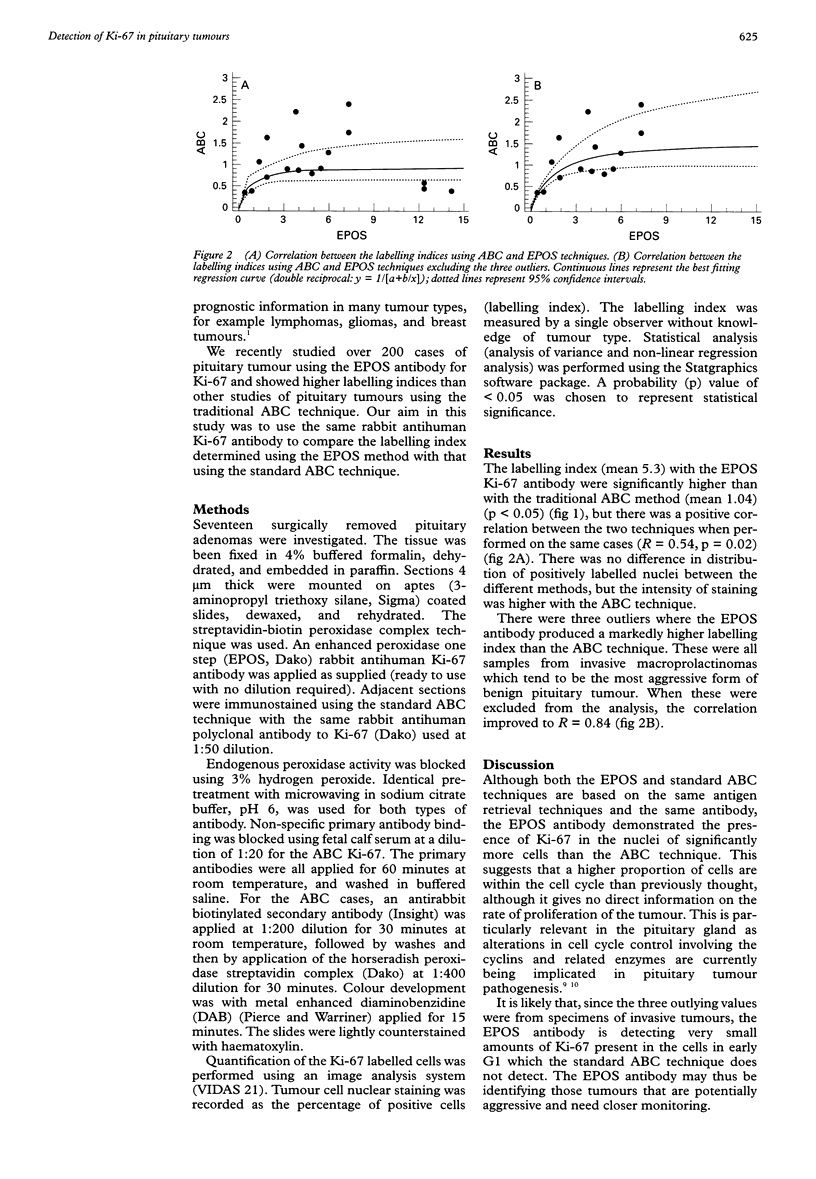
AIM: To compare the Ki-67 labelling index determined using the enhanced peroxidase one step (EPOS) method with that using the standard ABC technique in pituitary tumours. METHODS: Adjacent sections were immunostained using the EPOS and ABC techniques with the same Ki-67 antibody and same antigen retrieval method. RESULTS: The labelling index measurements with the EPOS Ki-67 antibody were significantly higher than when using the traditional ABC method, and there was a positive correlation between the two techniques when performed on the same cases. This suggests that a higher proportion of cells are within the cell cycle than previously thought, although it gives no direct information on the rate of proliferation of the tumour. CONCLUSIONS: It appears that the EPOS system is not only more convenient but may be more sensitive than traditional techniques for detecting Ki-67 in the nuclei of cells, thus demonstrating more accurately which cells have entered the cell cycle.
Full text
PDF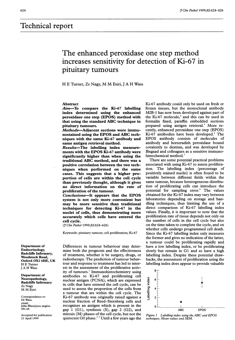

Selected References
These references are in PubMed. This may not be the complete list of references from this article.
- Brown D. C., Gatter K. C. Monoclonal antibody Ki-67: its use in histopathology. Histopathology. 1990 Dec;17(6):489–503. doi: 10.1111/j.1365-2559.1990.tb00788.x. [DOI] [PubMed] [Google Scholar]
- Gerdes J., Lemke H., Baisch H., Wacker H. H., Schwab U., Stein H. Cell cycle analysis of a cell proliferation-associated human nuclear antigen defined by the monoclonal antibody Ki-67. J Immunol. 1984 Oct;133(4):1710–1715. [PubMed] [Google Scholar]
- Gerdes J., Schwab U., Lemke H., Stein H. Production of a mouse monoclonal antibody reactive with a human nuclear antigen associated with cell proliferation. Int J Cancer. 1983 Jan 15;31(1):13–20. doi: 10.1002/ijc.2910310104. [DOI] [PubMed] [Google Scholar]
- Jin L., Qian X., Kulig E., Sanno N., Scheithauer B. W., Kovacs K., Young W. F., Jr, Lloyd R. V. Transforming growth factor-beta, transforming growth factor-beta receptor II, and p27Kip1 expression in nontumorous and neoplastic human pituitaries. Am J Pathol. 1997 Aug;151(2):509–519. [PMC free article] [PubMed] [Google Scholar]
- Key G., Becker M. H., Baron B., Duchrow M., Schlüter C., Flad H. D., Gerdes J. New Ki-67-equivalent murine monoclonal antibodies (MIB 1-3) generated against bacterially expressed parts of the Ki-67 cDNA containing three 62 base pair repetitive elements encoding for the Ki-67 epitope. Lab Invest. 1993 Jun;68(6):629–636. [PubMed] [Google Scholar]
- Qian X., Kulig E., Jin L., Lloyd R. V. Expression of D-type cyclins in normal and neoplastic rat pituitary. Endocrinology. 1998 Apr;139(4):2058–2067. doi: 10.1210/endo.139.4.5955. [DOI] [PubMed] [Google Scholar]
- Taylor C. R., Shi S. R., Chaiwun B., Young L., Imam S. A., Cote R. J. Strategies for improving the immunohistochemical staining of various intranuclear prognostic markers in formalin-paraffin sections: androgen receptor, estrogen receptor, progesterone receptor, p53 protein, proliferating cell nuclear antigen, and Ki-67 antigen revealed by antigen retrieval techniques. Hum Pathol. 1994 Mar;25(3):263–270. doi: 10.1016/0046-8177(94)90198-8. [DOI] [PubMed] [Google Scholar]
- Tsutsumi Y., Serizawa A., Kawai K. Enhanced polymer one-step staining (EPOS) for proliferating cell nuclear antigen (PCNA) and Ki-67 antigen: application to intra-operative frozen diagnosis. Pathol Int. 1995 Feb;45(2):108–115. doi: 10.1111/j.1440-1827.1995.tb03430.x. [DOI] [PubMed] [Google Scholar]
- Zuber P., Hamou M. F., de Tribolet N. Identification of proliferating cells in human gliomas using the monoclonal antibody Ki-67. Neurosurgery. 1988 Feb;22(2):364–368. doi: 10.1227/00006123-198802000-00015. [DOI] [PubMed] [Google Scholar]