

Abstract
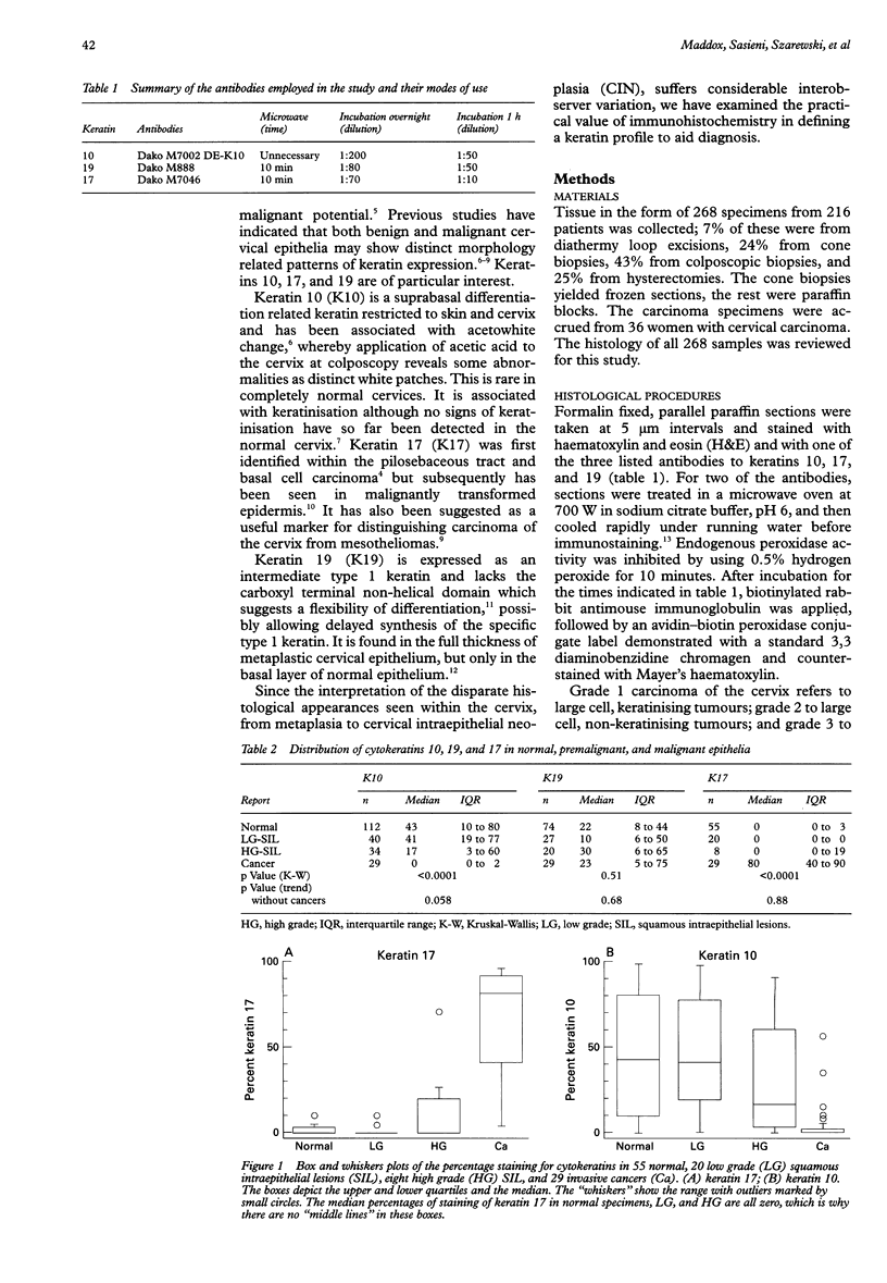
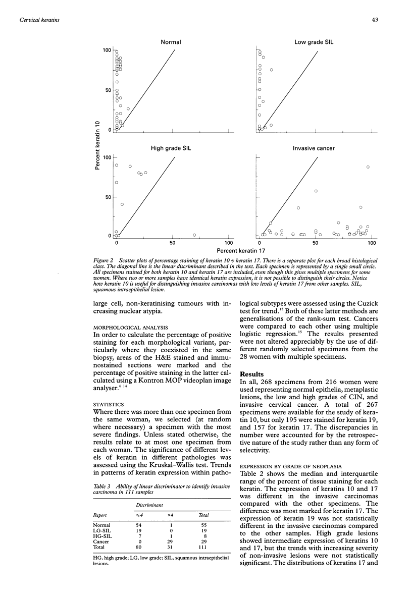
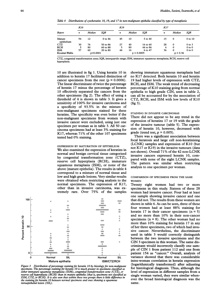
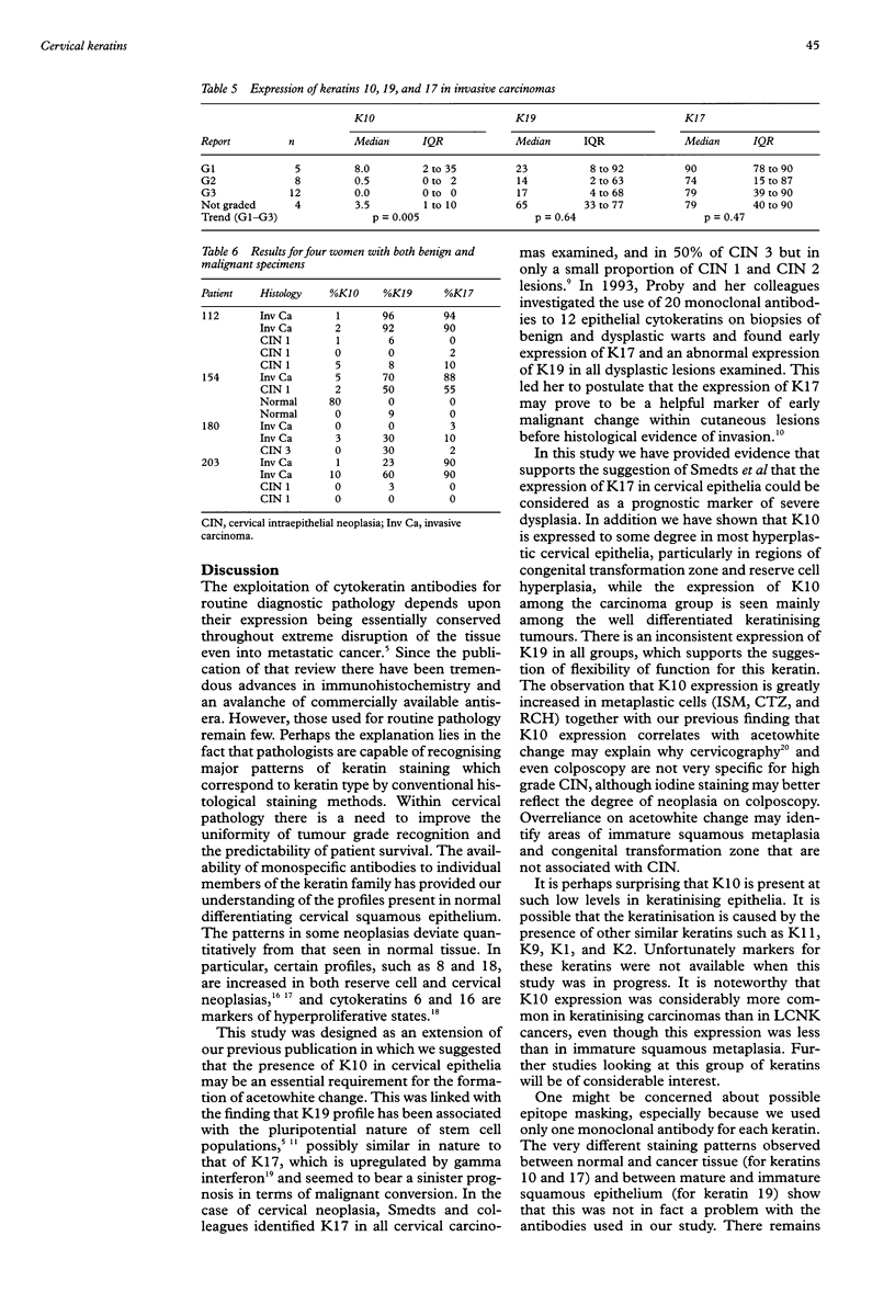
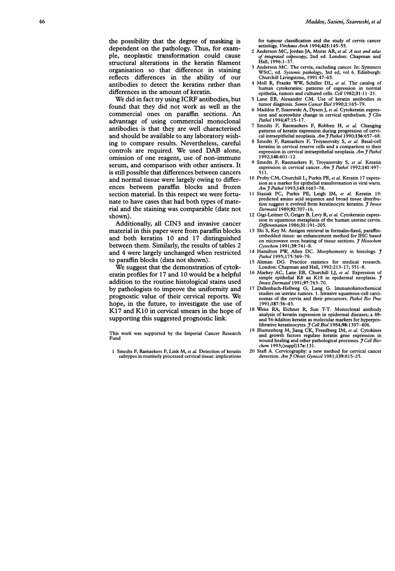
AIM: To examine the value of immunohistochemistry in defining a keratin profile to aid cervical histopathological diagnosis. METHODS: Immunohistochemical localisation of keratins 17, 10, and 19 was studied in 268 cervical biopsies from 216 women including normal epithelia (with and without human papilloma virus), low and high grade cervical intraepithelial neoplasia, and invasive carcinoma. The percentage of positive immunostaining was scored using a Kontron MOP videoplan image analyser. RESULTS: All major categories of cervical epithelia expressed these keratins to varying degrees. The median percentage of immunostaining for keratin 10 was 40% in normal tissue compared with just 1% in invasive carcinoma (p < 0.0001). The medians for keratin 17 were 0% in the normal group and 80% in carcinomas (p < 0.0001). By contrast, there was no significant difference in staining for keratin 19. Using a combination of the keratin 10 and 17 percentages, it was possible to separate the carcinomas from the benign conditions with a sensitivity of 100% and a specificity of 93%. Further analyses within the groups revealed more extensive staining for keratins 10 and 19 in reserve cell hyperplasia, immature squamous metaplasia, and congenital transformation zone. CONCLUSIONS: The morphological variety within the cervix is reflected, in part, by distinct keratin patterns. There are striking differences in the patterns of keratins 10 and 17 between infiltrating squamous carcinoma and normal cervical epithelia.
Full text
PDF
Selected References
These references are in PubMed. This may not be the complete list of references from this article.
- Dallenbach-Hellweg G., Lang G. Immunohistochemical studies on uterine tumors. I. Invasive squamous cell carcinomas of the cervix and their precursors. Pathol Res Pract. 1991 Jan;187(1):36–43. doi: 10.1016/S0344-0338(11)81042-3. [DOI] [PubMed] [Google Scholar]
- Gigi-Leitner O., Geiger B., Levy R., Czernobilsky B. Cytokeratin expression in squamous metaplasia of the human uterine cervix. Differentiation. 1986;31(3):191–205. doi: 10.1111/j.1432-0436.1986.tb00400.x. [DOI] [PubMed] [Google Scholar]
- Hamilton P. W., Allen D. C. Morphometry in histopathology. J Pathol. 1995 Apr;175(4):369–379. doi: 10.1002/path.1711750403. [DOI] [PubMed] [Google Scholar]
- Lane E. B., Alexander C. M. Use of keratin antibodies in tumor diagnosis. Semin Cancer Biol. 1990 Jun;1(3):165–179. [PubMed] [Google Scholar]
- Maddox P., Szarewski A., Dyson J., Cuzick J. Cytokeratin expression and acetowhite change in cervical epithelium. J Clin Pathol. 1994 Jan;47(1):15–17. doi: 10.1136/jcp.47.1.15. [DOI] [PMC free article] [PubMed] [Google Scholar]
- Markey A. C., Lane E. B., Churchill L. J., MacDonald D. M., Leigh I. M. Expression of simple epithelial keratins 8 and 18 in epidermal neoplasia. J Invest Dermatol. 1991 Nov;97(5):763–770. doi: 10.1111/1523-1747.ep12486607. [DOI] [PubMed] [Google Scholar]
- Proby C. M., Churchill L., Purkis P. E., Glover M. T., Sexton C. J., Leigh I. M. Keratin 17 expression as a marker for epithelial transformation in viral warts. Am J Pathol. 1993 Dec;143(6):1667–1678. [PMC free article] [PubMed] [Google Scholar]
- Shi S. R., Key M. E., Kalra K. L. Antigen retrieval in formalin-fixed, paraffin-embedded tissues: an enhancement method for immunohistochemical staining based on microwave oven heating of tissue sections. J Histochem Cytochem. 1991 Jun;39(6):741–748. doi: 10.1177/39.6.1709656. [DOI] [PubMed] [Google Scholar]
- Smedts F., Ramaekers F., Link M., Lauerova L., Troyanovsky S., Schijf C., Vooijs G. P. Detection of keratin subtypes in routinely processed cervical tissue: implications for tumour classification and the study of cervix cancer aetiology. Virchows Arch. 1994;425(2):145–155. doi: 10.1007/BF00230351. [DOI] [PubMed] [Google Scholar]
- Smedts F., Ramaekers F., Robben H., Pruszczynski M., van Muijen G., Lane B., Leigh I., Vooijs P. Changing patterns of keratin expression during progression of cervical intraepithelial neoplasia. Am J Pathol. 1990 Mar;136(3):657–668. [PMC free article] [PubMed] [Google Scholar]
- Smedts F., Ramaekers F., Troyanovsky S., Pruszczynski M., Link M., Lane B., Leigh I., Schijf C., Vooijs P. Keratin expression in cervical cancer. Am J Pathol. 1992 Aug;141(2):497–511. [PMC free article] [PubMed] [Google Scholar]
- Smedts F., Ramaekers F., Troyanovsky S., Pruszczynski M., Robben H., Lane B., Leigh I., Plantema F., Vooijs P. Basal-cell keratins in cervical reserve cells and a comparison to their expression in cervical intraepithelial neoplasia. Am J Pathol. 1992 Mar;140(3):601–612. [PMC free article] [PubMed] [Google Scholar]
- Stasiak P. C., Purkis P. E., Leigh I. M., Lane E. B. Keratin 19: predicted amino acid sequence and broad tissue distribution suggest it evolved from keratinocyte keratins. J Invest Dermatol. 1989 May;92(5):707–716. doi: 10.1111/1523-1747.ep12721500. [DOI] [PubMed] [Google Scholar]
- Weiss R. A., Eichner R., Sun T. T. Monoclonal antibody analysis of keratin expression in epidermal diseases: a 48- and 56-kdalton keratin as molecular markers for hyperproliferative keratinocytes. J Cell Biol. 1984 Apr;98(4):1397–1406. doi: 10.1083/jcb.98.4.1397. [DOI] [PMC free article] [PubMed] [Google Scholar]