

Abstract
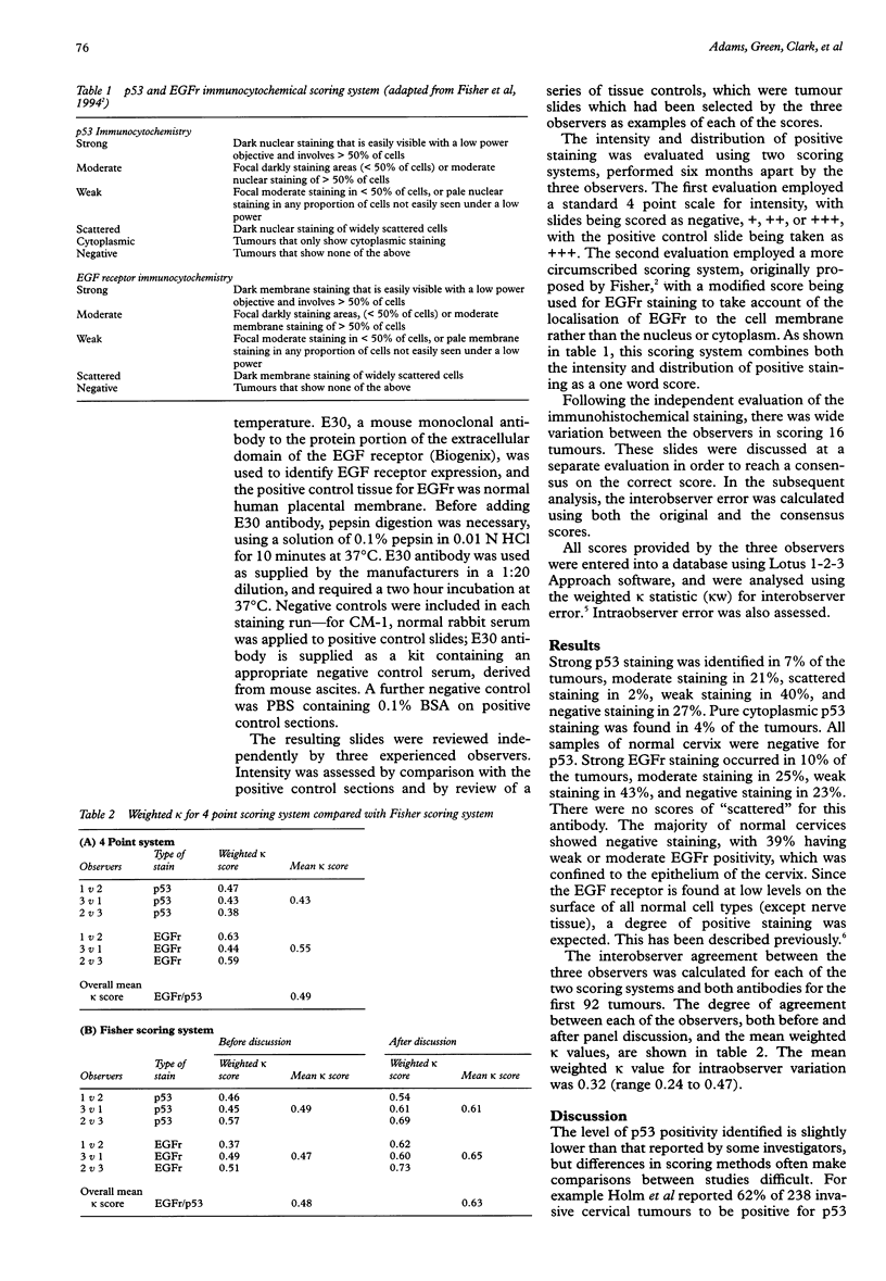
AIM: To carry out an objective assessment of two systems of scoring immunohistochemical staining, evaluating interobserver and intraobserver error. METHODS: 92 cervical tumours underwent immunohistochemical staining for p53 and epidermal growth factor receptor. Staining was assessed using two methods: a standard 4 point scale and a descriptive method, performed by three observers. Interobserver and intraobserver error were assessed for both scoring methods. RESULTS: In terms of interobserver error between three observers, no difference was found between a simple 4 point scale method of evaluation and the use of a highly circumscribed method. In all evaluations, interobserver error was scored as moderate (kappa w 0.48-0.49). However, evaluation of immunohistochemical staining by a panel of observers led to a marked improvement in the interobserver error scores (kappa w 0.63). CONCLUSIONS: There should be standardisation of immunohistochemical staining and scoring methods. More attention should be paid to measurement of interobserver and intraobserver error in studies. Use of a panel of tissue control slides and consensus scoring by several observers can lead to improvement in reproducibility.
Full text
PDF
Selected References
These references are in PubMed. This may not be the complete list of references from this article.
- Fisher C. J., Gillett C. E., Vojtesek B., Barnes D. M., Millis R. R. Problems with p53 immunohistochemical staining: the effect of fixation and variation in the methods of evaluation. Br J Cancer. 1994 Jan;69(1):26–31. doi: 10.1038/bjc.1994.4. [DOI] [PMC free article] [PubMed] [Google Scholar]
- Göppinger A., Wittmaack F. M., Wintzer H. O., Ikenberg H., Bauknecht T. Localization of human epidermal growth factor receptor in cervical intraepithelial neoplasias. J Cancer Res Clin Oncol. 1989;115(3):259–263. doi: 10.1007/BF00391699. [DOI] [PMC free article] [PubMed] [Google Scholar]
- Holm R., Skomedal H., Helland A., Kristensen G., Børresen A. L., Nesland J. M. Immunohistochemical analysis of p53 protein overexpression in normal, premalignant, and malignant tissues of the cervix uteri. J Pathol. 1993 Jan;169(1):21–26. doi: 10.1002/path.1711690105. [DOI] [PubMed] [Google Scholar]
- Hsu S. M., Raine L., Fanger H. A comparative study of the peroxidase-antiperoxidase method and an avidin-biotin complex method for studying polypeptide hormones with radioimmunoassay antibodies. Am J Clin Pathol. 1981 May;75(5):734–738. doi: 10.1093/ajcp/75.5.734. [DOI] [PubMed] [Google Scholar]
- Kohler M., Janz I., Wintzer H. O., Wagner E., Bauknecht T. The expression of EGF receptors, EGF-like factors and c-myc in ovarian and cervical carcinomas and their potential clinical significance. Anticancer Res. 1989 Nov-Dec;9(6):1537–1547. [PubMed] [Google Scholar]
- Landis J. R., Koch G. G. The measurement of observer agreement for categorical data. Biometrics. 1977 Mar;33(1):159–174. [PubMed] [Google Scholar]
- Oka K., Nakano T., Arai T. p53CM1 expression is not associated with prognosis in uterine cervical carcinoma. Cancer. 1993 Jul 1;72(1):160–164. doi: 10.1002/1097-0142(19930701)72:1<160::aid-cncr2820720130>3.0.co;2-c. [DOI] [PubMed] [Google Scholar]
- van Diest P. J., Weger D. R., Lindholm J. Reproducibility of subjective immunoscoring of steroid receptors in breast cancer. Anal Quant Cytol Histol. 1996 Oct;18(5):351–354. [PubMed] [Google Scholar]
- van Diest P. J., van Dam P., Henzen-Logmans S. C., Berns E., van der Burg M. E., Green J., Vergote I. A scoring system for immunohistochemical staining: consensus report of the task force for basic research of the EORTC-GCCG. European Organization for Research and Treatment of Cancer-Gynaecological Cancer Cooperative Group. J Clin Pathol. 1997 Oct;50(10):801–804. doi: 10.1136/jcp.50.10.801. [DOI] [PMC free article] [PubMed] [Google Scholar]