

Abstract
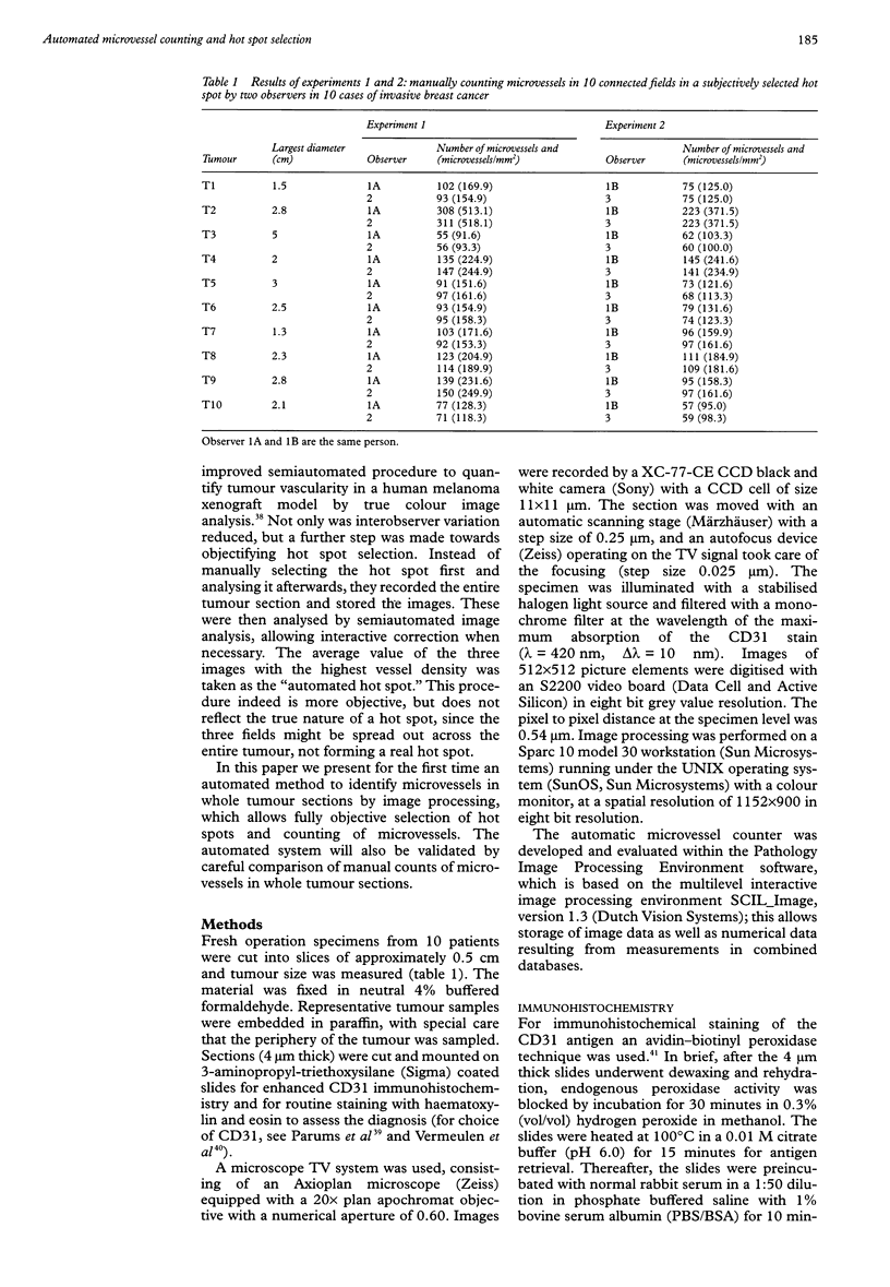
BACKGROUND: Manual counting of microvessels is subjective and may lead to unacceptable interobserver variability, which may explain conflicting results. AIMS: To develop and test an automated method for microvessel counting and objective selection of the hot spot, based on image processing of whole sections, and to compare this with manual selection of a hot spot and counting of microvessels. METHODS: Microvessels were stained by CD31 immunohistochemistry in 10 cases of invasive breast cancer. The number of microvessels was counted manually in a subjectively selected hot spot, and also in the same complete tumour sections by interactive and automated image processing methods. An algorithm identified the hot spots from microvessel maps of the whole tumour section. RESULTS: No significant difference in manual microvessel counts was found between two observers within the same hot spot, and counts were significantly correlated. However, when the hot spot was reselected, significantly different results were found between repeated counts by the same observer. Counting all microvessels manually within the entire tumour section resulted in significantly different hot spots than manual counts in selected hot spots by the same observer. Within the entire tumour section no significant differences were found between the hot spots of the manual and automated methods using an automated microscope. The hot spot was found using an eight connective path search algorithm, was located at or near the border of the tumour, and (depending on the size of the hot spot) did not always contain the field with the largest number of microvessels. CONCLUSIONS: The automated counting of microvessels is preferable to the manual method because of the reduction in measurement time when the complete tumour is scanned, the greater accuracy and objectivity of hot spot selection, and the possibility of visual inspection and relocation of each measurement field afterwards.
Full text
PDF
Images in this article
Selected References
These references are in PubMed. This may not be the complete list of references from this article.
- Adams J. C. Biotin amplification of biotin and horseradish peroxidase signals in histochemical stains. J Histochem Cytochem. 1992 Oct;40(10):1457–1463. doi: 10.1177/40.10.1527370. [DOI] [PubMed] [Google Scholar]
- Anderson R. E. Cholestasis: a retrospective. Hum Pathol. 1995 Jan;26(1):1–2. doi: 10.1016/0046-8177(95)90107-8. [DOI] [PubMed] [Google Scholar]
- Axelsson K., Ljung B. M., Moore D. H., 2nd, Thor A. D., Chew K. L., Edgerton S. M., Smith H. S., Mayall B. H. Tumor angiogenesis as a prognostic assay for invasive ductal breast carcinoma. J Natl Cancer Inst. 1995 Jul 5;87(13):997–1008. doi: 10.1093/jnci/87.13.997. [DOI] [PubMed] [Google Scholar]
- Beliën J. A., Baak J. P., van Diest P. J., van Ginkel A. H. Counting mitoses by image processing in Feulgen stained breast cancer sections: the influence of resolution. Cytometry. 1997 Jun 1;28(2):135–140. [PubMed] [Google Scholar]
- Bigler S. A., Deering R. E., Brawer M. K. Comparison of microscopic vascularity in benign and malignant prostate tissue. Hum Pathol. 1993 Feb;24(2):220–226. doi: 10.1016/0046-8177(93)90304-y. [DOI] [PubMed] [Google Scholar]
- Bosari S., Lee A. K., DeLellis R. A., Wiley B. D., Heatley G. J., Silverman M. L. Microvessel quantitation and prognosis in invasive breast carcinoma. Hum Pathol. 1992 Jul;23(7):755–761. doi: 10.1016/0046-8177(92)90344-3. [DOI] [PubMed] [Google Scholar]
- Brawer M. K., Deering R. E., Brown M., Preston S. D., Bigler S. A. Predictors of pathologic stage in prostatic carcinoma. The role of neovascularity. Cancer. 1994 Feb 1;73(3):678–687. doi: 10.1002/1097-0142(19940201)73:3<678::aid-cncr2820730329>3.0.co;2-6. [DOI] [PubMed] [Google Scholar]
- Charpin C., Devictor B., Bergeret D., Andrac L., Boulat J., Horschowski N., Lavaut M. N., Piana L. CD31 quantitative immunocytochemical assays in breast carcinomas. Correlation with current prognostic factors. Am J Clin Pathol. 1995 Apr;103(4):443–448. doi: 10.1093/ajcp/103.4.443. [DOI] [PubMed] [Google Scholar]
- Fidler I. J., Ellis L. M. The implications of angiogenesis for the biology and therapy of cancer metastasis. Cell. 1994 Oct 21;79(2):185–188. doi: 10.1016/0092-8674(94)90187-2. [DOI] [PubMed] [Google Scholar]
- Folkman J. Seminars in Medicine of the Beth Israel Hospital, Boston. Clinical applications of research on angiogenesis. N Engl J Med. 1995 Dec 28;333(26):1757–1763. doi: 10.1056/NEJM199512283332608. [DOI] [PubMed] [Google Scholar]
- Folkman J. Tumor angiogenesis: therapeutic implications. N Engl J Med. 1971 Nov 18;285(21):1182–1186. doi: 10.1056/NEJM197111182852108. [DOI] [PubMed] [Google Scholar]
- Fox S. B., Leek R. D., Weekes M. P., Whitehouse R. M., Gatter K. C., Harris A. L. Quantitation and prognostic value of breast cancer angiogenesis: comparison of microvessel density, Chalkley count, and computer image analysis. J Pathol. 1995 Nov;177(3):275–283. doi: 10.1002/path.1711770310. [DOI] [PubMed] [Google Scholar]
- Fox S. B. Tumour angiogenesis and prognosis. Histopathology. 1997 Mar;30(3):294–301. doi: 10.1046/j.1365-2559.1997.d01-606.x. [DOI] [PubMed] [Google Scholar]
- Gasparini G., Weidner N., Bevilacqua P., Maluta S., Dalla Palma P., Caffo O., Barbareschi M., Boracchi P., Marubini E., Pozza F. Tumor microvessel density, p53 expression, tumor size, and peritumoral lymphatic vessel invasion are relevant prognostic markers in node-negative breast carcinoma. J Clin Oncol. 1994 Mar;12(3):454–466. doi: 10.1200/JCO.1994.12.3.454. [DOI] [PubMed] [Google Scholar]
- Hall N. R., Fish D. E., Hunt N., Goldin R. D., Guillou P. J., Monson J. R. Is the relationship between angiogenesis and metastasis in breast cancer real? Surg Oncol. 1992 Jun;1(3):223–229. doi: 10.1016/0960-7404(92)90068-v. [DOI] [PubMed] [Google Scholar]
- Horak E. R., Leek R., Klenk N., LeJeune S., Smith K., Stuart N., Greenall M., Stepniewska K., Harris A. L. Angiogenesis, assessed by platelet/endothelial cell adhesion molecule antibodies, as indicator of node metastases and survival in breast cancer. Lancet. 1992 Nov 7;340(8828):1120–1124. doi: 10.1016/0140-6736(92)93150-l. [DOI] [PubMed] [Google Scholar]
- Kohlberger P. D., Obermair A., Sliutz G., Heinzl H., Koelbl H., Breitenecker G., Gitsch G., Kainz C. Quantitative immunohistochemistry of factor VIII-related antigen in breast carcinoma: a comparison of computer-assisted image analysis with established counting methods. Am J Clin Pathol. 1996 Jun;105(6):705–710. doi: 10.1093/ajcp/105.6.705. [DOI] [PubMed] [Google Scholar]
- Obermair A., Kurz C., Czerwenka K., Thoma M., Kaider A., Wagner T., Gitsch G., Sevelda P. Microvessel density and vessel invasion in lymph-node-negative breast cancer: effect on recurrence-free survival. Int J Cancer. 1995 Jul 17;62(2):126–131. doi: 10.1002/ijc.2910620203. [DOI] [PubMed] [Google Scholar]
- Ogawa Y., Chung Y. S., Nakata B., Takatsuka S., Maeda K., Sawada T., Kato Y., Yoshikawa K., Sakurai M., Sowa M. Microvessel quantitation in invasive breast cancer by staining for factor VIII-related antigen. Br J Cancer. 1995 Jun;71(6):1297–1301. doi: 10.1038/bjc.1995.251. [DOI] [PMC free article] [PubMed] [Google Scholar]
- Page D. L., Jensen R. A. Angiogenesis in human breast carcinoma: what is the question? Hum Pathol. 1995 Nov;26(11):1173–1174. doi: 10.1016/0046-8177(95)90188-4. [DOI] [PubMed] [Google Scholar]
- Parums D. V., Cordell J. L., Micklem K., Heryet A. R., Gatter K. C., Mason D. Y. JC70: a new monoclonal antibody that detects vascular endothelium associated antigen on routinely processed tissue sections. J Clin Pathol. 1990 Sep;43(9):752–757. doi: 10.1136/jcp.43.9.752. [DOI] [PMC free article] [PubMed] [Google Scholar]
- Rak J., Filmus J., Kerbel R. S. Reciprocal paracrine interactions between tumour cells and endothelial cells: the 'angiogenesis progression' hypothesis. Eur J Cancer. 1996 Dec;32A(14):2438–2450. doi: 10.1016/s0959-8049(96)00396-6. [DOI] [PubMed] [Google Scholar]
- Rucklidge G. J., Travis A. J. An automatic objective estimation of vascularization of normal and tumor-invaded brain tissue using image analysis. Anal Quant Cytol Histol. 1989 Aug;11(4):286–290. [PubMed] [Google Scholar]
- Siitonen S. M., Haapasalo H. K., Rantala I. S., Helin H. J., Isola J. J. Comparison of different immunohistochemical methods in the assessment of angiogenesis: lack of prognostic value in a group of 77 selected node-negative breast carcinomas. Mod Pathol. 1995 Sep;8(7):745–752. [PubMed] [Google Scholar]
- Simpson J. F., Ahn C., Battifora H., Esteban J. M. Endothelial area as a prognostic indicator for invasive breast carcinoma. Cancer. 1996 May 15;77(10):2077–2085. doi: 10.1002/(SICI)1097-0142(19960515)77:10<2077::AID-CNCR17>3.0.CO;2-S. [DOI] [PubMed] [Google Scholar]
- Smolle J., Soyer H. P., Hofmann-Wellenhof R., Smolle-Juettner F. M., Kerl H. Vascular architecture of melanocytic skin tumors. A quantitative immunohistochemical study using automated image analysis. Pathol Res Pract. 1989 Nov;185(5):740–745. doi: 10.1016/S0344-0338(89)80230-4. [DOI] [PubMed] [Google Scholar]
- Srivastava A., Laidler P., Davies R. P., Horgan K., Hughes L. E. The prognostic significance of tumor vascularity in intermediate-thickness (0.76-4.0 mm thick) skin melanoma. A quantitative histologic study. Am J Pathol. 1988 Nov;133(2):419–423. [PMC free article] [PubMed] [Google Scholar]
- Toi M., Kashitani J., Tominaga T. Tumor angiogenesis is an independent prognostic indicator in primary breast carcinoma. Int J Cancer. 1993 Sep 30;55(3):371–374. doi: 10.1002/ijc.2910550305. [DOI] [PubMed] [Google Scholar]
- Vermeulen P. B., Gasparini G., Fox S. B., Toi M., Martin L., McCulloch P., Pezzella F., Viale G., Weidner N., Harris A. L. Quantification of angiogenesis in solid human tumours: an international consensus on the methodology and criteria of evaluation. Eur J Cancer. 1996 Dec;32A(14):2474–2484. doi: 10.1016/s0959-8049(96)00379-6. [DOI] [PubMed] [Google Scholar]
- Visscher D. W., Smilanetz S., Drozdowicz S., Wykes S. M. Prognostic significance of image morphometric microvessel enumeration in breast carcinoma. Anal Quant Cytol Histol. 1993 Apr;15(2):88–92. [PubMed] [Google Scholar]
- Wakui S., Furusato M., Itoh T., Sasaki H., Akiyama A., Kinoshita I., Asano K., Tokuda T., Aizawa S., Ushigome S. Tumour angiogenesis in prostatic carcinoma with and without bone marrow metastasis: a morphometric study. J Pathol. 1992 Nov;168(3):257–262. doi: 10.1002/path.1711680303. [DOI] [PubMed] [Google Scholar]
- Weidner N., Folkman J., Pozza F., Bevilacqua P., Allred E. N., Moore D. H., Meli S., Gasparini G. Tumor angiogenesis: a new significant and independent prognostic indicator in early-stage breast carcinoma. J Natl Cancer Inst. 1992 Dec 16;84(24):1875–1887. doi: 10.1093/jnci/84.24.1875. [DOI] [PubMed] [Google Scholar]
- Weidner N. Intratumor microvessel density as a prognostic factor in cancer. Am J Pathol. 1995 Jul;147(1):9–19. [PMC free article] [PubMed] [Google Scholar]
- Wesseling P., van der Laak J. A., de Leeuw H., Ruiter D. J., Burger P. C. Quantitative immunohistological analysis of the microvasculature in untreated human glioblastoma multiforme. Computer-assisted image analysis of whole-tumor sections. J Neurosurg. 1994 Dec;81(6):902–909. doi: 10.3171/jns.1994.81.6.0902. [DOI] [PubMed] [Google Scholar]
- de Jong J. S., van Diest P. J., Baak J. P. Heterogeneity and reproducibility of microvessel counts in breast cancer. Lab Invest. 1995 Dec;73(6):922–926. [PubMed] [Google Scholar]
- ten Kate T. K., Beliën J. A., Smeulders A. W., Baak J. P. Method for counting mitoses by image processing in Feulgen stained breast cancer sections. Cytometry. 1993;14(3):241–250. doi: 10.1002/cyto.990140302. [DOI] [PubMed] [Google Scholar]
- van der Laak J. A., Westphal J. R., Schalkwijk L. J., Pahlplatz M. M., Ruiter D. J., de Waal R. M., de Wilde P. C. An improved procedure to quantify tumour vascularity using true colour image analysis. Comparison with the manual hot-spot procedure in a human melanoma xenograft model. J Pathol. 1998 Feb;184(2):136–143. doi: 10.1002/(SICI)1096-9896(199802)184:2<136::AID-PATH970>3.0.CO;2-9. [DOI] [PubMed] [Google Scholar]