

Abstract
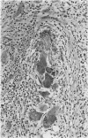
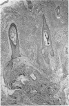
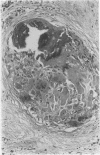
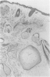
AIMS: To assess the incidence of foreign body giant cell reactions and ossification in benign/melanocytic naevi; and to examine their pathological features to gain an insight into their pathogenesis. METHODS: Intradermal (n = 185) and compound naevi (n = 110) from a routine histology service, together with 60 naevi submitted to an ophthalmic pathologist, were examined for foreign body reactions and ossification. Additional cases were identified prospectively in the course of routine reporting. The clinical and pathological features of positive cases were assessed. RESULTS: Foreign body reactions were identified in nine (4.9%) intradermal and four (3.6%) compound naevi, but in none of the naevi from around the eye. One intradermal naevus showed ossification. A further 11 naevi showing foreign body reaction and five showing ossification alone were identified prospectively. The 24 naevi showing a foreign body reaction had a similar age and sex distribution to controls but were more likely to occur on the head and neck. The reaction usually occurred deep to the naevus, sometimes in relation to a hair follicle, and fragments of hair or keratin were identified in most. Osteoid or bone was present within the reaction in five. In six other naevi, all from the head and neck of women, osteoid or mature bone was present deep to the naevus in the absence of a giant cell reaction. CONCLUSIONS: Foreign body giant cell reactions occur not uncommonly in relation to benign naevi, as a result of follicular damage, possibly due to trauma. The similar siting of foci of bone suggests that ossification occurs as a secondary phenomenon in these cases.
Full text
PDF


Images in this article
Selected References
These references are in PubMed. This may not be the complete list of references from this article.
- ANDERSON T. The search for primary causes. Glasgow Med J. 1955 Jan;36(1):1–14. [PMC free article] [PubMed] [Google Scholar]
- Burgdorf W., Nasemann T. Cutaneous osteomas: a clinical and histopathologic review. Arch Dermatol Res. 1977 Dec 12;260(2):121–135. doi: 10.1007/BF00561117. [DOI] [PubMed] [Google Scholar]
- DELACRETAZ J., FRENK E. ZUR PATHOGENESE DES OSTEO-NAEVUS NANTA. Hautarzt. 1964 Sep;15:487–489. [PubMed] [Google Scholar]
- HABER H. Some observations on common moles. Br J Dermatol. 1962 Jun;74:224–228. doi: 10.1111/j.1365-2133.1962.tb13496.x. [DOI] [PubMed] [Google Scholar]
- SAUNDERS T. S. Abscess formation in pigmented nevi; report of three cases. AMA Arch Derm. 1957 Aug;76(2):189–192. doi: 10.1001/archderm.1957.01550200033007. [DOI] [PubMed] [Google Scholar]
- Weedon D. Unusual features of nevocellular nevi. J Cutan Pathol. 1982 Oct;9(5):284–292. doi: 10.1111/j.1600-0560.1982.tb01065.x. [DOI] [PubMed] [Google Scholar]