Abstract
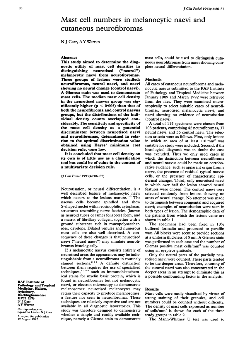
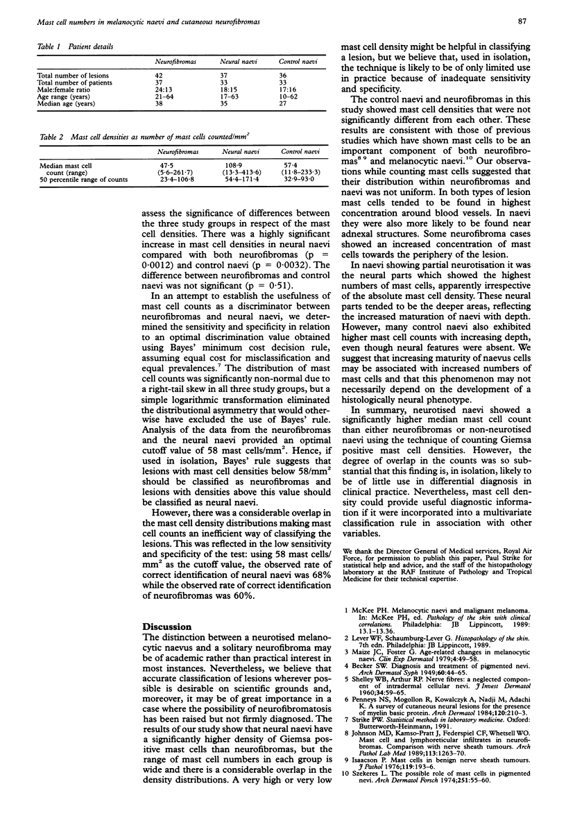
This study aimed to determine the diagnostic utility of mast cell densities in distinguishing neurotised ("neural") melanocytic naevi from neurofibromas. Three groups of lesions were studied: neurofibromas, neural naevi, and naevi showing no neural change (control naevi). A Giemsa stain was used to demonstrate mast cells. The median mast cell density in the neurotised naevus group was significantly higher (p < 0.005) than that of both the neurofibroma and control naevus groups, but the distributions of the individual density counts overlapped considerably. The sensitivity and specificity of the mast cell density as a potential discriminator between neurotised naevi and neurofibromas, determined in relation to the optimal discrimination value obtained using Bayes' minimum cost decision rule, were low. It is concluded that mast cell density on its own is of little use as a classification tool but could be of value in the context of a multivariate decision rule.
Full text
PDF

Selected References
These references are in PubMed. This may not be the complete list of references from this article.
- Isaacson P. Mast cells in benign nerve sheath tumours. J Pathol. 1976 Aug;119(4):193–196. doi: 10.1002/path.1711190402. [DOI] [PubMed] [Google Scholar]
- Johnson M. D., Kamso-Pratt J., Federspiel C. F., Whetsell W. O., Jr Mast cell and lymphoreticular infiltrates in neurofibromas. Comparison with nerve sheath tumors. Arch Pathol Lab Med. 1989 Nov;113(11):1263–1270. [PubMed] [Google Scholar]
- Maize J. C., Foster G. Age-related changes in melanocytic naevi. Clin Exp Dermatol. 1979 Mar;4(1):49–58. doi: 10.1111/j.1365-2230.1979.tb01590.x. [DOI] [PubMed] [Google Scholar]
- Penneys N. S., Mogollon R., Kowalczyk A., Nadji M., Adachi K. A survey of cutaneous neural lesions for the presence of myelin basic protein. An immunohistochemical study. Arch Dermatol. 1984 Feb;120(2):210–213. [PubMed] [Google Scholar]
- SHELLEY W. B., ARTHUR R. P. Nerve fibers, a neglected component of intradermal cellular nevi. J Invest Dermatol. 1960 Jan;34:59–65. [PubMed] [Google Scholar]
- Szekeres L. The possible role of mast cells in pigmented nevi. Arch Dermatol Forsch. 1974;251(1):55–60. doi: 10.1007/BF00561711. [DOI] [PubMed] [Google Scholar]


