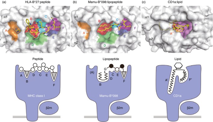
Figure 3.

Groove structures for accommodating peptide, lipopeptide, and lipid antigens. Top views of HLA‐B27 (a, PDB code 3B6S), Mamu‐B*098 (b, PDB code 4ZFZ), and CD1a (c, PDB code 1ONQ) molecules are shown in the upper panels. Six pockets (A–F) of HLA‐B27 and Mamu‐B*098 as well as two pockets (A′ and F′) of CD1a molecules are indicated, and bound ligands are shown in yellow sticks. Side views of each complex are illustrated in the corresponding lower panels.
