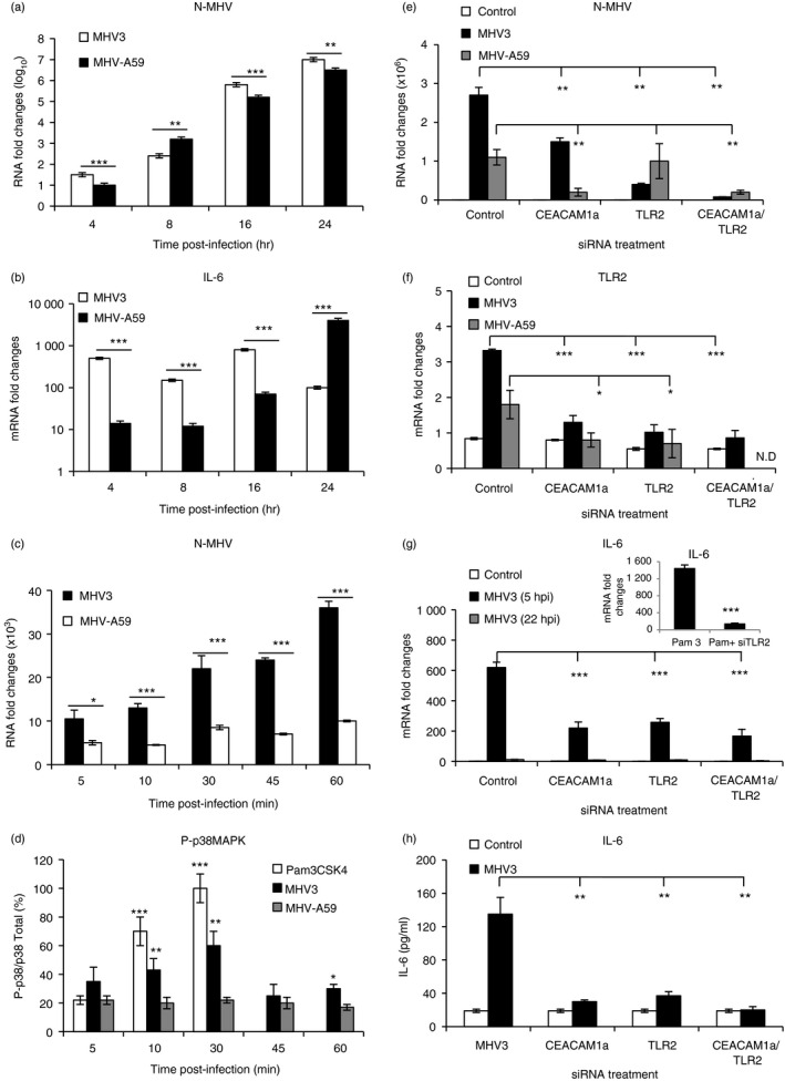
Figure 10.

In vitro murine hepatitis virus (MHV) 3 and MHV‐A59 infections of macrophages untreated or treated with small interfering (si) RNA for Toll‐like receptor 2 (TLR2) and CEACAM1a. J774A.1 macrophages were infected with 0·1–1 multiplicity of infection (m.o.i.) of MHV3 or MHV‐A59 and incubated for various times post‐infection (p.i.) according to experiments (a–d). Cells were also transfected with mouse CEACAM1 and/or TLR2 siRNA for at least 24 hr before infections in other experiments (e and f). Nucleocapsid RNA (a, c and e), TLR2 (f), and interleukin‐6 (IL‐6) (b and g) mRNA fold changes were analysed by quantitative RT‐PCR at various times p.i. Values represent fold change in gene expression relative to mock‐infected mice (arbitrary value of 1) after normalization with HPRT expression. Phosphorylated p38 mitogen‐activated protein kinase (MAPK) levels were evaluated by ELISA test and expressed as percentages of total p38 MAPK (d). The synthetic bacterial ligand for TLR2–TLR1 (Pam3CSK4) was used as TLR2 positive control for the detection of phosphorylated p38 MAPK and IL‐6 expression (d and g). IL‐6 secretion was quantified by ELISA test in MHV3‐infected cells at 5 hr p.i. (h). Results are representative of two different experiments. (*P < 0·05; **P < 0·01; ***P < 0·001). N.D. Not done.
