Abstract
The Pomacentridae (damselfish) and Apogonidae (cardinalfish) are among the most common fish families on coral reefs and in the aquarium trade. Members of both families undergo a pelagic larvae phase prior to settlement on the reef, where adults play key roles in benthic habitat structuring and trophic interactions. Fish-associated microbial communities (microbiomes) significantly influence fish health and ecology, yet little is known of how microbiomes change with life stage. We quantified the taxonomic (16S rRNA gene) composition of whole gut microbiomes from ten species of damselfish and two species of cardinalfish from Lizard Island, Australia, focusing specifically on comparisons between pelagic larvae prior to settlement on the reef versus post-settlement juvenile and adult individuals. On average, microbiome phylogenetic diversity increased from pre- to post-settlement, and was unrelated to the microbial composition in the surrounding water column. However, this trend varied among species, suggesting stochasticity in fish microbiome assembly. Pre-settlement fish were enriched with bacteria of the Endozoicomonaceae, Shewanellaceae, and Fusobacteriaceae, whereas settled fish harbored higher abundances of Vibrionaceae and Pasteurellaceae. Several individual operational taxonomic units, including ones related to Vibrio harveyi, Shewanella sp., and uncultured Endozoicomonas bacteria, were shared between both pre and post-settlement stages and may be of central importance in the intestinal niche across development. Richness of the core microbiome shared among pre-settlement fish was comparable to that of settled individuals, suggesting that changes in diversity with adulthood are due to the acquisition or loss of host-specific microbes. These results identify a key transition in microbiome structure across host life stage, suggesting changes in the functional contribution of microbiomes over development in two ecologically dominant reef fish families.
Keywords: Microbiome, Fish, Coral reef, Community structure, Vibrio, Endozoicomonas, Core diversity
Introduction
Animals harbor diverse microbial communities that influence key aspects of host health, development, and behavior. In most vertebrates, the majority of microorganisms inhabit the gastrointestinal (GI) tract at an abundance of potentially trillions of cells whose collective genome may be orders of magnitude larger than that of the host (Whitman, Coleman & Wiebe, 1998; Backhed et al., 2005; Gill et al., 2006). A wealth of studies in both fish and mammalian systems now confirm the importance of a gut microbiome to host health, fitness, and development (Rawls, Samuel & Gordon, 2004; Bates et al., 2006; Chung et al., 2012; Lee & Hase, 2014), with alterations of microbiome composition affecting such properties as host immunity, susceptibility to toxins, adiposity, efficiency of nutrient acquisition, behavior and mood, and chemical signaling among conspecifics (Turnbaugh et al., 2006; Round & Mazmanian , 2009; Heijtz et al., 2011; Greenblum, Turnbaugh & Borenstein, 2012; Theis et al., 2013b; Zheng et al., 2013; Wada-Katsumata et al., 2015).
Despite recognition of the important role of microbiomes in host ecology (Wong & Rawls, 2012), microbiomes remain unexplored for the vast majority of ecologically relevant taxa, including the most diverse of the vertebrate groups, the teleost fishes. Fish account for almost half of all vertebrate species on earth and span a wide spectrum of habitats, host ranges, physiologies, and ecological strategies. Studies of fish gut microbiomes have focused primarily on commercial or model species, with most targeting only a small number of host or microbial taxa (Roeselers et al., 2011; Clements et al., 2014; Llewellyn et al., 2014). These studies raise the possibility of a core set of fish gut microbes shared across diverse hosts, notably a dominance by bacteria of the Gammaproteobacteria and Firmicutes, including unique fish-associated strains of the Vibrionales and Clostridia (Spanggaard et al., 2000; Al-Harbi & Naim Uddin, 2004; Martin-Antonio et al., 2007; Sullam et al., 2012; Xing et al., 2013; Llewellyn et al., 2014) Significant variation in the fish gut microbiome has also been reported with changes in taxonomic composition shown to affect host immunity, nutrient acquisition, and epithelial differentiation (Rawls, Samuel & Gordon, 2004; Bates et al., 2006; Bates et al., 2007; Cheesman & Guillemin, 2007; Cheesman et al., 2010; Kanther & Rawls, 2010; Ghanbari, Kneifel & Domig, 2015). Fish microbiome composition has been linked to diverse factors including host type (Ye et al., 2014; Givens et al., 2015; Hennersdorf et al., 2016), trophic ecology and diet (Bolnick et al., 2014a; Bolnick et al., 2014b; Miyake, Ngugi & Stingl, 2015; Sullam et al., 2015), and environmental conditions (e.g., salinity; Sullam et al., 2012; Schmidt et al., 2015)
The composition of the fish microbiome can also change significantly over an individual’s lifespan (Bakke et al., 2015; Burns et al., 2015; Llewellyn et al., 2015). However, the timing, determinants, and magnitude of these shifts are unclear for most taxa, and likely highly variable among species with differing life histories. On coral reefs, for example, >90% of fish species undergo a dispersing or planktonic stage as either larvae or juveniles, followed by an adult phase marked by settlement into a constrained territory on the reef (Leis, 1991). For such species, microbiome structure is hypothesized to be shaped by a combination of factors that co-associate with host life stage, including diet shifts, immune system and GI tract development, and differences in the composition of the external microbial community between pre- and post-settlement habitats.
Comparing reef fish microbiomes over developmental stages will help identify shifts in the contribution of the microbiome to host health and behavior, thereby helping reach a holistic understanding of reef ecology. Collectively, coral reefs harbor over 2,500 fish species engaged in a complex web of interactions including predation, herbivory, corallivory, and symbiosis (Bellwood et al., 2004; Allen, 2014). These interactions together modulate material and energy transport and ecosystem structuring on coral reefs, with important consequences for reef-building corals that rely on fish waste for nutrients or fish herbivory to limit competition with benthic macroalgae (Mumby et al., 2006; Burkepile & Hay, 2008; McCauley et al., 2010). Such interactions may also be important vectors for moving microbes between reef habitats. A handful of studies have characterized gut microbiomes of coral reef fish, identifying a role for both diet type and host identity in shaping microbiome composition (Smriga, Sandin & Azam, 2010; Miyake, Ngugi & Stingl, 2015). However, the microbiomes of the vast majority of reef fishes, including some of the most abundant and ecologically relevant taxa, remain uncharacterized.
Damselfish (Pomacentridae) and cardinalfish (Apogonidae) are among the most diverse and abundant families of reef fishes and exhibit similar ecological strategies. Species within these families occupy critical positions in reef food webs as generalist planktivorous consumers, subsisting on a variety of algae, plankton, and benthic invertebrates (Emery, 1973; Emery & Thresher, 1980; Marnane & Bellwood, 2002). These fish also constitute major fractions of the diet of larger piscivorous reef fish (Beukers-Stewart & Jones, 2004). Most species of damselfish and cardinalfish spend their first 12–35 days of life as members of the off-reef plankton community before settling back onto the reef as juveniles (Wellington & Victor, 1989; Leis, 1991; Leis, Sweatman & Reader, 1996). Post-settlement, damselfishes (including members of the Chromis, Pomacentrus, and Dascyllus genera) occupy distinct microhabitats using various types of coral for habitat and cover (Wilson et al., 2008). Many Pomacentrids are highly territorial and influence turf algae composition through grazing (Hinds & Ballantine, 1987; Klumpp & Polunin, 1989). Apogonids also occupy coral microhabitats during the day when they are mostly inactive but leave coral cover at night to feed throughout the water column (Gardiner & Jones, 2005). Fish species of the Apogonidae often dominate nocturnal planktivore assemblages (Marnane & Bellwood, 2002). In this study we quantified microbiome taxonomic composition (16S rRNA gene diversity) in whole guts from replicate individuals from ten species of damselfish and two species of cardinalfish (Table S1). We focused on comparisons between pre-settlement planktonic larvae and post-settlement individuals (juveniles through to adults) to determine whether microbiome composition varied between ontogenetic stages and if so whether this variation was related to differences in the microbial composition of the surrounding water column.
Materials and Methods
Sample collection
Fishes were collected over a 3–4 day period from the fringing reefs surrounding Lizard Island, Australia (14°40.08′S 145°27.34′E) in February 2014. Water depths at collection sites were approximately 5–10 m. Larval pre-settlement individuals (8–16 mm total length, Table S1) were defined as samples collected from floating light traps deployed overnight at a single site. These traps sampled the upper meter of the water column above the reef. Post-settlement stage juveniles/adults (16–36 mm total length, Table S1) were individuals hand-collected on SCUBA at depth from nearby reefs using dip nets. Post-settlement fish in this study were likely sexually immature (juveniles) and recently settled given their small size (sexual maturity in most Pomacentrids typically occurs at lengths >60 mm, Schmale, Hensley & Udey, 1986; Kavanagh, 2000). Net capture of settled fish followed mild sedation with a clove oil/ethanol mixture dispensed via spray bottle. All fish were euthanized by immersion in a seawater-ice slurry combined with an overdose of clove oil and stored in ethanol for further analysis. Water column samples (n = 11) were collected on SCUBA at the same depth and location of post-settlement fish collection sites and the light trap site using sterile plastic 1L bottles. Bottles were first opened within the water column to ensure collection at the desired location and prevent contamination. Microbial biomass in the sampled water was concentrated onto 0.2 µm Polyvinylidene Flouride (PVDF) filters through filtration. Filters were immediately placed in ethanol in cryovials and stored at room temperature until processing. Fish were identified to the lowest possible taxonomic unit, typically either genus or species, using Indo-Pacific reef fish field guides based on morphology. In total, 49 pre-settlement individuals were sampled (Table S1), including individuals of the damselfish Pomacentrus moluccensis (n = 4), P. chrysurus (n = 4), P. nagasakiensis (n = 4), P. amboinensis (n = 4), P. wardii (n = 4), P. bankanensis (n = 4), P. coelestis (n = 4), Dascyllus aruanus (n = 3) and an unidentified Chromis species (n = 7). Two species of pre-settlement stage cardinalfish, Ostorhinchus doederleini (n = 4) and an unidentified Apogon species (n = 7), were also collected. A total of 24 post-settlement individuals were sampled (Table S1), including individuals of 5 species of damselfish (P. moluccensis (n = 6), P. chrysurus (n = 3), A. polyacanthus (n = 3), Dascyllus aruanus (n = 5) and an unidentified Chromis sp. (n = 3)) and 1 cardinalfish (O. doederleini (n = 4)). Note that the individuals of the unidentified Chromis species (collected both pre and post-settlement) were almost certainly either Chromis viridis or Chromis atripectoralis, both of which are similar ecologically and closely related. All research was reviewed and conducted under the guidelines of Georgia Tech IACUC #A14063 and Great Barrier Reef Marine Park Authority collection permits G13.36166.1 and animal ethics permit A1920. These methods were approved by Georgia Tech IACUC and Great Barrier Reef Marine Park Authority for use in this study.
DNA extractions
Whole fish samples were removed from storage containers and rinsed thoroughly with fresh, filter-sterilized ethanol to remove (potentially) surface attached microbes. As dissection of only the intestinal contents was not possible due to the small size of most pre-settlement individuals, whole gut contents (stomach + intestines) were excised by dissection with a sterile razor from each individual. Excised contents were placed in a sterile 1.5 ml centrifuge tube and frozen until extraction. Bulk DNA was extracted from gut contents using the Qiagen DNeasy blood and tissue kit according to manufacturer instructions. To process water samples, each collection filter was transferred from ethanol into a clean, 1.5 ml centrifuge tube. The ethanol remaining in the original storage tube was vacuumfiltered onto a new 0.2 µm PVDF filter, which was then dried at room temperature and pooled with the initial collection filter. DNA was extracted from filters using a phenol:chloroform method. Briefly, cells were lysed by adding lysozyme (2 mg in 40 μl of lysis buffer per sample) directly to the pooled filters, capping, and incubating for 45 min at 37°C. Proteinase K (1 mg in 100 μl lysis buffer, with 100 μl 20% SDS) was added and samples incubated for an additional 2 h at 55°C. The lysate was removed, and DNA was extracted once with phenol:chloroform:isoamyl alcohol (25:24:1) and once with chloroform:isoamyl alcohol (24:1). Extracted DNA was concentrated by spin dialysis using Ultra-4 (100 kDa, Amicon) centrifugal filters and stored frozen.
PCR amplification and sequencing
High-throughput sequencing of dual-indexed PCR amplicons spanning the V3-V4 hypervariable regions of the 16S rRNA gene was used to assess gut microbiome taxonomic composition. Amplicons were synthesized using Platinum® PCR SuperMix (Life Technologies) with primers F515 (5’-GTGCCAGCMGCCGCGGTAA-3’) and R806 (5’-GGACTACHVGGGTWTCTAAT-3’, Caporaso et al., 2011), with both primers modified to include sample-specific barcodes and Illumina sequencing adapters according to Kozich et al. (2013). Ten nanograms of starting DNA was used as template for each PCR reaction. Amplification was performed using denaturation at 94°C (3 min), followed by 30 cycles of denaturation at 94°C (45 sec), primer annealing at 55°C (45 sec), primer extension at 72°C (90 sec), and a final extension at 72°C for 10 min. Amplicon products were verified using gel electrophoresis, purified using Diffinity RapidTip2 PCR purification tips (Diffinity Genomics, NY), and quantitated fluorometrically using the Qubit (Life Technologies). Amplicons from different samples were pooled at equimolar concentrations and sequenced on an Illumina MiSeq using a 500 cycle kit with 30% PhiX added to increase sequence diversity. All raw sequences are available in the NCBI Sequence Read Archive under BioProject ID PRJNA290348.
Sequence analysis
Barcoded sequences were de-multiplexed and trimmed (length cutoff: 100 bp) and filtered to remove low quality reads (cutoff: Phred score of 25, averaged over all bases) using Trim Galore (http://www.bioinformatics.babraham.ac.uk/projects/trim_galore/). Paired-end reads were then merged using FLASH (Magoč & Salzberg, 2011), with an average read length threshold of 250 bp, and a fragment length threshold of 300 bp. Merged reads were analyzed using the QIIME pipeline (Caporaso et al., 2010). In QIIME, chimeric sequences were identified using USEARCH (Edgar, 2010) and removed from the input dataset. Merged non-chimeric sequences were clustered into Operational Taxonomic Units (OTUs) at 97% sequence similarity using open-reference picking with the UCLUST algorithm (Edgar, 2010). Singleton OTUs (those represented by only one sequence read in the combined dataset) were also removed at this step. Taxonomy was assigned to each OTU by comparison with the Greengenes database 13_5 release (DeSantis et al., 2006). Cyanobacterial and chloroplast sequences were manually removed from the OTU tables as these sequences can be assumed to reflect non-host-associated taxa, potentially those brought into the gut through food passage. Selected OTUs were queried via BLASTN against the NCBI-nr database to identify closest relatives.
OTU counts were rarefied (10 iterations) to 4,262 sequences per sample and the rarified OTU table was used in downstream analysis. A Bonferroni-corrected ANOVA was used to identify microbial families significantly enriched in pre- vs. post-settlement individuals with family composition data expressed as percentage of total reads. Alpha and beta diversity statistics for pre- and post-settlement individuals were calculated in QIIME using the core_diversity_analyes.py command. The unweighted Unifrac metric was used to conduct principal coordinates analysis to visualize beta diversity. Significant differences in beta diversity were identified using a two-sided t-test (Bonferroni-corrected). Core microbiome analysis was performed in QIIME using the compute_core_microbiome.py script to identify individual OTUs shared by at least 70% of all individuals per sample grouping (all pre-settlement individuals vs. all post-settlement individuals). This script was also used to estimate the average number of OTUs shared between random pairs of individuals from each life stage, based on 100 random iterations per life stage. Indicator analysis was performed with the indicspecies package in R to identify genera (indicator taxa) significantly enriched according to life stage, taking into consideration both the extent to which an OTU is exclusive to a life stage (A component), as well as the relative frequency at which the OTU occurs in all individuals within a stage (B component)(Dufrene & Legendre, 1997; Fortunato et al., 2013). Only those taxa occurring in more than half of all samples (B component > 0.5) and for which permutation testing yielded p-values <0.05 are included in the output. Indicator values range from 0 to 1 with higher values reflecting stronger relationships between indicator taxa and the tested sample groupings (pre- vs. post-settlement).
Results and Discussion
General microbiome characteristics
A total of 49 pre-settlement and 24 post-settlement individuals representing ten Pomacentridae species and two Apogonidae species were collected from Lizard Island, Australia. Of these, five species—the pomacentrids Pomacentrus moluccensis, P. chrysurus, Dascillus aruanus, and an unidentified Chromis sp. (either C. viridis or C. atripectoralis) and the apogonid Ostorhinchus doederleini—were represented by both pre- and post-settlement individuals, with the remaining species represented only in the pre-settlement sample set (n = 6 species) or only in the post-settlement set (n = 1).
A total of 3,503,605 16S rRNA gene sequences were obtained after quality control filtering (average: 41,709 per sample; range: 4262–238,858). After rarefaction, OTU counts per sample were highly variable, from 44 to 557 OTUs, with averages of 163 (±59 standard deviation (SD)) and 245 (±102 SD) for pre- and post-settlement stages, respectively. As observed in other fish species (Spanggaard et al., 2000; Al-Harbi & Naim Uddin, 2004; Martin-Antonio et al., 2007; Sullam et al., 2012; Xing et al., 2013), Pomacentrid and Apogonid gut microbiomes were dominated by Gammaproteobacteria of the Pseudoaltermonadaceae, Endozoicimonaceae, Vibrionaceae, and Shewanellaceae with these Families constituting 80% (±10.3 SD) and 67% (±6.7 SD) of all sequence reads in pre- and post-settlement fishes, respectively (Fig. S1), although key differences in the relative abundance of microbial families were also evident (discussed below, Table 1).
Table 1. Median % abundance of bacterial Families* in pre- versus post-settlement damselfish and cardinalfish (pooled).
| Pre | Post | ||||
|---|---|---|---|---|---|
| Family | Med | MAD | Med | MAD | Fold |
| Vibrionaceae | 18.12 | 14.20 | 41.91 | 28.69 | 2.31 |
| Endozoicimonaceae** | 11.02 | 9.50 | 0.14 | 0.14 | 76.40 |
| Pseudoalteromonadaceae | 1.28 | 1.15 | 0.45 | 0.42 | 2.83 |
| Rhodobacteraceae | 1.24 | 1.01 | 1.35 | 1.22 | 1.09 |
| Flavobacteriaceae | 1.07 | 0.89 | 0.61 | 0.60 | 1.75 |
| Alteromonadaceae | 0.71 | 0.63 | 0.85 | 0.75 | 1.20 |
| Shewanellaceae | 0.68 | 0.65 | 0.11 | 0.11 | 5.99 |
| Oceanospirillaceae | 0.56 | 0.51 | 0.15 | 0.15 | 3.74 |
| Pseudomonadaceae | 0.29 | 0.24 | 0.49 | 0.45 | 1.70 |
| Moraxellaceae | 0.26 | 0.23 | 0.02 | 0.02 | 10.31 |
| Pirellulaceae** | 0.01 | 0.01 | 0.83 | 0.81 | 81.97 |
| Chromatiales, Unknown | 0.10 | 0.08 | 0.21 | 0.20 | 2.03 |
Notes.
only includes Families with median abundance >0.2% in either pre or post datasets.
significant change pre vs. post (P < 0.05, ANOVA, Bonferroni corrected) MAD, median absolute deviation. Fold, fold increase in median (pre to post); underlined indicates post to pre.
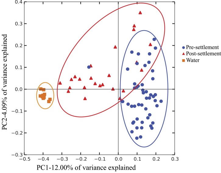
It is important to note that our data do not differentiate communities based on location in the GI tract, as the small size of individuals prohibited separation of intestine from stomach contents. Thus, we cannot distinguish between resident gut microbiome members, which might be relatively more common in the intestine, versus members derived from food contents or passively from seawater intake, which might be relatively enriched in the stomach. Both host-adapted as well as transient microbiome members may nonetheless exert effects on host health, as either symbionts or potentially as inocula for resident populations. However, gut microbiome composition (weighted Unifrac; Pomacentridae + Apogonidae combined) of both pre- and post-settlement stages differed significantly from that of the surrounding seawater (p = 0.028; Fig. 1), which was instead enriched in Gammaproteobacteria of the Halomonadaceae family, Alphaproteobacteria, and Bacteroidetes (Fig. S2). Moreover, microbiome composition did not differ significantly between water collection sites (Fig. S3), suggesting fine-scale variation in local environments alone should not influence microbiome composition and an overall minimal effect of source water on gut microbiome structure.
Figure 1. Compositional relatedness of microbiome samples.
Principal coordinate analysis based on unweighted Unifrac distance with sequence data rarified to 4,262 sequences per sample (49 pre-settlement, 24 post-settlement, 11 seawater samples). According to a two-sided t-test (Bonferroni-corrected), pre-settlement and post-settlement individuals are significantly different from one another (p < 0.05) and both are significantly different from the water column (p < 0.05).
Microbiome diversity and composition
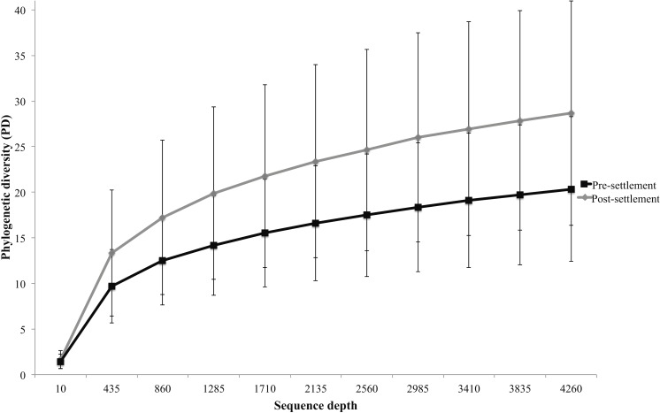
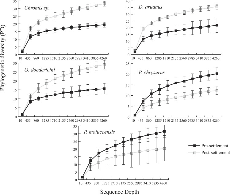
Microbiomes showed a general trend toward greater complexity following settlement, although this trend was variable among species. The phylogenetic composition of the combined (Pomacentrid + Apogonid) microbiomes changed significantly from pre- to post-settlement groups (weighted Unifrac metric; p = 0.028), with pre-settlement microbiomes showing less intra-individual variation compared to those of post-settlement fish (Fig. 1) and overall lower phylogenetic diversity (PD) (average: 20.3 ± 7 SD, versus 28.7 ± 12 SD post-settlement; p = 0.001, t-stat = 3.5; Fig. 2). Both pre- and post-settlement microbiomes exhibited substantially lower PD than the surrounding water column community (PD: 79.4 ± 23.5 SD; p = 0.001). Of the five species for which both pre- and post-settlement samples were available, three - Chromis sp., O. doederleini, and D. aruanus - exhibited significantly higher PD post-settlement (Fig. 3, p = 0.001). Greater microbiome diversity with age has been observed in other fish species, with suggested linkages to increases in diet complexity (Bolnick et al., 2014a; Bolnick et al., 2014b; Miyake, Ngugi & Stingl, 2015; Sullam et al., 2015) or potentially to differentiation of the GI tract into distinct niches with development. In contrast, elevated microbiome diversity in early life was observed in P. chrysurus and P. moluccensis (Fig. 3, p = 0.001), consistent with data from lab-reared zebrafish (Stephens et al., 2016). This pattern suggests that pre-settlement microbiomes in these species are influenced primarily by transient associations with bacteria from the exterior environment or by higher diet complexity at the planktonic stage. As an individual matures, its microbiome may become more specialized in response to diverse factors, including diet shifts, immune system development, physiological and chemical changes along the GI tract, or decreased connectivity with the external environment. Indeed, species-specific microhabitat usage (i.e., selection of distinct coral types by different species) has been observed in some of the hosts examined here, including P. moluccensis and P. nagasakiensis. However, all settled hosts in our dataset are assumed to be generalist planktivores with fairly similar post-settlement diets based on prior work (Emery, 1973; Emery & Thresher, 1980; Marnane & Bellwood, 2002). Although this study does not allow us to quantify the influence of each of the factors discussed above on developmental shifts in microbiome composition, our data, combined with results from prior studies, suggest that microbiome diversity is dynamic over life stages and assembly processes vary on a per-species basis.
Figure 2. Microbiome phylogenetic diversity (PD) as a function of sequence depth in pre- and post-settlement fish (all species combined, based on data rarified to 4262 sequences per sample).
Error bars are ± 1 standard error.
Figure 3. Microbiome phylogenetic diversity (PD) as a function of sequence depth in five species represented in both pre- and post-settlement datasets.
Error bars are ±1 standard error.
Key microbial taxa differed in abundance between pre- and post-settlement microbiomes (Table 1), although microbiome composition was relatively consistent within hosts per age group (Fig. S3). At the microbial family level, pre-settlement microbiomes were significantly enriched (>70-fold) in bacteria of the Endozoicimonaceae, while settled fish harbored over 2-fold higher median abundances of the Vibrionaceae and an 80-fold enrichment of planctomycetes of the Pirellulaceae (Table 1). Differences in the relative abundance of microbes at the family level did not appear to be driven by a single host. For example, all pre-settlement individuals were enriched in bacteria of the Endozoicomonaceae compared to post-settlement hosts (Fig. S4). At the microbial genus level, both life stages were marked by sets of indicator taxa (Table 2). Indicator taxa represent individual OTUs showing significant enrichment (as a percent of total sequence reads) in one life stage vs. the other according to permutation testing (p < 0.05, Dufrene & Legendre, 1997 Fortunato et al., 2013). The number of indicators was considerably higher for settled fish (38) compared to larvae (5; Table 2), consistent with the increase in diversity associated with post-settlement, perhaps reflective of an overall expansion in microbial niche breadth as discussed above. The marine bacterial genera Kordia (Flavobacteriia) and Halomonas (Gammaproteobacteria, Oceanospirillales) were the strongest indicators of pre-settlement stage (Table 2), followed by members of the Arcobacter (Epsilonproteobacteria, Campylobacterales), Oceanospirillum (Gammaproteobacteria, Oceanospirillales), and Idiomarina (Gammaproteobacteria, Alteromonadales), all taxa common to the marine environment but not often observed in fish microbiomes. Pre-settlement enrichment implicates these taxa as important during early development, more successful at colonizing and growing in young fishes, or commonly associated with pre-settlement food sources. Indicators of post-settlement spanned a wide phylogenetic breadth, with several classifiable only to the order or family level, and the strongest indicators including diverse members of the Gammaproteobacteria (Vibrionaceae, Portiera), Firmicutes (Epulopiscium), Alphaproteobacteria (Kiloniellales, Hyphomicrobiaceae), Verrucomicrobia (Coraliomargarita), and Planctomycetes (Pirellulaceae) (Table 2). Enrichment of these taxa in fully developed, settled fish suggests the host GI tract as a specific niche for members of these groups.
Table 2. Indicator genera associated with pre- and post-settlement damselfish and cardinalfish (pooled).
| Indicator genera | Indicator for pre or post settlement fish | Indicator value | p-value |
|---|---|---|---|
| Kordia | Pre | 0.943 | 0.001 |
| Halomonas | Pre | 0.896 | 0.001 |
| Arcobacter | Pre | 0.869 | 0.001 |
| Oceanospirillum | Pre | 0.823 | 0.001 |
| Idiomarina | Pre | 0.751 | 0.002 |
| unclassified Vibrionaceae | Post | 0.997 | 0.01 |
| unclassified Pirellulaceae | Post | 0.922 | 0.001 |
| unclassified Kiloniellales | Post | 0.904 | 0.001 |
| unclassified Gammaproteobacteria | Post | 0.9 | 0.001 |
| Coraliomargarita | Post | 0.863 | 0.001 |
| Portiera | Post | 0.857 | 0.001 |
| Epulopiscium | Post | 0.857 | 0.001 |
| unclassified Hyphomicrobiaceae | Post | 0.851 | 0.001 |
| Verrucomicrobium | Post | 0.816 | 0.001 |
| unclassified Rhizobiales | Post | 0.786 | 0.001 |
| Rhodospirillaceae | Post | 0.779 | 0.001 |
| unclassified Acidimicrobiales OCS155 | Post | 0.778 | 0.001 |
| unclassified Oceanospirillales | Post | 0.771 | 0.001 |
| unclassified Peptostreptococcaceae | Post | 0.771 | 0.001 |
| unclassified Myxococcales | Post | 0.768 | 0.001 |
| unclassified Altermonadales OM60 | Post | 0.763 | 0.013 |
| unclassified Phyllobacteriaceae | Post | 0.761 | 0.001 |
| Ferrimonas | Post | 0.75 | 0.001 |
| unclassified Thiohalorhabdales | Post | 0.746 | 0.001 |
| unclassified Flavobacteriales | Post | 0.743 | 0.001 |
| unclassified Cryomorphaceae | Post | 0.733 | 0.038 |
| unclassified Altermonadales | Post | 0.733 | 0.038 |
| unclassified Myxococcales OM27 | Post | 0.728 | 0.001 |
| Balneola | Post | 0.724 | 0.001 |
| Crocinitomix | Post | 0.723 | 0.001 |
| unclassified Flammeovirgaceae | Post | 0.722 | 0.002 |
| unclassified Altermonadaceae | Post | 0.722 | 0.035 |
| unclassified Francisellaceae | Post | 0.712 | 0.001 |
| Saprospria | Post | 0.692 | 0.003 |
| Clostridium | Post | 0.692 | 0.016 |
| unclassified Rickettsiales | Post | 0.688 | 0.001 |
| Turicibacter | Post | 0.682 | 0.001 |
| unclassified Deltaproteobacteria GMD14H09 | Post | 0.678 | 0.006 |
| unclassified Pasteurellaceae | Post | 0.676 | 0.002 |
| unclassified Actinomycetales | Post | 0.675 | 0.001 |
| Altermonadaceae HTCC2207 | Post | 0.67 | 0.001 |
| unclassified Comamonadaceae | Post | 0.664 | 0.004 |
| Flavobacterium | Post | 0.656 | 0.003 |
Comparison of the core microbiome between pre and post-settlement individuals
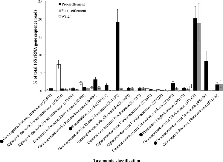
Although egg-tending behavior is common in many pomacentrids, fish are presumed sterile at birth and the fish gut microbiome is assumed to be inoculated primarily from the environment and via food items early in life (Nayak, 2010). If the pre-settlement, pelagic diet is similar across species, it is reasonable to predict that larval individuals of different fish species will share a greater number of microbial taxa (i.e., larger core microbiome). The core microbiome might then be expected to shrink post-settlement as species adapt to specific diets and microhabitats. We explored this prediction in two ways. First, we determined the average number of OTUs shared between randomly sampled pairs of pre-settlement individuals compared to post-settlement individuals. Contrary to our expectation, the average count of shared OTUs did not vary with life stage (22.8 vs 23.2 for pre- and post-settlement, respectively). Second, we compared the richness and composition of the core microbiome between life stages, following normalization to a uniform sample size (# of host individuals) per stage. Consistent with a recent analysis of microbiomes from 15 coastal fish species (Givens et al., 2015), no single microbial OTU was present in all individuals of either group (pre or post) in our study. Contrary to our prediction, the richness of the core microbiome was similar for both pre and post-settlement stages; for example, 17 OTUs were shared across 70% of pre-settlement individuals, compared to 15 for post-settlement (Fig. 4). Furthermore, many of the same OTUs were present in the core microbiomes identified at each stage. For example, of the OTUs occurring in 70% of total microbiome samples (pre- and post combined), 12 (of 16 total) were present in both the pre- and post-settlement core sets (70% threshold) when evaluated separately (Fig. 4), indicating conservation of a subset of key microbial members across settlement. Together, these results suggest processes affecting assembly and diversity of the core microbiome, when defined at the OTU level, are relatively constant from pre- to post-settlement.
Figure 4. Taxonomic identity and relative abundance of OTUs detected in greater than 70% of all fish microbiome samples (pre-settlement (n = 49) and post-settlement (n = 24) samples) combined for analysis.
• indicates that an OTU was shared among 70% of the pre-settlement samples when this sample set was evaluated independently, but was not detected in >70% of samples in the post-settlement set when evaluated independently. The relative abundances of these OTUs in the water column samples (n = 11) are included for comparison. Taxonomic classifications are at the level of genus whenever possible. Error bars are ±1 standard error.
Many of these core OTUs were absent from or at much lower abundances in seawater samples (with the exception of 3 OTUs), indicating selective accumulation in the fish gut (Fig. 4). Notably a single OTU with high sequence similarity to Vibrio harveyi (Vibrionaceae, Genbank accession KX380754.1) comprised an average of 18%–20% of the total sequence reads in both pre- and post-settlement fish, but only 2% of the seawater community (Fig. 4). Vibrios are often described as opportunistic pathogens (Karunasagar et al., 1994; Austin & Zhang, 2006). For example, metagenome sequences related to Vibrio species, including V. harveyi, from the guts of farmed adult turbot were enriched in genes encoding potential virulence functions (Xing et al., 2013). However, the wide distribution of V. harveyi in healthy marine hosts, observed here and in other studies of diverse invertebrates and fish (Onarheim et al., 1994; Guerrero-Ferreira et al., 2013), suggests that the primary niche of this bacterium may be commensal or even mutualistic with the host, potentially with a role in protein degradation and digestion (Ray, Ghosh & Ringø, 2012). Interestingly, the Vibrionaceae family as a whole (multiple OTUs) was over twice as abundant in post-settlement fish (Table 1) and therefore may be more typical of mature gut microbiome communities.
The proportional abundance of other core microbiome taxa varied significantly between life stages. The 16 taxa detected in 70% of all microbiome samples, a core set dominated by OTUs belonging to the Gammaproteobacteria (Fig. 4), constituted ∼60% of the total gut microbiome sequence reads in pre-settlement fish but only ∼25% in settled fish, indicating that these OTUs may be important early in life. Notably, OTUs closely related to Shewanella putrafaciens (Genbank accession KP967510.1) and to an uncultured bacterium of the family Endozoicomonaceae (Genbank accession LN626318.1) constituted 8% and 18% of the pre-settlement microbiome, respectively, but less than 1% of the post-settlement community (Fig. 4). To our knowledge, this is the first report of Endozoicomonaceae occupying a large proportion of the fish gut microbiome. Bacteria of this family, within the widely distributed Oceanospiralles group, exhibit diverse heterotrophic metabolisms (Neave et al., 2014) and are commonly identified as symbionts in marine invertebrates (Bayer et al., 2013a; Bayer et al., 2013b; Forget & Kim Juniper, 2013; Nishijima et al., 2013; Beinart et al., 2014; Hyun et al., 2014), suggesting invertebrate prey as a potential vector of transport into the fish microbiome. The functional properties and potential for long-term residency of these bacteria in reef fishes remain to be verified. In contrast, Gammaproteobacteria of the genus Shewanella, have been detected widely in fish. Indeed, Shewanella putrefaciens, the species most closely related (97%) to the core OTU in this study, has been used as a probiotic to increase growth and pathogen resistance in juvenile sole (Lobo et al., 2014).
Other members of the core OTU set spanned diverse bacterial divisions, but occurred at lower abundances (Fig. 4). These included members of genera containing known fish pathogens, such as Photobacterium (Chabrillón et al., 2005), Pseudoalteromonas (Pujalte Domarco et al., 2004), Pseudomonas (Wakabayashi et al., 1996), and Halomonas (Austin, 2005). However, these groups also contain potentially beneficial members, as certain Pseudoalteromonas and Alteromonas strains can inhibit bacterial pathogens in fish (Gatesoupe, 1999) and Photobacterium species are known bioluminescent symbionts in diverse fish hosts (Ruby & Nealson, 1976; Ast et al., 2007). Core OTUs also included a Chromatiales-affiliated taxon with 100% similarity to the uncultured gill symbiont of Ifremeria nautilei (Beinart et al., 2015; Genbank accession KF780855.1), a member of the Kordia genus resembling a heterotrophic isolate from the gut of marine polychaetes (Choi et al., 2011, Genbank accession NR_117471.1), and Salinivibrio costicola, a halotolerant, facultatively anaerobic bacterium first isolated from a hypersaline pond (Huang et al., 2000). Three OTUS matching marine Rhodobacteraceae, a broadly distributed aquatic group that has also been detected in reef surgeonfish (Miyake, Ngugi & Stingl, 2015), were also prevalent across samples. However, two of these OTUs were more abundant in water samples, suggesting these taxa may be transient members of the gut community. Together, these data identify a relatively species-poor (e.g., 16 OTUs at 70%) but abundant (35%–65% of total 16S sequences) core microbiome across the damselfish and cardinalfish sample set, including potentially pathogenic or beneficial bacteria, and a subset of core OTUs (12 of 16) that persist across pre- and post-settlement life stages. Persistence over development may be an important indicator of commensal or mutualistic taxa ubiquitous to reef fish.
Conclusion
These results describe a diverse gut microbiome in two abundant reef fish families and highlight the importance of life stage in structuring microbiome composition. A trend toward greater microbiome diversity in settled (older) individuals was observed in the pooled dataset, potentially explained by the hypothesis that pre-settlement fish of diverse species are more similar to each other physiologically compared to adults, occupy the same niche (pelagic zone), and acquire gut microbes from a common environmental pool, perhaps due to similarities in diet. These factors could homogenize the pre-settlement microbiome across diverse host species, with microbiomes diversifying after fish settle on the reef and transition to adult feeding roles. However, this pattern is not uniform across host species, suggesting a need for discretion in concluding general trends in microbiome succession over development. Our data also identify core microbes common to pre- and post-settlement fish of several host species. Persistence of these microbes over major life transitions, regardless of variable trends in bulk microbiome complexity, may implicate these microbes as particularly important to host health and physiology. Differences in microbiome assembly between pre and post-settlement fish are likely driven by a combination of factors, including physiological changes associated with development, changes in diet and potentially feeding frequency, and varying connectivity with microbes from the external environment. More research is needed to disentangle the relative contributions of these determinants over the settlement transition.
Supplemental Information
Principal coordinate analysis based on unweighted Unifrac distance with sequence data rarified to 4262 sequences per sample (49 pre-settlement, 24 post-settlement, 11 seawater samples). Samples color-coded according to host.
Outlined host group represents pre-settlement individuals.
Acknowledgments
We thank the staff of Lizard Island Research Station for logistical support.
Funding Statement
This work was enabled by support from the Simons Foundation (Grant 346253 to FJS). The funders had no role in study design, data collection and analysis, decision to publish, or preparation of the manuscript.
Additional Information and Declarations
Competing Interests
The authors declare there are no competing interests.
Author Contributions
Darren J. Parris conceived and designed the experiments, performed the experiments, analyzed the data, wrote the paper, prepared figures and/or tables, reviewed drafts of the paper.
Rohan M. Brooker conceived and designed the experiments, performed the experiments, reviewed drafts of the paper.
Michael A. Morgan performed the experiments.
Danielle L. Dixson conceived and designed the experiments, performed the experiments, contributed reagents/materials/analysis tools, reviewed drafts of the paper.
Frank J. Stewart conceived and designed the experiments, analyzed the data, contributed reagents/materials/analysis tools, wrote the paper, prepared figures and/or tables, reviewed drafts of the paper.
Animal Ethics
The following information was supplied relating to ethical approvals (i.e., approving body and any reference numbers):
All research was reviewed and conducted under the guidelines of Georgia Tech IACUC #A14063 and Great Barrier Reef Marine Park Authority collection permits G13.36166.1 and animal ethics permits A1920. These methods were approved by Georgia Tech IACUC and Great Barrier Reef Marine Park Authority for use in this study.
Data Availability
The following information was supplied regarding data availability:
All raw data (sequences) are available in the NCBI Sequence Read Archive (GenBank) under BioProject ID PRJNA290348.
References
- Al-Harbi & Naim Uddin (2004).Al-Harbi AH, Naim Uddin M. Seasonal variation in the intestinal bacterial flora of hybrid tilapia (Oreochromis niloticus×Oreochromis aureus) cultured in earthen ponds in Saudi Arabia. Aquaculture. 2004;229:37–44. doi: 10.1016/S0044-8486(03)00388-0. [DOI] [Google Scholar]
- Allen (2014).Allen GR. Review of Indo-Pacific coral reef fish systematics: 1980 to 2014. Ichthyological Research. 2014;62(1):2–8. [Google Scholar]
- Ast et al. (2007).Ast JC, Cleenwerck I, Engelbeen K, Urbanczyk H, Thompson FL, De Vos P, Dunlap PV. Photobacterium kishitanii sp. nov., a luminous marine bacterium symbiotic with deep-sea fishes. International Journal of Systematic and Evolutionary Microbiology. 2007;57(9):2073–2078. doi: 10.1099/ijs.0.65153-0. [DOI] [PubMed] [Google Scholar]
- Austin (2005).Austin B. Oceans and health: pathogens in the marine environment. Springer; New York: 2005. Bacterial pathogens of marine fish; pp. 381–413. [Google Scholar]
- Austin & Zhang (2006).Austin B, Zhang XH. Vibrio harveyi: a significant pathogen of marine vertebrates and invertebrates. Letters in Applied Microbiology. 2006;43(2):119–124. doi: 10.1111/j.1472-765X.2006.01989.x. [DOI] [PubMed] [Google Scholar]
- Backhed et al. (2005).Backhed F, Ley R, Sonnenburg J, Peterson D, Gordon J. Host-Bacterial mutualism in the human intestine. Science. 2005;307:1915–1920. doi: 10.1126/science.1104816. [DOI] [PubMed] [Google Scholar]
- Bakke et al. (2015).Bakke I, Coward E, Andersen T, Vadstein O. Selection in the host structures the microbiota associated with developing cod larvae (Gadus morhua) Environmental Microbiology. 2015;17(10):3914–3924. doi: 10.1111/1462-2920.12888. [DOI] [PubMed] [Google Scholar]
- Bates et al. (2007).Bates JM, Akerlund J, Mittge E, Guillemin K. Intestinal alkaline phosphatase detoxifies lipopolysaccharide and prevents inflammation in zebrafish in response to the gut microbiota. Cell Host Microbe. 2007;2:371–382. doi: 10.1016/j.chom.2007.10.010. [DOI] [PMC free article] [PubMed] [Google Scholar]
- Bates et al. (2006).Bates JM, Mittge E, Kuhlman J, Baden KN, Cheesman SE, Guillemin K. Distinct signals from the microbiota promote different aspects of zebrafish gut differentiation. Developmental Biology. 2006;297:374–386. doi: 10.1016/j.ydbio.2006.05.006. [DOI] [PubMed] [Google Scholar]
- Bayer et al. (2013b).Bayer T, Arif C, Ferrier-Pagès C, Zoccola D, Aranda M, Voolstra CR. Bacteria of the genus Endozoicomonas dominate the microbiome of the Mediterranean gorgonian coral Eunicella cavolini. Marine Ecology Progress Series. 2013b;479:75–84. [Google Scholar]
- Bayer et al. (2013a).Bayer T, Neave MJ, Alsheikh-Hussain A, Aranda M, Yum LK, Mincer T, Hughen K, Apprill A, Voolstra CR. The microbiome of the Red Sea coral Stylophora pistillata is dominated by tissue-associated Endozoicomonas bacteria. Applied and Environmental Microbiology. 2013a;79(15):4759–4762. doi: 10.1128/AEM.00695-13. [DOI] [PMC free article] [PubMed] [Google Scholar]
- Beinart et al. (2015).Beinart RA, Gartman A, Sanders JG, Luther GW, Girguis PR. The uptake and excretion of partially oxidized sulfur expands the repertoire of energy resources metabolized by hydrothermal vent symbioses. Proceedings of the Royal Society B. 2015;282(1806):20142811. doi: 10.1098/rspb.2014.2811. [DOI] [PMC free article] [PubMed] [Google Scholar]
- Beinart et al. (2014).Beinart RA, Nyholm SV, Dubilier N, Girguis PR. Intracellular Oceanospirillales inhabit the gills of the hydrothermal vent snail Alviniconcha with chemosynthetic, γ-Proteobacterial symbionts. Environmental Microbiology Reports. 2014;6(6):656–664. doi: 10.1111/1758-2229.12183. [DOI] [PubMed] [Google Scholar]
- Bellwood et al. (2004).Bellwood DR, Hughes TP, Folke C, Nyström M. Confronting the coral reef crisis. Nature. 2004;429:827– 833. doi: 10.1038/nature02691. [DOI] [PubMed] [Google Scholar]
- Beukers-Stewart & Jones (2004).Beukers-Stewart BD, Jones GP. The influence of prey abundance on the feeding ecology of two piscivorous species of coral reef fish. Journal of Experimental Marine Biology and Ecology. 2004;299(2):155–184. doi: 10.1016/j.jembe.2003.08.015. [DOI] [Google Scholar]
- Bolnick et al. (2014a).Bolnick DI, Snowberg LK, Hirsch PE, Lauber CL, Knight R, Caporaso JG, Svanbäck R. Individuals’ diet diversity influences gut microbial diversity in two freshwater fish (threespine stickleback and Eurasian perch) Ecology Letters. 2014a;17(8):979–987. doi: 10.1111/ele.12301. [DOI] [PMC free article] [PubMed] [Google Scholar]
- Bolnick et al. (2014b).Bolnick DI, Snowberg LK, Hirsch PE, Lauber CL, Org E, Parks B, Lusis AJ, Knight R, Gregory Caporaso J, Svanbäck R. Individual diet has sex-dependent effects on vertebrate gut microbiota. Nature Communications. 2014b;5:4500. doi: 10.1038/ncomms5500. [DOI] [PMC free article] [PubMed] [Google Scholar]
- Burkepile & Hay (2008).Burkepile DE, Hay ME. Herbivore species richness and feeding complementarity affect community structure and function on a coral reef. Proceedings of the National Academy of Sciences of the United States of America. 2008;105:16201–16206. doi: 10.1073/pnas.0801946105. [DOI] [PMC free article] [PubMed] [Google Scholar]
- Burns et al. (2015).Burns AR, Zac Stephens W, Stagaman K, Wong S, Rawls JF, Guillemin K, Bohannan BJ. Contribution of neutral processes to the assembly of gut microbial communities in the zebrafish over host development. The ISME Journal. 2015;10:655–664. doi: 10.1038/ismej.2015.142. [DOI] [PMC free article] [PubMed] [Google Scholar]
- Caporaso et al. (2010).Caporaso JG, Kuczynski J, Stombaugh J, Bittinger K, Bushman FD, Costello EK, Fierer N, Peña AG, Goodrich JK, Gordon JI, Huttley GA, Kelley ST, Knights D, Koenig JE, Ley RE, Lozupone CA, McDonald D, Muegge BD, Pirrung M, Reeder J, Sevinsky JR, Turnbaugh PJ, Walters WA, Widmann J, Yatsunenko T, Zaneveld J, Knight R. QIIME allows analysis of high-throughput community sequencing data. Nature Methods. 2010;7(5):335–336. doi: 10.1038/nmeth.f.303. [DOI] [PMC free article] [PubMed] [Google Scholar]
- Caporaso et al. (2011).Caporaso JG, Lauber CL, Walters WA, Berg-Lyons D, Lozupone CA, Turnbaugh PJ, Fierer N, Knight R. Global patterns of 16S rRNA diversity at a depth of millions of sequences per sample. Proceedings of the National Academy of Sciences of the United States of America. 2011;108(Supplement 1):4516–4522. doi: 10.1073/pnas.1000080107. [DOI] [PMC free article] [PubMed] [Google Scholar]
- Chabrillón et al. (2005).Chabrillón M, Rico RM, Balebona MC, Moriñigo MA. Adhesion to sole, Solea senegalensis Kaup, mucus of microorganisms isolated from farmed fish, and their interaction with Photobacterium damselae subsp Piscicida. Journal of Fish Diseases. 2005;28(4):229–237. doi: 10.1111/j.1365-2761.2005.00623.x. [DOI] [PubMed] [Google Scholar]
- Cheesman & Guillemin (2007).Cheesman S, Guillemin K. We know you are in there: conversing with the indigenous gut microbiota. Research in Microbiology. 2007;158:2–9. doi: 10.1016/j.resmic.2006.10.005. [DOI] [PubMed] [Google Scholar]
- Cheesman et al. (2010).Cheesman SE, Neal JT, Mittge E, Seredick BM, Guillemin Ka. Microbes and Health Sackler Coloquiium: epethelial cell proliferation in the developing zebrafish intestine is regulated by the Wnt pathway and microbial signaling via Myd88. Proceedings of the National Academy of Sciences of the United States of America. 2010;108:4570–4577. doi: 10.1073/pnas.1000072107. [DOI] [PMC free article] [PubMed] [Google Scholar]
- Choi et al. (2011).Choi A, Oh HM, Yang SJ, Cho JC. Kordia periserrulae sp. nov., isolated from a marine polychaete Periserrula leucophryna, and emended description of the genus Kordia. International Journal of Systematic and Evolutionary Microbiology. 2011;61(4):864–869. doi: 10.1099/ijs.0.022764-0. [DOI] [PubMed] [Google Scholar]
- Chung et al. (2012).Chung H, Pamp SJ, Hill JA, Surana NK, Edelman SM, Troy EB, Reading NC, Villablanca EJ, Wang S, Mora JR, Umesaki Y, Mathis D, Benoist C, Relman DA, Kasper DL. Gut immune maturation depends on colonization with a host-specific microbiota. Cell. 2012;149(7):1578–1593. doi: 10.1016/j.cell.2012.04.037. [DOI] [PMC free article] [PubMed] [Google Scholar]
- Clements et al. (2014).Clements KD, Angert ER, Montgomery WL, Choat JH. Intestinal microbiota in fishes: what’s known and what’s not. Molecular Ecology. 2014;23:1891–1898. doi: 10.1111/mec.12699. [DOI] [PubMed] [Google Scholar]
- DeSantis et al. (2006).DeSantis TZ, Hugenholtz P, Larsen N, Rojas M, Brodie EL, Keller K, Huber T, Dalevi D, Hu P, Andersen GL. Greengenes, a chimera-checked 16S rRNA gene database and workbench compatible with ARB. Applied and Environmental Microbiology. 2006;72(7):5069–5072. doi: 10.1128/AEM.03006-05. [DOI] [PMC free article] [PubMed] [Google Scholar]
- Dufrene & Legendre (1997).Dufrene M, Legendre P. Species assemblages and indicator species: the need for a flexible assymetrical approach. Ecological Monographs. 1997;67:345–366. [Google Scholar]
- Edgar (2010).Edgar RC. Search and clustering orders of magnitude faster than BLAST. Bioinformatics. 2010;26:2460–2461. doi: 10.1093/bioinformatics/btq461. [DOI] [PubMed] [Google Scholar]
- Emery (1973).Emery AR. Comparative ecology and functional osteology of fourteen species of damselfish (Pisces: pomacentridae) at Alligator Reef, Florida Keys. Bulletin of Marine Science. 1973;23(3):649–770. [Google Scholar]
- Emery, Thresher(1980).Emery AR, Thresher RE. Biology of the damselfishes. Bulletin of Marine Science. 1980;30:145–328. [Google Scholar]
- Forget & Kim Juniper (2013).Forget NL, Kim Juniper S. Free-living bacterial communities associated with tubeworm (Ridgeia piscesae) aggregations in contrasting diffuse flow hydrothermal vent habitats at the Main Endeavour Field, Juan de Fuca Ridge. MicrobiologyOpen. 2013;2:259–275. doi: 10.1002/mbo3.70. [DOI] [PMC free article] [PubMed] [Google Scholar]
- Fortunato et al. (2013).Fortunato CS, Eiler A, Herfort L, Needoba JA, Peterson TD, Crump BC. Determining indicator taxa across spatial and seasonal gradients in the Columbia River coastal margin. The ISME Journal. 2013;7(10):1899–1911. doi: 10.1038/ismej.2013.79. [DOI] [PMC free article] [PubMed] [Google Scholar]
- Gardiner & Jones (2005).Gardiner NM, Jones GP. Habitat specialisation and overlap in a guild of coral reef cardinalfishes (Apogonidae) Marine Ecology Progress Series. 2005;305:163–175. doi: 10.3354/meps305163. [DOI] [Google Scholar]
- Gatesoupe (1999).Gatesoupe FJ. The use of probiotics in aquaculture. Aquaculture. 1999;180(1):147–165. doi: 10.1016/S0044-8486(99)00187-8. [DOI] [Google Scholar]
- Ghanbari, Kneifel & Domig (2015).Ghanbari M, Kneifel W, Domig KJ. A new view of the fish gut microbiome: advances from next-generation sequencing. Aquaculture. 2015;448:464–475. doi: 10.1016/j.aquaculture.2015.06.033. [DOI] [Google Scholar]
- Gill et al. (2006).Gill SR, Pop M, Deboy RT, Eckburg PB, Turnbaugh PJ, Samuel BS, Gordon JI, Relman DA, Fraser-Liggett CM, Nelson KE. Metagenomic analysis of the human distal gut microbiome. Science. 2006;312:1355–1359. doi: 10.1126/science.1124234. [DOI] [PMC free article] [PubMed] [Google Scholar]
- Givens et al. (2015).Givens CE, Ransom B, Bano N, Hollibaugh JT. Comparison of the gut microbiomes of 12 bony fish and 3 shark species. Marine Ecology Progress Series. 2015;518:209–223. doi: 10.3354/meps11034. [DOI] [Google Scholar]
- Greenblum, Turnbaugh & Borenstein (2012).Greenblum S, Turnbaugh P, Borenstein E. Metagenomic systems biology of the human gut microbiome reveals topological shifts associated with obesity and inflammatory bowel disease. Proceedings of the National Academy of Sciences of the United States of America. 2012;109:594–599. doi: 10.1073/pnas.1116053109. [DOI] [PMC free article] [PubMed] [Google Scholar]
- Guerrero-Ferreira et al. (2013).Guerrero-Ferreira R, Gorman C, Chavez AA, Willie S, Nishiguchi MK. Characterization of the bacterial diversity in Indo-West Pacific loliginid and sepiolid squid light organs. Microbial Ecology. 2013;65(1):214–226. doi: 10.1007/s00248-012-0099-6. [DOI] [PMC free article] [PubMed] [Google Scholar]
- Heijtz et al. (2011).Heijtz R, Wang S, Anuar F, Qian Y, Björkholm B, Samuelsson A, Hibberd M, Forssberg H, Pettersson S. Normal gut microbiota modulates brain development and behavior. Proceedings of the National Academy of Sciences of the United States of America. 2011;108:3047–3052. doi: 10.1073/pnas.1010529108. [DOI] [PMC free article] [PubMed] [Google Scholar]
- Hennersdorf et al. (2016).Hennersdorf P, Kleinertz S, Theisen S, Abdul-Aziz MA, Mrotzek G, Palm HW, Saluz HP. Microbial diversity and parasitic load in tropical fish of different environmental conditions. PLoS ONE. 2016;11(3):e0151594. doi: 10.1371/journal.pone.0151594. [DOI] [PMC free article] [PubMed] [Google Scholar]
- Hinds & Ballantine (1987).Hinds PA, Ballantine DL. Effects of the Caribbean threespot damselfish, Stegastes planifrons (Cuvier), on algal lawn composition. Aquatic Botany. 1987;27(4):299–308. [Google Scholar]
- Huang et al. (2000).Huang CY, Garcia JL, Patel BK, Cayol JL, Baresi L, Mah RA. Salinivibrio costicola subsp. vallismortis subsp. nov., a halotolerant facultative anaerobe from Death Valley, and emended description of Salinivibrio costicola. International Journal of Systematic and Evolutionary Microbiology. 2000;50(2):615–622. doi: 10.1099/00207713-50-2-615. [DOI] [PubMed] [Google Scholar]
- Hyun et al. (2014).Hyun DW, Shin NR, Kim MS, Oh SJ, Kim PS, Whon TW, Bae JW. Endozoicomonas atrinae sp. nov., a bacterium isolated from the intestine of a comb pen shell Atrina pectinata. International Journal of Systematic and Evolutionary Microbiology. 2014;64:2312–2318. doi: 10.1099/ijs.0.060780-0. [DOI] [PubMed] [Google Scholar]
- Kanther & Rawls (2010).Kanther M, Rawls JF. Host-microbe interactions in the developing zebrafish. Current Opinion in Immunology. 2010;22:10–19. doi: 10.1016/j.coi.2010.01.006. [DOI] [PMC free article] [PubMed] [Google Scholar]
- Karunasagar et al. (1994).Karunasagar I, Pai R, Malathi GR, Karunasagar I. Mass mortality of Penaeusmonodon larvae due to antibiotic-resistant Vibrio harveyi infection. Aquaculture. 1994;128(3):203–209. doi: 10.1016/0044-8486(94)90309-3. [DOI] [Google Scholar]
- Kavanagh (2000).Kavanagh KD. Larval brooding in the marine damselfish Acanthochromis polyacanthus (Pomacentridae) is correlated with highly divergent morphology, ontogeny and life-history traits. Bulletin of Marine Science. 2000;66(2):321–337. [Google Scholar]
- Klumpp & Polunin (1989).Klumpp DW, Polunin NVC. Partitioning among grazers of food resources within damselfish territories on a coral reef. Journal of Experimental Marine Biology and Ecology. 1989;125(2):145–169. doi: 10.1016/0022-0981(89)90040-3. [DOI] [Google Scholar]
- Kozich et al. (2013).Kozich JJ, Westcott SL, Baxter NT, Highlander SK, Schloss PD. Development of a dual-index sequencing strategy and curation pipeline for analyzing amplicon sequence data on the MiSeq Illumina sequencing platform. Applied and Environmental Microbiology. 2013;79:5112– 5120. doi: 10.1128/AEM.01043-13. [DOI] [PMC free article] [PubMed] [Google Scholar]
- Lee & Hase (2014).Lee WJ, Hase K. Gut microbiota-generated metabolites in animal health and disease. Nature Chemical Biology. 2014;10(6):416–424. doi: 10.1038/nchembio.1535. [DOI] [PubMed] [Google Scholar]
- Leis, Sweatman & Reader (1996).Leis JM, Sweatman HP, Reader SE. What the pelagic stages of coral reef fishes are doing out in blue water: daytime field observations of larval behavioural capabilities. Marine and Freshwater Research. 1996;47(2):401–411. doi: 10.1071/MF9960401. [DOI] [Google Scholar]
- Llewellyn et al. (2014).Llewellyn MS, Boutin S, Hoseinifar SH, Derome N. Teleost microbiomes: the state of the art in their characterization, manipulation and importance in aquaculture and fisheries. Frontiers in Microbiology. 2014;5:207. doi: 10.3389/fmicb.2014.00207. [DOI] [PMC free article] [PubMed] [Google Scholar]
- Llewellyn et al. (2015).Llewellyn MS, McGinnity P, Dionne M, Letourneau J, Thonier F, Carvalho GR, Creer S, Derome N. The biogeography of the atlantic salmon (Salmo salar) gut microbiome. The ISME Journal. 2015;10(5):1280–1284. doi: 10.1038/ismej.2015.189. [DOI] [PMC free article] [PubMed] [Google Scholar]
- Lobo et al. (2014).Lobo C, Moreno-Ventas X, Tapia-Paniagua S, Rodríguez C, Moriñigo MA, De La Banda IG. Dietary probiotic supplementation (Shewanella putrefaciens Pdp11) modulates gut microbiota and promotes growth and condition in Senegalese sole larviculture. Fish Physiology and Biochemistry. 2014;40(1):295–309. doi: 10.1007/s10695-013-9844-0. [DOI] [PubMed] [Google Scholar]
- Leis (1991).Leis JM. The pelagic stage of reef fishes: the larval biology of coral reef fishes. In: Sale PF, editor. The ecology of fishes on coral reefs. Academic Press; San Diego: 1991. pp. 183–230. [Google Scholar]
- Magoč & Salzberg (2011).Magoč T, Salzberg SL. FLASH: fast length adjustment of short reads to improve genome assemblies. Bioinformatics. 2011;27:2957–2963. doi: 10.1093/bioinformatics/btr507. [DOI] [PMC free article] [PubMed] [Google Scholar]
- Marnane & Bellwood (2002).Marnane MJ, Bellwood DR. Diet and nocturnal foraging in cardinalfishes (Apogonidae) at one tree reef, great barrier reef, Australia. Marine Ecology progress Series. 2002;231:261–268. doi: 10.3354/meps231261. [DOI] [Google Scholar]
- Martin-Antonio et al. (2007).Martin-Antonio B, Manchado M, Infante C, Zerolo R, Labella A, Alonso C, Borrego JJ. Intestinal microbiota variation in Senegalese sole (Solea senegalensis) under different feeding regimes. Aquaculture Research. 2007;38:1213–1222. doi: 10.1111/j.1365-2109.2007.01790.x. [DOI] [Google Scholar]
- McCauley et al. (2010).McCauley DJ, Micheli F, Young HS, Tittensor DP, Brumbaugh DR, Madin EM, Holmes KE, Smith JE, Lotze HK, DeSalles PA, Arnold SN, Worm B. Acute effects of removing large fish from a near-pristine coral reef. Marine Biology. 2010;157:2739–2750. doi: 10.1007/s00227-010-1533-2. [DOI] [PMC free article] [PubMed] [Google Scholar]
- Miyake, Ngugi & Stingl (2015).Miyake S, Ngugi DK, Stingl U. Diet strongly influences the gut microbiota of surgeonfishes. Molecular Ecology. 2015;24:656–672. doi: 10.1111/mec.13050. [DOI] [PubMed] [Google Scholar]
- Mumby et al. (2006).Mumby PJ, Dahlgren CP, Harborne AR, Kappel CV, Micheli F, Brumbaugh DR, Holmes KE, Mendes JM, Broad K, Sanchirico JN, Buch K, Box S, Stoffle RW, Gill AB. Fishing, trophic cascades, and the process of grazing on coral reefs. Science. 2006;311:98–101. doi: 10.1126/science.1121129. [DOI] [PubMed] [Google Scholar]
- Nayak (2010).Nayak SK. Role of gastrointestinal microbiota in fish. Aquaculture Research. 2010;41:1553–1573. doi: 10.1111/j.1365-2109.2010.02546.x. [DOI] [Google Scholar]
- Neave et al. (2014).Neave MJ, Michell CT, Apprill A, Voolstra CR. Whole-genome sequences of three symbiotic Endozoicomonas strains. Genome Announcements. 2014;2(4):e00802–14. doi: 10.1128/genomeA.00802-14. [DOI] [PMC free article] [PubMed] [Google Scholar]
- Nishijima et al. (2013).Nishijima M, Adachi K, Katsuta A, Shizuri Y, Yamasato K. Endozoicomonas numazuensis sp. nov., a gammaproteobacterium isolated from marine sponges, and emended description of the genus Endozoicomonas Kurahashi and Yokota 2007. International Journal of Systematic and Evolutionary Microbiology. 2013;63:709–714. doi: 10.1099/ijs.0.042077-0. [DOI] [PubMed] [Google Scholar]
- Onarheim et al. (1994).Onarheim AM, Wiik R, Burghardt J, Stackebrandt E. Characterization and identification of two Vibrio species indigenous to the intestine of fish in cold sea water; description of Vibrio iliopiscarius sp. nov. Systematic and Applied Microbiology. 1994;17(3):370–379. doi: 10.1016/S0723-2020(11)80053-6. [DOI] [Google Scholar]
- Pujalte Domarco et al. (2004).Pujalte Domarco MJ, Sitjà Bobadilla A, Álvarez Pellitero P, Garay Auban E. Carriage of potentially fish-pathogenic bacteria in Sparus aurata cultured in Mediterranean fish farms. Diseases of Aquatic Organisms. 2004;54:119–126. doi: 10.3354/dao054119. [DOI] [PubMed] [Google Scholar]
- Rawls, Samuel & Gordon (2004).Rawls J, Samuel BS, Gordon JI. Gnotobiotic zebrafish reveal evolutionarily conserved responses to the gut microbiota. Proceedings of the National Academy of Sciences of the United States of America. 2004;101:4596–4601. doi: 10.1073/pnas.0400706101. [DOI] [PMC free article] [PubMed] [Google Scholar]
- Ray, Ghosh & Ringø, E. (2012).Ray AK, Ghosh K, Ringø E. Enzyme-producing bacteria isolated from fish gut: a review. Aquaculture Nutrition. 2012;18(5):465–492. doi: 10.1111/j.1365-2095.2012.00943.x. [DOI] [Google Scholar]
- Roeselers et al. (2011).Roeselers G, Mittge EK, Stephens WZ, Parichy DM, Cavanaugh CM, Guillemin K, Rawls JF. Evidence for a core gut microbiota in the zebrafish. The ISME Journal. 2011;5(10):1595–1608. doi: 10.1038/ismej.2011.38. [DOI] [PMC free article] [PubMed] [Google Scholar]
- Round & Mazmanian (2009).Round J, Mazmanian S. The gut microbiota shapes intestinal immune responses during health and disease. Nature Reviews Immunology. 2009;9:313–323. doi: 10.1038/nri2515. [DOI] [PMC free article] [PubMed] [Google Scholar]
- Ruby & Nealson (1976).Ruby EG, Nealson KH. Symbiotic association of Photobacterium fischeri with the marine luminous fish Monocentris japonica: a model of symbiosis based on bacterial studies. The Biological Bulletin. 1976;151(3):574–586. doi: 10.2307/1540507. [DOI] [PubMed] [Google Scholar]
- Schmale, Hensley & Udey (1986).Schmale MC, Hensley GT, Udey LR. Neurofibromatosis in the Bicolor Damselfish (Pomacentrus partitus) as a Model of von Recklinghausen Neurofibromatosisa. Annals of the New York Academy of Sciences. 1986;486(1):386–402. doi: 10.1111/j.1749-6632.1986.tb48092.x. [DOI] [PubMed] [Google Scholar]
- Schmidt et al. (2015).Schmidt VT, Smith KF, Melvin DW, Amaral-Zettler LA. Community assembly of a euryhaline fish microbiome during salinity acclimation. Molecular Ecology. 2015;24:2537–2550. doi: 10.1111/mec.13177. [DOI] [PubMed] [Google Scholar]
- Smriga, Sandin & Azam (2010).Smriga S, Sandin S, Azam F. Abundance, diversity, and activity of microbial assemblages associated with coral reef fish guts and feces. FEMS Microbiology Ecology. 2010;73:31–42. doi: 10.1111/j.1574-6941.2010.00879.x. [DOI] [PubMed] [Google Scholar]
- Spanggaard et al. (2000).Spanggaard B, Huber I, Nielsen J, Nielsen T, Appel KF, Gram L. The microflora of rainbow trout intestine: a comparison of traditional and molecular identification. Aquaculture. 2000;182:1–15. doi: 10.1016/S0044-8486(99)00250-1. [DOI] [Google Scholar]
- Stephens et al. (2016).Stephens WZ, Burns AR, Stagaman K, Wong S, Rawls JF, Guillemin K, Bohannan BJ. The composition of the zebrafish intestinal microbial community varies across development. The ISME Journal. 2016;10(3):644–654. doi: 10.1038/ismej.2015.140. [DOI] [PMC free article] [PubMed] [Google Scholar]
- Sullam et al. (2012).Sullam KE, Essinger SD, Lozupone CA, O’Connor MP, Rosen GL, Knight ROB, Kilham SS, Russell JA. Environmental and ecological factors that shape the gut bacterial communities of fish: a meta-analysis. Molecular Ecology. 2012;21:3363–3378. doi: 10.1111/j.1365-294X.2012.05552.x. [DOI] [PMC free article] [PubMed] [Google Scholar]
- Sullam et al. (2015).Sullam KE, Rubin BE, Dalton CM, Kilham SS, Flecker AS, Russell JA. Divergence across diet, time and populations rules out parallel evolution in the gut microbiomes of Trinidadian guppies. The ISME Journal. 2015;9(7):1508–1522. doi: 10.1038/ismej.2014.231. [DOI] [PMC free article] [PubMed] [Google Scholar]
- Theis et al. (2013).Theis KR, Venkataraman A, Dycus JA, Koonter KD, Schmitt-Matzen EN, Wagner AP, Holekamp KE, Schmidt TM. Symbiotic bacteria appear to mediate hyena social odors. Proceedings of the National Academy of Sciences of the United States of America. 2013;110(49):19832–19837. doi: 10.1073/pnas.1306477110. [DOI] [PMC free article] [PubMed] [Google Scholar]
- Turnbaugh et al. (2006).Turnbaugh PJ, Ley RE, Mahowald MA, Magrini V, Mardis ER, Gordon JI. An obesity-associated gut microbiome with increased capacity for energy harvest. Nature. 2006;444:1027–1031. doi: 10.1038/nature05414. [DOI] [PubMed] [Google Scholar]
- Wada-Katsumata et al. (2015).Wada-Katsumata A, Zurek L, Nalyanya G, Roelofs WL, Zhang A, Schal C. Gut bacteria mediate aggregation in the German cockroach. Proceedings of the National Academy of Sciences of the United States of America. 2015;112(51):15678–15683. doi: 10.1073/pnas.1504031112. [DOI] [PMC free article] [PubMed] [Google Scholar]
- Wakabayashi et al. (1996).Wakabayashi H, Sawada K, Ninomiya K, Nishimori E. Bacterial hemorrhagic ascites of ayu caused by Pseudomonas sp. Fish Pathology. 1996;31:239–240. doi: 10.3147/jsfp.31.239. [DOI] [Google Scholar]
- Wellington & Victor (1989).Wellington GM, Victor BC. Planktonic larval duration of one hundred species of Pacific and Atlantic damselfishes (Pomacentridae) Marine Biology. 1989;101(4):557–567. doi: 10.1007/BF00541659. [DOI] [Google Scholar]
- Whitman, Coleman & Wiebe (1998).Whitman WB, Coleman DC, Wiebe WJ. Prokaryotes: the unseen majority. Proceedings of the National Academy of Sciences of the United States of America. 1998;95:6578–6583. doi: 10.1073/pnas.95.12.6578. [DOI] [PMC free article] [PubMed] [Google Scholar]
- Wilson et al. (2008).Wilson SK, Burgess SC, Cheal AJ, Emslie M, Fisher R, Miller I, Polunin NV, Sweatman H. Habitat utilization by coral reef fish: implications for specialists vs. generalists in a changing environment. Journal of Animal Ecology. 2008;77(2):220–228. doi: 10.1111/j.1365-2656.2007.01341.x. [DOI] [PubMed] [Google Scholar]
- Wong & Rawls (2012).Wong S, Rawls JF. Intestinal microbiota composition in fishes is influenced by host ecology and environment. Molecular Ecology. 2012;21(13):3100–3102. doi: 10.1111/j.1365-294X.2012.05646.x. [DOI] [PMC free article] [PubMed] [Google Scholar]
- Xing et al. (2013).Xing M, Hou Z, Yuan J, Liu Y, Qu Y, Liu B. Taxonomic and functional metagenomic profiling of gastrointestinal tract microbiome of the farmed adult turbot (Scophthalmus maximus) FEMS Microbiology Ecology. 2013;86(3):432–443. doi: 10.1111/1574-6941.12174. [DOI] [PubMed] [Google Scholar]
- Ye et al. (2014).Ye L, Amberg J, Chapman D, Gaikowski M, Liu WT. Fish gut microbiota analysis differentiates physiology and behavior of invasive Asian carp and indigenous American fish. The ISME Journal. 2014;8:541–551. doi: 10.1038/ismej.2013.181. [DOI] [PMC free article] [PubMed] [Google Scholar]
- Zheng et al. (2013).Zheng X, Zhao A, Xie G, Chi Y, Zhao L, Li H, Wang C, Bao Y, Jia W, Luther M, Su M, Nicholson JK, Jia W. Melamine-induced renal toxicity is mediated by the gut microbiota. Science Translational Medicine. 2013;5 doi: 10.1126/scitranslmed.3005114. 172ra22. [DOI] [PubMed] [Google Scholar]
Associated Data
This section collects any data citations, data availability statements, or supplementary materials included in this article.
Supplementary Materials
Principal coordinate analysis based on unweighted Unifrac distance with sequence data rarified to 4262 sequences per sample (49 pre-settlement, 24 post-settlement, 11 seawater samples). Samples color-coded according to host.
Outlined host group represents pre-settlement individuals.
Data Availability Statement
The following information was supplied regarding data availability:
All raw data (sequences) are available in the NCBI Sequence Read Archive (GenBank) under BioProject ID PRJNA290348.


