Abstract
The microsporidian parasite designated here as Nosema sp. Isolate YNPr was isolated from the cabbage butterfly Pieris rapae collected in Honghe Prefecture, Yunnan Province, China. The genome was sequenced by Illumina sequencing and compared to those of two related members of the Nosema/Vairimorpha clade, Nosema ceranae and Nosema apis. Based upon assembly statistics, the Nosema sp. YNPr genome is 3.36 x 10⁶bp with a G+C content of 23.18% and 2,075 protein coding sequences. An “ACCCTT” motif is present approximately 50-bp upstream of the start codon, as reported from other members of the clade and from Encephalitozoon cuniculi, a sister taxon. Comparative small subunit ribosomal DNA (SSU rDNA) analysis as well as genome-wide phylogenetic analysis confirms a closer relationship between N. ceranae and Nosema sp. YNPr than between the two honeybee parasites N. ceranae and N. apis. The more closely related N. ceranae and Nosema sp. YNPr show similarities in a number of structural characteristics such as gene synteny, gene length, gene number, transposon composition and gene reduction. Based on transposable element content of the assemblies, the transposon content of Nosema sp. YNPr is 4.8%, that of N. ceranae is 3.7%, and that of N. apis is 2.5%, with large differences in the types of transposons present among these 3 species. Gene function annotation indicates that the number of genes participating in most metabolic activities is similar in all three species. However, the number of genes in the transcription, general function, and cysteine protease categories is greater in N. apis than in the other two species. Our studies further characterize the evolution of the Nosema/Vairimorpha clade of microsporidia. These organisms maintain variable but very reduced genomes. We are interested in understanding the effects of genetic drift versus natural selection on genome size in the microsporidia and in developing a testable hypothesis for further studies on the genomic ecology of this group.
Introduction
Microsporidia are unicellular eukaryotes that are obligate intracellular parasites, entering host cells by injection of the sporoplasm through a polar filament [1,2,3]. Microsporidial infections in insects are thought to be responsible for naturally occurring low to moderate insect mortality and have promise for biological control [4,5,6]. At present, more than 1,500 microsporidial species belonging to 187 genera have been reported [7,8]. The greatest numbers of species have been described from insects and fish, but microsporidia appear to infect all animal species which have been carefully examined. Microsporidia cause economic damage in sericulture [9,10], apiculture [11,12,13] and aquaculture [14,15], and as opportunistic infections are an important consideration in the AIDS epidemic as numerous microsporidial infections have been reported from immunocompromised humans [16,17]. Some groups of microsporidia, such as the Amblyosporidae, have complex life cycles [18,19] and appear to be relatively specific regarding their definitive host [20,21].
Nosema sp. YNPr is considered to be a parasite primarily of the "cabbage butterfly" (Pieris rapae), an economic pest of cruciferous crops, such as cabbage, rape, cauliflower and broccoli [4,22]. In addition, there are a number of other microsporidian species from the Nosema/Vairimorpha clade that have been isolated from P. rapae [23,24,25,26].
Based upon comparative small subunit ribosomal DNA (SSU rDNA) and large subunit ribosomal DNA (LSU rDNA) analyses, the genera Nosema and Vairimorpha are paraphyletic taxa clustering together in what is now referred to as the Nosema/Vairimorpha clade [27,28]. There are over 100 reported species from this clade and they appear to be generalist parasites with simple single-host life cycles, able to switch hosts through cross-infection and adaptation [28,29]. It has been known for some time that, in addition to the domesticated silkworm Bombyx mori, Nosema bombycis can also infect various other lepidopteran insects [29,30,31]. In fact, it appears that N. bombycis, the first described microsporidian species, isolated from B. mori [32], was an opportunistic infection originating from other Lepidoptera living in or near the mulberry fields. Host-range virulence feeding studies revealed that many of the members of the Nosema/Vairimorpha clade seem to be switching hosts relatively rapidly with varying degrees of infection in various tissues making such host range studies an important part of microsporidian ecology [6,33,34].
The genomes of some microsporidia are extremely compact. The genomes of Encephalitozoon species (a sister taxon to the Nosema/Vairimorpha clade) range in size from 2.3 to 2.9 x 10⁶ bp. The genomes of Encephalitozoon species encode roughly 2,000 genes with few introns, very short intergenic regions, and no transposable elements [35,36,37,38]. Phylogenetic analysis of the microsporidia shows that microsporidial genomes expand and contract over relatively short evolutionary time [39]. The highly compact nature of the microsporidial genome has been discussed in terms of minimal genes necessary for the survival of an obligate intracellular parasite and the origins of the microsporidia [40,37]. The reduced genome of Enterocytozoon bieneusi lacks some of the genes for core carbon metabolism [41] and contains highly compacted, overlapping genes [42]. The microsporidia are considered to have rapidly evolving genes while maintaining a high degree of genome synteny. The "shrinking" of the genome has been attributed to the obligate intracellular life style which "has permitted the loss of many genes whose functions can be provided by the host cell" and to the loss of introns[43]. Organism complexity is not thought to be related to genome size but perhaps to other factors such as metabolic rate, body size, population size, cell size and nucleus size [44].
The idea that genome size is a limiting factor in the rate of reproduction in prokaryotes is an attractive one, however an analysis of 214 species of bacteria and archaea suggests that in prokaryotes growth optimization is correlated with traits such as codon usage biases and shows no correlation between genome size and reproductive rate. It has been suggested that generation times may be determined by factors such as environment stability and nutrient availability [45].
Streamlining, the reduction of genome size, has been examined in a number of prokaryotic species. For most species that have been studied, streamlining appears to occur through genetic drift [46]. In thermophilic bacteria, however, it has been shown that the proportion of genomic DNA in intergenic regions decreases with smaller genome size, that there is a correlation between genome size and generation time and that the genome-wide selective constraints (dN/dS measurements) do not decrease with smaller genomes, suggesting that in thermophiles genome reduction is due to natural selection [46]. It has been suggested that cell size, which correlates with genome size, may be the direct target of natural selection; large cells may suffer fitness costs at higher temperatures [46]. Examination of bacterial genome size reduction through experimental selection suggests that genome reduction can occur over a short evolutionary time [47].
Because of their speciose nature and their presence in a wide range of insects, members of the Nosema/Vairimorpha clade present a good system in which to study ecological genomics of intracellular parasites. In this study we compare the genome of Nosema. sp. isolate YNPr from P. rapae collected in Honghe Prefecture, Yunnan Province, China with the genomes of two microsporidial honeybee parasites, Nosema ceranae and Nosema apis. We hypothesize that genome size is a limiting factor in the rate of reproduction of the Microsporidia and that there is an evolutionary tradeoff between having a small genome and reproducing rapidly versus having a larger genome and giving the parasite more genetic options with which to challenge a host.
Materials and Methods
Isolation of spores and genomic DNA extraction
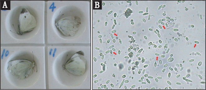
Wild-caught cabbage butterflies (P. rapae) infected with microsporidia were field collected in Honghe Prefecture, Yunnan Province, China (Fig 1A), and no specific permissions were required for these locations. The wings were removed and the infected P. rapae were then disrupted with a glass homogenizer. The homogenates were filtered through three layers of cheesecloth and centrifuged at 10,000 x g for 30 seconds. The pellets were re-suspended in PBS buffer and purified by centrifugation at 1,500 x g for 30 min on a Percoll gradient. The spore band was collected and washed several times with sterile water and checked for purity with a phase contrast microscope (Fig 1B).
Fig 1. Images of host and parasite.
A. Micrograph of the adult of Pieris rapae Linne. B. Spores of Nosema sp. YNPr at 400X.
Approximately 1×10⁹ spores of Nosema sp. YNPr were extracted directly using the cetyl trimethylammonium bromide (CTAB) method [48]. The concentration and purity of the DNA were determined spectrophotometrically based on absorption readings and ratios at 260 nm and 280 nm by using a NanoDrop spectrophotometerND-1000
Ribosomal DNA sequencing
PCR amplification was performed under the following conditions:
Samples were heated to 94°C for 5 min to denature the DNA followed by 30 cycles of: 1 min of denaturation at 94°C, 30 s of annealing at 55–58°C, and 2 min of extension at 72°C. A final extension step was carried out at 72°C for 10 min. Primers used are as follows:
SSU rRNA: 5'-CACCAGGTTGATTCTGCC-3'
5'-TTATGATCCTGCTAATGGTTC-3'
ITS: 5'-TGAATGTGTCCCTGTTCTTTG-3'
5'-GTTAGTTTCTTTTCCTCC-3'
PCR products were run on agarose gels, excised and purified using a gel extraction kit (E.Z.N.A. ® Gel Extraction Kit) following the manufacturer’s instructions. The purified PCR products were then cloned into a pMD19-T vector using a TA Cloning Kit (TaKaRa Biotechnology, Dalian, China) and bacterial colonies were sent to Beijing Genomics Institute (BGI) Shenzhen, China for Sanger sequencing.
Comparative rDNA analysis
The SSU rDNA and ITS rDNA sequences from Nosema sp. YNPr were compared to those downloaded from GenBank. Sequence alignments were performed by MUSCLE [49].Phylogenetic trees were constructed using neighbor-joining (NJ) analysis with MEGA version 5 [50]. The Maximum Composite Likelihood model was used and bootstrap support was evaluated based on 1,000 replicates.
Genome sequencing and assembly
A genomic DNA library was prepared for Illumina HiSeq2500sequencing following the manufacturer’s instructions (Illumina). The genomic DNA was fragmented by nebulization with compressed nitrogen gas at 32psi for 9 minutes. Any overhangs were converted to blunt ends using T4 DNA polymerase and Klenow polymerase, after which an adenine nucleotide was added to the ends of double-stranded DNA using Klenow Exo-Minus polymerase (Qiagen). DNA adaptors with a single “T” base overhang at the 3’ end were ligated to the above products. The resulting DNA was then separated on a 2% agarose gel and fragments of approximately 800 bp were excised from the gel and purified (Qiagen Gel Extraction Kit). The adapter-modified DNA fragments were amplified using Illumina primers 1.1 and 2.1. The concentration of the DNA library was determined by measuring the absorbance at 260nm and the DNA was then sequenced (BGI). The paired-end reads were processed by removing adaptor and low quality (Q<30) sequences. As a result, 3,154,248 high-quality reads were obtained for a total of 1.57 million base pairs. Each read was about 100 bp in length. The paired-end reads were assembled de novo using the Velvet1.2.10 assembly program [51], with the standard option as following: insert size of 800 ± 160 bp and a Hash length of 31. The resulting scaffolds were further processed with GapFiller1.11[52] using the default settings.
Gene prediction and annotation
The Nosema sp. YNPr protein-coding genes were annotated with Glimmer 3.0 software using the lower eukaryote settings [53,54]. We identified 2075 ORFs. To avoid the over-prediction of small genes, the ORFs were re-annotated by searching for transcriptional signals (CCC or GGG-like motifs) within 50 nucleotides upstream of the first or successive downstream AUG codons within the ORF [48,55]. We identified 1425 CDS by this method. The remaining 650 ORFs were further examined by searching for AT-rich regions (AT content > = 80%) within 50 nucleotides upstream of the first or successive AUG codons [55] within the ORFs. We identified a further 275 CDS using this method.
The MEME 3.0 software program was used for motif exhibition of putative transcription regulators by searching for over-represented motifs in the 50-bp upstream region of the start codon of protein-coding genes. Gene annotation was accomplished utilizing NCBI BLASTP against the GenBank non-redundant database (nr), Swiss-Prot database with a cut-off e-value of 1e-6. Gene ontology classification of the three Nosema species was performed using InterProScan5 software and then visualized through on-line WEGO tools (http://wego.genomics.org.cn/cgi-bin/wego/index.pl).
Comparison of genes based upon function among N. ceranae, N. apis and Nosema sp. YNPr was accomplished using the Clusters of Orthologous Groups (COG) protein database (http://www.ncbi.nlm.nih.gov/COG/). BLAST searches against the local COG database were performed with a cut-off e-value of 1e-6, and the best hit which contained the protein identity was used to assign the functional categories of COG based on the list of COG annotations. Similarly, proteases were identified using a BLAST search of all predicted genes against the MEROPS database (http://merops.sanger.ac.uk/cgi-bin/batch_blast) [56]. Genes with e-values less than 1e-6 were further verified as proteases by searching against the Genbank database. The best hits in the MEROPS database were used to assign the protease classes. BLASTCLUST 2.2 was used to identify homologous genes among Nosema sp. YNPr and three other species (N. ceranae, N. apis and E. cuniculi) with 30% identity and 50% coverage. Differences in gene length among homologous genes and the number of unique genes in each of the species were calculated using custom PERL scripts written in our lab.
The final genome and predicted protein-coding genes for Nosema sp. YNPr are deposited in NCBI (Project PRJNA325422) and the Silkworm Pathogen Database (SilkPathDB, http://silkpathdb.swu.edu.cn/). The Illumina data generated for Nosema sp. YNPr is available in the NCBI SRA (accession number SRR3673305).
Transposon and signal peptide analysis
The assembled genomes of Nosema sp. YNPr, N. ceranae, N. apis and N. bombycis were checked for interspersed repeats and low complexity DNA sequences using RepeatMasker 4.05(http://www.repeatmasker.org)[57]. The transposons were identified and their lengths were calculated. The conserved reverse transcriptase domain sequences (RVT) from LTR retrotransposons were obtained through the NCBI Conserved Domains search site (http://www.ncbi.nlm.nih.gov/Structure/cdd/wrpsb.cgi) to predict the conserved domains.
Signal peptide sequences of genes for each microsporidia species were predicted by SignalP 4.1 under the default D-cutoff values (http://www.cbs.dtu.dk/services/SignalP/).
Results
Phylogenetic analysis
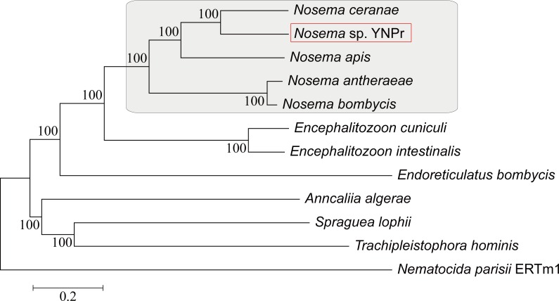
Neighbor Joining analysis using homologous genes shows a closer relationship between N. ceranae and Nosema sp. YNPr than between N. ceranae and N. apis (both honeybee parasites) and a close relationship between the two silkworm parasites N. antheraeae and N. bombycis (Fig 2). The same phylogenetic relationships were obtained using the SSU rDNA and LSU rDNA genes only (S1A and S1B Fig). The arrangement of the functional ribosomal RNA operon from Nosema sp. YNPr (5′-SSU-ITS-LSU-3′) is the same as that found in most members of the Nosema/Vairimorpha clade [58,59,60]. The ITS sequence of Nosema sp.YNPr from Yunnan Province is identical to that of a previously reported isolate, Nosema sp. MPr from the same host species (P. rapae) but from Jiangsu province (S1C Fig)[61]. It is clear, as shown in S1A Fig, that genome size changes quite rapidly even between closely related species.
Fig 2. The evolution of microsporidia at the genome level.
The maximum likelihood phylogeny of 12 microsporidia species based on a concatenation of common orthologous genes.
Genomic architecture of Nosema sp. YNPr
Sequencing and assembly statistics are summarized in Table 1. The random genomic Nosema sp. YNPr library of 800 bp inserts yielded 3,154,248 reads and was assembled into 462 scaffolds. The combined scaffold length was 3.36 Mb. The transposable element (TE) content of the assembly was used to approximate the total TE content of the genome and the inferred genome size is contingent on the accuracy of this assumption. The N50 of the Nosema sp. YNPr genome is 12,222 bp; in comparison the N50 of N. apis is 24,309 bp and that of N. ceranae is 2,902 bp. The longest scaffold size is 45,514 bp and the mean scaffold length is 7,874 bp. The mean sequence coverage of scaffolds was 90 X. The G+C content of the Nosema sp. YNPr genome is 23.19%, which is lower than those of N. ceranae (26%), N. bombycis (31%) and N. antheraeae (28%) but higher than that of N. apis (18.78%) [54,55,56]. The protein coding regions have a significantly higher G+C content (25.53%) than does the overall genome.
Table 1. Genome assembly statistics for Nosema sp. YNPr.
| General characteristics | Nosema sp. YNPr |
|---|---|
| Clean reads | 3,154,248 |
| Assembled (Mb) | 3.36 |
| Genome coverage | 90X |
| G+C content (%) | 23.19 |
| Total number of scaffold | 462 |
| Length of scaffold (bp) | 654~45,514 |
| Scaffold N50 (bp) | 12,222 |
| No.of CDS | 2,075 |
| Mean CDS length (bp) | 969 |
| Mean size of scaffold (bp) | 7,874 |
We identified 2075 ORFs in the Nosema sp. YNPr genome, with a mean length of 969 bp (Table 1). Based on our analysis there are 1,425 ORFs containing the CCC or GGG motifs in the genome. and An additional 275 CDS are predicted based on an an AT content >80% in the region 50 nucleotides upstream of an AUG codon within the ORF. (S1 Table, S2 Fig). All of the genes involved in core carbon metabolic pathways previously published from microsporidial genomes have been identified in Nosema sp. YNPr. The inability to identify some of the hypothetical genes using BLAST can be explained either by the highly divergent nature of the microsporidia resulting in low similarity with known genes or as genes specific to the microsporidia.
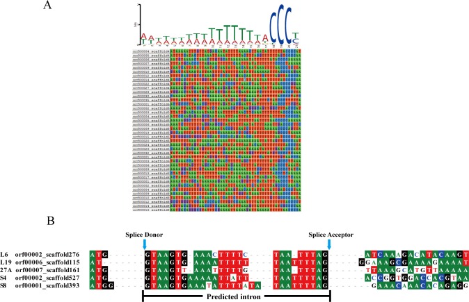
Results of the analysis for the ACCCTT motif approximately 50 bp upstream of the start codon in Nosema sp. YNPr is shown in Fig 3A. This motif is conserved in N. ceranae, N. apis and E. cuniculi and was present in 60% of the predicted genes for Nosema sp. YNPr [62,63,35]. A search for homology with the genes of N. ceranae identified five intron-containing ribosomal protein genes in Nosema sp. YNPr. They all contain GTAAGT at the donor site and TTAG at the acceptor site (Fig 3B). Two of the proteins (L19, S4) have orthologous intron-containing genes in N. apis and N. bombycis while homologues of the other three proteins (L6, 27A, S8) do not contain introns in N. apis and N.bombycis.
Fig 3. Noncoding regions of selected Nosema sp. YNPr genes.
A. Putative regulatory C-rich motif in the region upstream of the start codon in Nosema sp. YNPr. B. Comparison of the regions flanking the introns for five Nosema sp. YNPr genes.
Comparison of functional genes
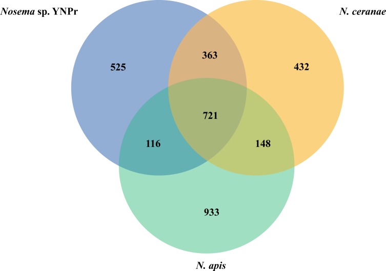
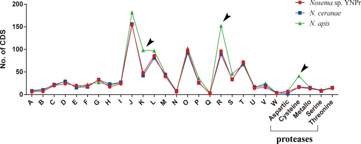
Comparison of genes homologous among Nosema sp. YNPr, N. ceranae and N. apis revealed that there are 721 genes shared among all three species, 525 genes that are Nosema sp. YNPr-specific, 432 that are N. ceranae -specific, and 933 that are N. apis-specific (Fig 4). Results of the COG protein database analysis indicate that the various cellular functions are similar in almost all categories except for Transcription (K) and General function prediction (R) which are substantially more numerous in N. apis than in the other two species (Fig 5). Cysteine proteases are also more abundant in N. apis than in N. ceranae and Nosema sp. YNPr (Fig 5).
Fig 4. Venn diagram comparing the genes of three species of microsporidia.
The number of homologous genes and species-specific genes among Nosema sp. YNPr, Nosema ceranae and Nosema apis are shown.
Fig 5. Comparison of coding sequence (CDS) numbers based on COG annotation and protease class among Nosema sp. YNPr, N. ceranae and N. apis.
Black arrows indicate gene function categories for which Nosema sp. YNPr and N. ceranae differ in number of coding sequences from N. apis. A: RNA processing and modification; B: Chromatin structure and dynamics; C: Energy production and conversion; D: Cell cycle control, cell division, chromosome partitioning; E: Amino acid transport and metabolism; F: Nucleotide transport and metabolism; G: Carbohydrate transport and metabolism; H: Coenzyme transport and metabolism; I: Lipid transport and metabolism; J: Translation, ribosomal structure and biogenesis; K: Transcription; L: Replication, recombination and repair; M: Cell wall/membrane/envelope biogenesis; N: Cell motility; O: Posttranslational modification, protein turnover, chaperones; P: Inorganic ion transport and metabolism; Q: Secondary metabolites biosynthesis, transport and catabolism; R: General function prediction only; S: Function unknown; T: Signal transduction mechanisms; U: Intracellular trafficking, secretion, and vesicular transport; V: Defense mechanisms; W: Extracellular structures.
Variation in Genome Composition
Decrease in gene length
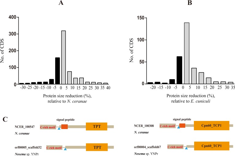
We compared the lengths of 1084 genes common to Nosema sp. YNPr and N. ceranae and found that 75% of the genes were shorter in Nosema sp. YNPr (Fig 6A). The total coding region of the genome was 364,065 amino acids in Nosema sp. YNPr and 368,901 amino acids in N. ceranae. Similarly, when 340 genes common to Nosema sp. YNPr and E. cuniculi, the closest sister taxon to the Nosema/Vairimorpha clade, were compared, it was found that 74% of the genes were smaller in Nosema sp. YNPr than in E. cuniculi (Fig 6B). The total coding region of the genome was 126,156 amino acids in Nosema sp. YNPr and 131,659 amino acids in E. cuniculi. An analysis of the common genes that are shorter in Nosema sp. YNPr than in N. ceranae shows a loss of signal sequences in Nosema sp. YNPr (Fig 6C) rather than a loss of individual amino acids.
Fig 6. Comparison of CDS size between Nosema sp. YNPr and two other microsporidia.
A. Degrees of reduction in length of Nosema sp. YNPr (Np) CDS relative to those of N. ceranae (Nc). X-axis value is expressed as percentage: 100 (Nc CDS length–Np CDS length)/(Nc CDS length). B. Degrees of reduction in length of Nosema sp. YNPr CDS relative to those of E.cuniculi (Ec). X-axis value is expressed as percentage: 100 (Ec CDS length–Np CDS length)/(Ec protein length). The positive classes representative of shorter Nosema sp.YNPr CDS are in grey. C. Two cases showing the signal peptide deletions in Nosema sp. YNPr relative to those of N. ceranae.
Signal Peptides
The number of genes with signal peptides varies widely among species in the Nosema/Vairimorpha clade (S2 Table). Closely related species show similar numbers and percentages of genes containing signal peptides. Nosema bombycis and Nosema antheraeae have 431 and 394 genes with signal peptides respectively, while Nosema sp. YNPr and Nosema ceranae have109 and 159 respectively. S3 Table compares homologous genes of Nosema sp. YNPr and Nosema ceranae that contain predicted signal peptides in one or both species. Twenty-three genes in Nosema sp. YNPr lack the signal peptide seen in Nosema ceranae, while 5 genes in Nosema ceranae lack the signal peptide seen in Nosema sp. YNPr.
Decrease in size of intergenic regions
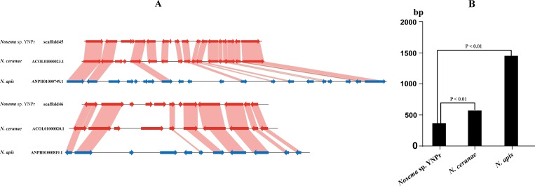
Syntenic comparisons among Nosema sp. YNPr, N. ceranae and N. apis indicate that genes are more tightly arranged (compacted) in Nosema sp. YNPr and N. ceranae than in N. apis (Fig 7A). The average length of the intergenic regions for the two scaffolds shown is significantly greater in N. apis (1438 nucleotides) than in Nosema sp. YNPr (357 nucleotides) and N. ceranae (607 nucleotides) (Fig 7B).
Fig 7. Syntenic, intergenic and overall size comparisons of Nosema sp. YNPr scaffolds with those of N. ceranae and N. apis.
A. Syntenic and intergenic comparisons of two Nosema sp. YNPr scaffolds with those of N. ceranae and N. apis. B. Comparison of average intergenic lengths in Nosema sp. YNPr, N. ceranae and N. apis.
Transposon numbers
A search of the assembled Nosema sp. YNPr genome for repetitive DNA yielded a number of transposon types including LTR, Merlin, Tc1/mariner, LINE and Helitron (Table 2). The transposon content is 4.8% in Nosema sp. YNPr, 3.7% in N. ceranae, and 2.5% in N.apis. Nosema sp. YNPr and N. ceranae possess all 5 of the above mentioned transposon types but the Merlin, Tc1/mariner and Helitron classes are missing from N. apis. For the transposon types shared among these species there is a high variability in copy number. Analysis of the reverse transcriptase of the long terminal repeat (LTR) transposons shows that they cluster into 4 major groups (S3 Fig). The LTR copy number in N. bombycis, a sister taxon to the three Nosema species analyzed here, is much higher indicating that LTR transposons account for some of the differences in genome size in closely related taxa. Of note also is the absence of Group II LTR sequences in N. apis.
Table 2. Transposon types in three Nosema species.
| Type | Nosema sp. YNPr | N. ceranae | N. apis |
|---|---|---|---|
| LTR | 107103 | 99104 | 52531 |
| Merlin | 829 | 1165 | 0 |
| Tc1/mariner | 1983 | 36357 | 0 |
| LINE | 9756 | 27203 | 13496 |
| Helitron | 1190 | 453 | 0 |
| Others | 42596 | 126319 | 143533 |
| Total | 163457(4.8%) | 290601(3.7%) | 209560(2.5%) |
Discussion
Microsporidia have highly reduced genomes [36,39]. These obligate intracellular parasites can use host metabolites for their own cellular processes [41]. Genome compaction in the microsporidia has been studied extensively in terms of biochemical pathways and minimal genome sizes [37,39]. These studies show that the microsporidia have a core set of genes and an expanded set of cell surface transporters which allow them to import metabolic precursors from the host instead of producing these molecules themselves. The highly dynamic nature of genome evolution in the microsporidia is discussed in terms of gene compaction, size of intergenic regions, introns and overlapping genes [39].
Microsporidial genomes, though small, change in a dynamic fashion and have been shown in some cases to expand and in others to contract over time (S1 Fig) [39]. The fact that closely related microsporidial species have a number of unique genes (Fig 4) suggests that genes may be gained and lost during the process of host switching. Studies are needed to elucidate the functions of these species-specific genes
Our results show that Nosema sp. YNPr has shorter genes (Fig 6), shorter intergenic regions (Fig 7) and fewer transposons than do N. apis and N. ceranae. Gene size appears to be decreasing through loss of domains, including signal peptides (Fig 6). In a comparison of homologous genes between Nosema sp. YNPr and N. ceranae, 23 of the Nosema sp. YNPr genes lacked the signal peptide present in the Nosema ceranae homologue(S3 Table), indicating that the signal peptide content of homologous genes in closely related species can change rapidly. However, the total number of genes (both homologous and unique) containing signal peptides is similar in closely related species (S2 Table).
Phylogenetic analysis shows Nosema sp. YNPr and N.ceranae to be more closely related than are the two honeybee parasites, N. ceranae and N. apis. This relationship provides evidence of host switching across the insect orders Lepidoptera and Hymenoptera in the Nosema/Vairimorpha clade. A search of the protease database shows that N. apis has substantially more cysteine proteases than do either N. ceranae or Nosema sp. YNPr (Fig 5). Cysteine proteases are among the main proteolytic enzymes found in many protozoan parasites, and cysteine protease inhibitors have been shown to be effective against a variety of protozoans including Trypanosoma cruzi [64], Entamoeba histolytica [65] and Plasmodium falciparum [66].
The transposon makeup of these Nosema species varies widely and would seem likely to play a large role in genome evolution. N. apis appears to have no Merlin, Tc1/mariner or Helitron transposons, all of which are present in both Nosema sp. YNPr and N. ceranae (Table 2). These differences in transposon content among closely related species indicate that transposons can move in and out of genomes rapidly over relatively short evolutionary time periods. Determining the roles of these transposons in the adaptation of a microsporidial parasite to its host and in genome expansion and contraction in the microsporidia would be illuminating.
Conclusion
Microsporidia comprise over 1,300 species and infect hosts from every animal phylum from marine, freshwater and terrestrial habitats. Because of their rich host diversity they are an excellent model system for the study of interactions between obligate single-celled parasites and their hosts at many levels. The Nosema/Vairimorpha clade encompasses a wide-ranging group of parasites from a diverse collection of hosts in which host-parasite co-evolutionary principles can be tested. Members of the Nosema/Vairimorpha clade have been reported from Lepidoptera, Coleoptera, Hymenoptera and other invertebrates including mites [27,67,68]. From our phylogenetic analyses and those of others [39,69,70] it appears that host switching occurs relatively rapidly over evolutionary time in the microsporidia. We hypothesize that natural selection plays a role in the evolution of the small yet dynamically changing microsporidial genomes and suggest that there may be a trade-off between smaller genomes for rapid reproduction and larger genomes with more genetic options with which to challenge a host. However, in order to examine the role of genetic drift versus natural selection in microsporidial genome evolution it will be necessary to sequence and analyze additional genomes from the Nosema/Vairimorpha clade. Analyses would include determining dN/dS ratios, functions of genes unique to closely related species [39] in different hosts, genome size versus host persistence, and the searching for convergence in microsporidia from different genera that infect the same host.
Supporting Information
A: Phylogenetic tree of SSU rDNA; B: Phylogenetic tree of LSU rDNA tree; C: Multiple sequence alignments of ITS between Nosema sp. YNPr and Nosema sp. MPr.
(TIF)
(TIF)
(TIF)
(XLS)
(DOC)
(DOC)
Acknowledgments
This study is supported by National High-tech R&D Program (863 Program, No.2013AA102507), National Natural Science Foundation of China (No.31270138, 31302037, 31402142, 31001037), Natural Science Foundation Project of Chongqing Science&technology Commission (No.cstc2015jcyjA80020).
Data Availability
All the genomic data that used in this study were produced by the third party and can be freely downloaded from not only public Silkworm Pathogen Database (SilkPathDB, http://silkpathdb.swu.edu.cn/), but also GenBank database (accession no.PRJNA325422) without any restriction. All other data have been provided as part of Supporting Information in the paper.
Funding Statement
This study is supported by National High-tech R&D Program (863 Program, No.2013AA102507), National Natural Science Foundation of China (No.31270138, 31302037, 31402142, 31001037), Natural Science Foundation Project of Chongqing Science & technology Commission (No.cstc2015jcyjA80020).
References
- 1.Lom J, Vavra J. The mode of sporoplasm extrusion in microsporidian spores. Acta Protozool. 1963; 1:81–89. [Google Scholar]
- 2.Weidner E Ultrastructural study of microsporidian invasion into cells. Z Parasitenk. 1972; 40: 227–242. [DOI] [PubMed] [Google Scholar]
- 3.Keohane EM, Weiss LM. Characterization and function of the microsporidian polar tube: a review. Folia Parasitol. 1998; 45: 117–127. [PubMed] [Google Scholar]
- 4.Tanada Y. Microbial Control of Insect Pests. Annu Rev Entomology. 1959; 4: 277–302. [Google Scholar]
- 5.Sweeney AW, Becnel JJ. Potential of microsporidia for the biological control of mosquitoes. Parasitol Today. 1991; 7: 217–220. [DOI] [PubMed] [Google Scholar]
- 6.Solter LF, Maddox JV. Physiological host specifiy of microsporidia as an indicator of ecological host specificity. J Invertebr Pathol. 1998; 71: 207–216. [DOI] [PubMed] [Google Scholar]
- 7.Sprague V. Systematics of the Microsporidia In: Bulla LA,Cheng TC, editors. Comparative Pathobiology, vol. 2 New York: Plenum Press; 1977. [Google Scholar]
- 8.Becnel JJ, Takvorian PM, Cali A. Checklist of available generic names for Microsporidia with type species and type host In: Weiss LM, Becnel JJ, editors. Microsporidia: Pathogens of opportunity. John Wiley & Sons, Inc. 2014. pp. 671–686. [Google Scholar]
- 9.Bhat SA, Bashir I, Kamili AF. Microsporidiosis of silkworm, Bombyx mori L. (Lepidoptera-Bombycidae): a review. Afr J Agric Res. 2009; 4: 1519–1523. [Google Scholar]
- 10.Kawarabata T. Biology of microsporidians infecting the silkworm, Bombyx mori, in Japan. J Biotechnol Sericol. 2003; 72: 1–32. [Google Scholar]
- 11.Higes M, Martin R, Meana A. Nosema ceranae, a new microsporidian parasite in honeybees in Europe. J In. Pathol. 2006; 92: 93–95. [DOI] [PubMed] [Google Scholar]
- 12.Cox-Foster DL, Conlan S, Holmes EC, Palacios G, Evans JD, Moran NA, et al. A metagenomic survey of microbes in honey bee colony collapse disorder. Science. 2007; 318: 283–286. [DOI] [PubMed] [Google Scholar]
- 13.Natsopoulou ME, McMahon DP, Doublet V, Bryden J, Paxton RJ. Interspecific competition in honeybee intracellular gut parasites is asymmetric and favours the spread of an emerging infectious disease. Proceedings of the Royal Society of London B: Biological Sciences. 2015. January 7; 282(1798):20141896. [DOI] [PMC free article] [PubMed] [Google Scholar]
- 14.Shaw RW, Kent ML. Fish microsporidia In: Wittner M, Weiss LM, editors. The Microsporidia and Microsporidiosis. ASM Press; 1999. pp. 418–446. [Google Scholar]
- 15.Kent ML, Shaw RW, Sanders JL. Microsporidia in fish In: Weiss LM, Becnel JJ, editors. Microsporidia: Pathogens of opportunity. John Wiley & Sons, Inc. 2014. pp. 493–520. [Google Scholar]
- 16.Didier ES, Vossbrinck CR, Baker MD, Rogers LB, Bertucci DC, Shadduck JA. Identification and characterization of three Encephalitozoon cuniculi strains. Parasitology. 1995; 111: 411–421. [DOI] [PubMed] [Google Scholar]
- 17.Didier ES. Microsporidiosis: an emerging and opportunistic infection in humans and animals. Acta Trop. 2005; 94: 61–76. [DOI] [PubMed] [Google Scholar]
- 18.Andreadis TG. Experimental transmission of a microsporidian pathogen from mosquitoes to an alternate copepod host. Proceedings of the National Academy of Sciences. 1985; 82 (16): 5574–5577. [DOI] [PMC free article] [PubMed] [Google Scholar]
- 19.Sweeney AW, Hazard EI, Graham MF. Intermediate host for an Amblyospora sp. (microspora) infecting the mosquito, Culex annulirostris. J Invertebr Pathol. 1985; 46: 98–102. [DOI] [PubMed] [Google Scholar]
- 20.Andreadis TG. Host specificity of Amblyosporaconnecticus (Microsporida: Amblyosporidae), a polymorphic microsporidian parasite of Aedes cantator (Diptera: Culicidae). J Med Entomol. 1986; 26: 140–145. [DOI] [PubMed] [Google Scholar]
- 21.Andreadis TG. Host range tests with Edhazardia aedis (Microsporida: Culicosporidae) against northern Nearctic mosquitoes. J Invertebr Pathol.1994; 64: 46–51. [DOI] [PubMed] [Google Scholar]
- 22.Kingsolver JG. Feeding, growth, and the thermal environment of cabbage white caterpillars, Pieris rapae L. Physiol Biochem Zool. 2000;73: 621–628. [DOI] [PubMed] [Google Scholar]
- 23.Watanabe H. First Report of the Microsporidian Infection of the Cabbageworm, Pieris rapaecrucivora Boisduval (Lepitoptera: Pieridae) in Japan. Appl Ent Zool. 1974; 3: 133–142. [Google Scholar]
- 24.Chen D, Shen Z, Zhu F, Guan R, Hou J, Zhang J, et al. Phylogenetic characterization of a microsporidium (Nosema sp. MPr) isolated from the Pieris rapae. Parasitol Res. 2012; 111: 263–269. 10.1007/s00436-012-2829-6 [DOI] [PubMed] [Google Scholar]
- 25.Choi J, Kim J, Choi Y, Nam SH, Russell J, Kim W, et al. Structure of ribosomal RNA gene and phylogeny of Nosema isolates in Korea. Genes & Genomics. 2009; 31: 443–450. [Google Scholar]
- 26.Choi Y, Lee Y, Cho KS, Lee S, Russell J, Choi J et al. Chimerical nature of the ribosomal RNA gene of a Nosema species. J Invertebr Pathol. 2011; 107: 86–89. 10.1016/j.jip.2011.02.005 [DOI] [PubMed] [Google Scholar]
- 27.Vossbrinck CR, Debrunner-Vossbrinck BA. Molecular phylogeny of the Microsporidia: ecological, ultrastructural and taxonomic considerations. Folia Parasitol. 2005; 52(1–2): 131–142. [DOI] [PubMed] [Google Scholar]
- 28.Kyei-Poku G, Gauthier D, van Frankenhuyzen K. Molecular data and phylogeny of Nosema infecting lepidopteran forest defoliators in the genera Choristoneura and Malacosoma. Jounal of Eukaryot Microbiol. 2008; 55: 51–58. [DOI] [PubMed] [Google Scholar]
- 29.Hayasaka S, Yonemura N. Infection and development of Nosema sp. NIS H5 (Microsporida: Protozoa) in several lepidopteran insects. JARQ. 1999; 33: 65–68. [Google Scholar]
- 30.Kashkarova L, Khakhanov A. Range of the hosts of the causative agent of pebrine (Nosema bombycis) in the mulberry silkworm. Parazitologiia. 1979; 14: 164–167. [PubMed] [Google Scholar]
- 31.Kudo R, DeCoursey J. Experimental infection of Hyphantria cunea with Nosema bombycis. The Journal of Parasitology. 1940; 26: 123–125. [Google Scholar]
- 32.Nageli K. Uber die neue Krankheit der Seidenraupe und verwandte Organismen. Bot Z. 1857; 15: 760–761. [Google Scholar]
- 33.Solter LF, Maddox JV, McManus ML. Host Specificity of Microsporidia (Protista: Microspora) from European Populations of Lymantria dispar (Lepidoptera: Lymantriidae) to Indigenous North American Lepidoptera. J Invertebr Pathol. 1997; 69(2): 135–150. [DOI] [PubMed] [Google Scholar]
- 34.Solter LF, Pilarska DK, Vossbrinck CR. Host specificity of microsporidia pathogenic to forest Lepidoptera. Biological control. 2000; 19: 48–56. [Google Scholar]
- 35.Katinka MD, Duprat S, Cornillot E, Méténier G, Thomarat F, Prensier G, et al. Genome sequence and gene compaction of the eukaryote parasite Encephalitozoon cuniculi. Nature. 2001; 414: 450–453. [DOI] [PubMed] [Google Scholar]
- 36.Selman M, Pombert JF, Solter L, Farinelli L, Weiss LM, Keeling P, Corradi N Acquisition of ananimal gene by microsporidian intracellular parasites. Curr Biol. 2011;21(15):R576–7. 10.1016/j.cub.2011.06.017 [DOI] [PMC free article] [PubMed] [Google Scholar]
- 37.Corradi N, Pombert JF, Farinelli L, Didier ES, Keeling PJ. The complete sequence of the smallest known nuclear genome from the microsporidian Encephalitozoon intestinalis. Nature communications. 2010; 1: 77 10.1038/ncomms1082 [DOI] [PMC free article] [PubMed] [Google Scholar]
- 38.Pombert JF, Selman M, Burki F, Bardell FT, Farinelli L, Solter LF, et al. Gain and loss of multiple functionally related, horizontally transferred genes in the reduced genomes of two microsporidian parasites. PNAS. 2012; 109; 12638–12643. 10.1073/pnas.1205020109 [DOI] [PMC free article] [PubMed] [Google Scholar]
- 39.Nakjang S, Williams TA, Heinz E, Watson AK, Foster PG, Sendra KM. Reduction and expansion in microsporidian genome evolution: new insights from comparative genomics. Genome Biol Evol. 2013; 5: 2285–2303. 10.1093/gbe/evt184 [DOI] [PMC free article] [PubMed] [Google Scholar]
- 40.Keeling PJ, Fast NM. (2002). Microsporidia: biology and evolution of highly reduced intracellular parasites. Annual Reviews in Microbiology, 56(1), 93–116. [DOI] [PubMed] [Google Scholar]
- 41.Keeling PJ, Corradi N, Morrison HG, Haag KL, Ebert D, Weiss LM. The reduced genome of the parasitic microsporidian Enterocytozoon bieneusi lacks genes for core carbon metabolism. Genome Biol Evol. 2010; 2: 304–9. 10.1093/gbe/evq022 [DOI] [PMC free article] [PubMed] [Google Scholar]
- 42.Williams BAP, Slamovits CH, Patron JJ, Fast NM, Keeling PJ. A high frequency of overlapping gene expression in compacted eukaryotic genomes. PNAS (2005) 102:10936–10941. [DOI] [PMC free article] [PubMed] [Google Scholar]
- 43.Corradi N, Akiyoshi D E, Morrison HG, Feng X, Weiss LM, Tzipori S, Keeling P J. Patterns of genome evolution among the microsporidian parasites Encephalitozoon cuniculi, Antonospora locustae and Enterocytozoon bieneusi. PLoS One, (2007)2(12), e1277 [DOI] [PMC free article] [PubMed] [Google Scholar]
- 44.Keeling Patrick J, Slamovits Claudio H. "Causes and effects of nuclear genome reduction." Current opinion in genetics & development 156 (2005): 601–608. [DOI] [PubMed] [Google Scholar]
- 45.Vieira-Silva S, Rocha EP. (2010). The systemic imprint of growth and its uses in ecological (meta) genomics. PLoS Genet, 6(1), e1000808 10.1371/journal.pgen.1000808 [DOI] [PMC free article] [PubMed] [Google Scholar]
- 46.Sabath N, Ferrada E, Barve A, Wagner A. Growth temperature and genome size in bacteria are negatively correlated, suggesting genomic streamlining during thermal adaptation. Genome biology and evolution. 2013. May 1;5(5):966–77. 10.1093/gbe/evt050 [DOI] [PMC free article] [PubMed] [Google Scholar]
- 47.Nilsson AI, Koskiniemi S, Eriksson S, Kugelberg E, Hinton JCD, Andersson DI. (2005). Bacterial genome size reduction by experimental evolution. Proceedings of the National Academy of Sciences of the United States of America, 102(34), 12112–12116. [DOI] [PMC free article] [PubMed] [Google Scholar]
- 48.He Q, Ma Z, Dang X, Xu J, Zhou Z. Identification, Diversity and Evolution of MITEs in the Genomes of Microsporidian Nosema Parasites. PloS one. 2015; 10(4): e0123170 10.1371/journal.pone.0123170 [DOI] [PMC free article] [PubMed] [Google Scholar]
- 49.Edgar RC. MUSCLE: a multiple sequence alignment method with reduced time and space complexity. BMC bioinformatics. 2004; 5: 113 [DOI] [PMC free article] [PubMed] [Google Scholar]
- 50.Tamura K, Dudley J, Nei M, Kumar S. MEGA4: molecular evolutionary genetics analysis (MEGA) software version 4.0. Molecular biology and evolution. 2007. August 1; 24(8): 1596–9. [DOI] [PubMed] [Google Scholar]
- 51.Zerbino DR, Birney E. Velvet: algorithms for de novo short read assembly using de Bruijn graphs. Genome Res. 2008; 18: 821–829. 10.1101/gr.074492.107 [DOI] [PMC free article] [PubMed] [Google Scholar]
- 52.Boetzer M, Pirovano W. Toward almost closed genomes with GapFiller. Genome Biol. 2012; 13: R56 10.1186/gb-2012-13-6-r56 [DOI] [PMC free article] [PubMed] [Google Scholar]
- 53.Delcher AL, Harmon D, Kasif S, White O, Salzberg SL. Improved microbial gene identification with GLIMMER. Nucleic Acids Res. 1999; 27: 4636–4641. [DOI] [PMC free article] [PubMed] [Google Scholar]
- 54.Delcher AL, Bratke KA, Powers, EC, Salzberg SL. Identifying bacterial genes and endosymbiont DNA with Glimmer. Bioinformatics. 2007; 23: 673–679. [DOI] [PMC free article] [PubMed] [Google Scholar]
- 55.Peyretaillade E, Parisot N, Polonais V, Terrat S, Denonfoux J, Dugat-Bony E, et al. Annotation of microsporidian genomes using transcriptional signals. Nat Commun. 2012; 3: 1137 10.1038/ncomms2156 [DOI] [PubMed] [Google Scholar]
- 56.Rawlings ND, Waller M, Barrett AJ, Bateman A. MEROPS: the database of proteolytic enzymes, their substrates and inhibitors. Nucleic Acids Res. 2014; 42: D503–509. 10.1093/nar/gkt953 [DOI] [PMC free article] [PubMed] [Google Scholar]
- 57.Smit AF, Hubley R, Green P. Available: http://www.repeatmasker.org.RepeatMasker Open.1996; 3: 1996-2004.
- 58.Gatehouse HS, Malone LA. The Ribosomal RNA Gene Region of Nosema apis (Microspora): DNA Sequence for Small and Large Subunit rRNA Genes and Evidence of a Large Tandem Repeat Unit Size. J Invertebr Pathol. 1998;71: 97–105. [DOI] [PubMed] [Google Scholar]
- 59.Huang WF, Tsai SJ, Lo CF, Soichi Y, Wang CH. The novel organization and complete sequence of the ribosomal RNA gene of Nosema bombycis. Fungal Genetics and Biology. 2004; 41: 473–481. [DOI] [PubMed] [Google Scholar]
- 60.Tsai SJ, Wei FH, Chung HW. Complete sequence and gene organization of the Nosema spodopterae rRNA gene. J Euk Microbiol. 2005; 52: 52–54. [DOI] [PubMed] [Google Scholar]
- 61.Chen D, Shen Z, Zhu F, Guan R, Hou J, Zhang J. Phylogenetic characterization of a microsporidium (Nosema sp. MPr) isolated from the Pieris rapae. Parasitol Res. 2012; 111: 263–269. 10.1007/s00436-012-2829-6 [DOI] [PubMed] [Google Scholar]
- 62.Chen YP, Pettis J, Zhao Y, Liu X, Tallon L, Sadzewicz L, et al. Genome sequencing and comparative genomics of honey bee microsporidia, Nosema apis reveal novel insights into host-parasite interactions. BMC genomics. 2013; 14: 451 10.1186/1471-2164-14-451 [DOI] [PMC free article] [PubMed] [Google Scholar]
- 63.Cornman RS, Chen YP, Schatz MC, Street C, Zhao Y, Desany B. Genomic analyses of the microsporidian Nosema ceranae, an emergent pathogen of honey bees. PLoS pathogens. 2009; 5: e1000466 10.1371/journal.ppat.1000466 [DOI] [PMC free article] [PubMed] [Google Scholar]
- 64.Eakin AE, Mills AA, Harth G, McKerrow JH, Craik CS. The sequence, organization, and expression of the major cysteine protease (cruzain) from Trypanosoma cruzi. J Biol Chem. 1992; 267(11): 7411–7420. [PubMed] [Google Scholar]
- 65.Bruchhaus I, Loftus BJ, Hall N, Tannich E. The intestinal protozoan parasite Entamoeba histolytica contains 20 cysteine protease genes, of which only a small subset is expressed during in vitro cultivation. Eukaryot cell. 2003; 2(3): 501–509. [DOI] [PMC free article] [PubMed] [Google Scholar]
- 66.Sijwali PS, Rosenthal PJ. Gene disruption confirms a critical role for the cysteine protease falcipain-2 in hemoglobin hydrolysis by Plasmodium falciparum. PNAS. 2004; 101(13): 4384–4389. [DOI] [PMC free article] [PubMed] [Google Scholar]
- 67.Bjørnson S, Keddie BA. Effects of Microsporidium phytoseiuli (Microsporidia) on the performance of the predatory mite, Phytoseiulus persimilis (Acari: Phytoseiidae). Biological Control. 1999; 15(2): 153–161. [Google Scholar]
- 68.Becnel JJ, Jeyaprakash A, Hoy MA, Shapiro A. Morphological and molecular characterization of a new microsporidian species from the predatory mite Metaseiulus occidentalis (Nesbitt) (Acari, Phytoseiidae). J Invertebr pathol, 2002; 79(3): 163–172. [DOI] [PubMed] [Google Scholar]
- 69.Cuomo CA, Desjardins CA, Bakowski MA, Goldberg J, Ma AT, Becnel JJ, et al. Microsporidian genome analysis reveals evolutionary strategies for obligate intracellular growth. Genome research. 2012; 22(12): 2478–88. 10.1101/gr.142802.112 [DOI] [PMC free article] [PubMed] [Google Scholar]
- 70.Pan G, Xu J, Li T, Xia Q, Liu SL, Zhang G. Comparative genomics of parasitic silkworm microsporidia reveal an association between genome expansion and host adaptation. BMC genomics. 2013; 14: 186 10.1186/1471-2164-14-186 [DOI] [PMC free article] [PubMed] [Google Scholar]
Associated Data
This section collects any data citations, data availability statements, or supplementary materials included in this article.
Supplementary Materials
A: Phylogenetic tree of SSU rDNA; B: Phylogenetic tree of LSU rDNA tree; C: Multiple sequence alignments of ITS between Nosema sp. YNPr and Nosema sp. MPr.
(TIF)
(TIF)
(TIF)
(XLS)
(DOC)
(DOC)
Data Availability Statement
All the genomic data that used in this study were produced by the third party and can be freely downloaded from not only public Silkworm Pathogen Database (SilkPathDB, http://silkpathdb.swu.edu.cn/), but also GenBank database (accession no.PRJNA325422) without any restriction. All other data have been provided as part of Supporting Information in the paper.


