

Abstract
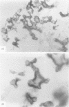
AIM--To investigate the hypothesis that complement mediates the recruitment of mononuclear osteoclast precursors to the exposed mineralised bone surface. METHODS--Synthetic hydroxyapatite was incubated in vitro with fresh human serum, with and without complement activation inhibitors. Assays for complement components and the generation of the C3 breakdown product C3d were done. C3 deposition in human fetal tibia primary spongiosa was localised immunohistochemically and complement receptors CR1, CR2, CR3, and CR4 were localised cellularly. Immunohistochemical and enzyme histochemical characterisation of the mononuclear and multinuclear osteoclasts was made with emphasis on their association with complement C3 deposition. RESULTS--Components of complement bind to synthetic hydroxyapatite crystals and, at lower concentrations, C3d was generated in the fluid phase. C3 was deposited in a focal and linear distribution on newly formed bone trabecular surfaces in the primary spongiosa. In a similar distribution CD61, CD68, and tartrate resistant acid phosphatase positive mononuclear osteoclasts were shown in close apposition to the bone trabecular surface. These mononuclear osteoclasts, unlike multinucleate osteoclasts, expressed the complement receptors CR3 and CR4. CR1 and CR2, however, could not be shown on either mononuclear or multinuclear osteoclasts. CONCLUSION--It is suggested that C3 deposition on mineralised bone surfaces mediates the recruitment of mononuclear osteoclasts to this site. As the mononuclear osteoclasts fuse to form the multinucleate osteoclast, complement receptor expression is lost.
Full text
PDF


Images in this article


Selected References
These references are in PubMed. This may not be the complete list of references from this article.
- Ash P., Loutit J. F., Townsend K. M. Osteoclasts derive from hematopoietic stem cells according to marker, giant lysosomes of beige mice. Clin Orthop Relat Res. 1981 Mar-Apr;(155):249–258. [PubMed] [Google Scholar]
- Athanasou N. A., Puddle B., Quinn J., Woods C. G. Use of monoclonal antibodies to recognise osteoclasts in routinely processed bone biopsy specimens. J Clin Pathol. 1991 Aug;44(8):664–666. doi: 10.1136/jcp.44.8.664. [DOI] [PMC free article] [PubMed] [Google Scholar]
- Athanasou N. A., Quinn J. Immunophenotypic differences between osteoclasts and macrophage polykaryons: immunohistological distinction and implications for osteoclast ontogeny and function. J Clin Pathol. 1990 Dec;43(12):997–1003. doi: 10.1136/jcp.43.12.997. [DOI] [PMC free article] [PubMed] [Google Scholar]
- Chambers T. J., Fuller K. Bone cells predispose bone surfaces to resorption by exposure of mineral to osteoclastic contact. J Cell Sci. 1985 Jun;76:155–165. doi: 10.1242/jcs.76.1.155. [DOI] [PubMed] [Google Scholar]
- Chambers T. J. Fusion of macrophages following simultaneous attempted phagocytosis of glutaraldehyde-fixed red cells. J Pathol. 1977 Jun;122(2):71–80. doi: 10.1002/path.1711220204. [DOI] [PubMed] [Google Scholar]
- Chambers T. J. Multinucleate giant cells. J Pathol. 1978 Nov;126(3):125–148. doi: 10.1002/path.1711260302. [DOI] [PubMed] [Google Scholar]
- Göthlin G., Ericsson J. L. On the histogenesis of the cells in fracture callus. Electron microscopic autoradiographic observations in parabiotic rats and studies on labeled monocytes. Virchows Arch B Cell Pathol. 1973 Mar 30;12(4):318–329. [PubMed] [Google Scholar]
- Hamilton J. A., Lingelbach S. R., Partridge N. C., Martin T. J. Stimulation of plasminogen activator in osteoblast-like cells by bone-resorbing hormones. Biochem Biophys Res Commun. 1984 Jul 18;122(1):230–236. doi: 10.1016/0006-291x(84)90464-9. [DOI] [PubMed] [Google Scholar]
- Heath J. K., Meikle M. C., Atkinson S. J., Reynolds J. J. A factor synthesized by rabbit periosteal fibroblasts stimulates bone resorption and collagenase production by connective tissue cells in vitro. Biochim Biophys Acta. 1984 Aug 21;800(3):301–305. doi: 10.1016/0304-4165(84)90409-4. [DOI] [PubMed] [Google Scholar]
- Jin C. H., Shinki T., Hong M. H., Sato T., Yamaguchi A., Ikeda T., Yoshiki S., Abe E., Suda T. 1 alpha,25-dihydroxyvitamin D3 regulates in vivo production of the third component of complement (C3) in bone. Endocrinology. 1992 Nov;131(5):2468–2475. doi: 10.1210/endo.131.5.1425444. [DOI] [PubMed] [Google Scholar]
- Jotereau F. V., Le Douarin N. M. The development relationship between osteocytes and osteoclasts: a study using the quail-chick nuclear marker in endochondral ossification. Dev Biol. 1978 Apr;63(2):253–265. doi: 10.1016/0012-1606(78)90132-x. [DOI] [PubMed] [Google Scholar]
- Kahn A. J., Simmons D. J. Investigation of cell lineage in bone using a chimaera of chick and quial embryonic tissue. Nature. 1975 Nov 27;258(5533):325–327. doi: 10.1038/258325a0. [DOI] [PubMed] [Google Scholar]
- NILSSON U. R., MUELLER-EBERHARD H. J. ISOLATION OF BETA IF-GLOBULIN FROM HUMAN SERUM AND ITS CHARACTERIZATION AS THE FIFTH COMPONENT OF COMPLEMENT. J Exp Med. 1965 Aug 1;122:277–298. doi: 10.1084/jem.122.2.277. [DOI] [PMC free article] [PubMed] [Google Scholar]
- Nijweide P. J., Burger E. H., Feyen J. H. Cells of bone: proliferation, differentiation, and hormonal regulation. Physiol Rev. 1986 Oct;66(4):855–886. doi: 10.1152/physrev.1986.66.4.855. [DOI] [PubMed] [Google Scholar]
- Zambonin-Zallone A., Teti A., Grano M., Rubinacci A., Abbadini M., Gaboli M., Marchisio P. C. Immunocytochemical distribution of extracellular matrix receptors in human osteoclasts: a beta 3 integrin is colocalized with vinculin and talin in the podosomes of osteoclastoma giant cells. Exp Cell Res. 1989 Jun;182(2):645–652. doi: 10.1016/0014-4827(89)90266-8. [DOI] [PubMed] [Google Scholar]