

Abstract
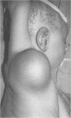
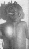
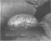
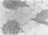
AIM--To study the clinicopathological features of fibrous hamartoma of infancy in Nigerian children. METHODS--Six children aged between 6 months and 10 years were studied. All specimens were stained with haemotoxylin and eosin and examined routinely. The children were followed up for between one and three years. RESULTS--In four of the children lesions were present at birth; in the other two they appeared by the age of 1 year. Some of the children had had the lesion for between three and 10 years. All lesions were located in the subcutis. They were solitary and varied in size and shape. They had grown rapidly up to the age of 5, after which growth decelerated, but did not stop or regress. The younger the child the less clearly demarcated was the tumour on the deep surface. In the older children the capsule was more developed. CONCLUSIONS--Fibrous hamartoma of infancy is rare, but it is important for clinicians to know that it is benign and readily amenable to treatment.
Full text
PDF


Images in this article
Selected References
These references are in PubMed. This may not be the complete list of references from this article.
- Chung E. B. Pitfalls in diagnosing benign soft tissue tumors in infancy and childhood. Pathol Annu. 1985;20(Pt 2):323–386. [PubMed] [Google Scholar]
- ENZINGER F. M. FIBROUS HAMARTOMA OF INFANCY. Cancer. 1965 Feb;18:241–248. doi: 10.1002/1097-0142(196502)18:2<241::aid-cncr2820180216>3.0.co;2-c. [DOI] [PubMed] [Google Scholar]
- Lee J. T., Girvan D. P., Armstrong R. F. Fibrous hamartoma of infancy. J Pediatr Surg. 1988 Aug;23(8):759–761. doi: 10.1016/s0022-3468(88)80420-2. [DOI] [PubMed] [Google Scholar]
- Paller A. S., Gonzalez-Crussi F., Sherman J. O. Fibrous hamartoma of infancy. Eight additional cases and a review of the literature. Arch Dermatol. 1989 Jan;125(1):88–91. doi: 10.1001/archderm.125.1.88. [DOI] [PubMed] [Google Scholar]