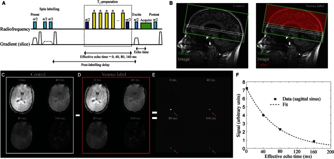
Figure 2.
The TRUST MRI approach for quantifying whole-brain OEF. The pulse-sequence is shown in A and a conceptual representation of the method is shown in B. Briefly, the method consists of a presaturation pulse followed by an inversion pulse and corresponding gradient for labelling venous blood water in a label condition or inversion pulse with no gradient for a control condition. Following the venous blood water labelling (B; red), a post-labelling delay (PLD = 1022 ms) is prescribed during which a T2-preparation module with varying duration (duration = effective echo time) allows for variable T2-weighting. An image (B; white) is acquired at the level of the supratentorial sagittal sinus, ∼ 20 mm above the foramen magnum. The difference in signal between control (C) and label (D) acquisitions yields only venous signal in the superior sagittal sinus (E), which allows for venous T2 and corresponding oxygenation level to be determined upon application of appropriate models.

