Abstract
Von Hippel-Lindau (VHL) disease is a familial cancer syndrome characterized by the development of a variety of malignant and benign tumors, including epididymal cystadenomas. We report a case of a VHL patient with bilateral epididymal cystadenomas who was evaluated with 68Ga-DOTA-TATE-PET/CT, showing intensely increased activity (SUVmax:21.6) associated with the epididymal cystadenomas, indicating cell-surface over-expression of somatostatin receptors (SSTRs). The presented case supports the usefulness of SSTR imaging using 68Ga-DOTA-conjugated-peptides for detection and follow-up of VHL manifestations, as well as surveillance of asymptomatic gene carriers.
FIGURE LEGEND
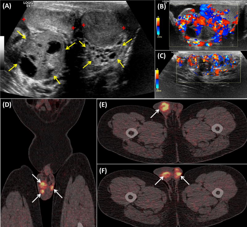
A 46-year-old male with known history of von Hippel-Lindau (VHL) disease underwent whole-body PET/CT scan using 68Ga-DOTA-TATE as part of his evaluation for the detection of neuroendocrine lesions. Consecutive ultrasound (US) tests over a period of 4 years had shown bilateral lobulated extra-testicular masses with mixed echotexture consisting of solid tissue and multiple small cystic spaces (Figure 1A: yellow arrows) behind otherwise normal testes (Figure 1A: red crosses), typical of bilateral epididymal cystadenomas. Vascularity of the cystadenomas was intense and elevated (Doppler US: transverse images of the right Fig. 1B and the left Fig. 1C epididymis). The 68Ga-DOTA-TATE PET/CT study showed intensely increased activity (SUVmax: 21.6) in the scrotum corresponding to the epididymal cystadenomas (Figures 1D, 1E, 1F: coronal and axial PET/CT images of the scrotum respectively). VHL is an autosomal dominantly inherited familial cancer syndrome with a prevalence of 1 in 39,000–53,000 associated with inactivation of a tumor suppression gene located on the short arm of chromosome 3 and characterized by the development of a variety of benign and malignant tumors [1]. VHL’s spectrum of manifestations is broad with 40 different lesions in 14 different organs including retinal and central nervous system hemangioblastomas, endolymphatic sac tumors, renal cysts and tumors, pancreatic cysts and tumors, pheochromocytomas, as well as epididymal and broad ligament cystadenomas [1,2]. Papillary epididymal cystadenomas are rare benign tumors mainly occurring in young adult males, with one-third of the cases reported in the literature to having occurred in VHL patients [3]. Cystadenomas of the epididymis are encountered as unilateral lesions in the general population, but when bilateral they are virtually considered pathognomonic of VHL disease [4]. Although genetic testing for VHL syndrome is available, imaging with multiple modalities contributes to the early detection and follow-up of the various manifestations of this multisystem disorder, as well as to the screening and long-term surveillance of asymptomatic gene carriers. Since, many VHL lesions such as hemangioblastomas or pancreatic neuroendocrine tumors (NETs) are known to over-express SSTRs they can be effectively targeted and localized using radiolabed SST analogues [5]. The introduction of 68Ga-DOTA-conjugated-peptides (SST-analogues) into clinical practice allowed SSTR imaging with PET, and is evolving as the new imaging standard of reference for the detection and characterization of NETs and other SSTR-positive tumors [6,7], with promising applications in VHL patients [8–10]. The presented case of a VHL patient with bilateral epididymal cystadenomas showing intensely increased activity on 68Ga-DOTA-TATE PET/CT, suggests cell-surface over-expression of STTRs by these tumors and particularly SSTR-2 for which 68Ga-DOTA-TATE has a predominant affinity. Considering the wide spectrum of VHL manifestations, the presented data supports the usefulness of SSTR imaging with 68Ga-DOTA-conjugated-peptides in diagnosing not only neuroendocrine lesions, but also various SSTR-overexpressing tumors. Furthermore, the ability of 68Ga-DOTA-TATE-PET/CT to directly demonstrate whole-body SSTR expression, allows selection of VHL patients for hormonal therapy with cold octreotide or theranostic application of peptide receptor radionuclide therapy (PRRT).
Figure 1.

Footnotes
Disclosure: All authors have nothing to disclose
References
- 1.Lonser RR, Glenn GM, Walther M, et al. Von Hippel-Lindau disease. Lancet. 2003;361:2059–2067. doi: 10.1016/S0140-6736(03)13643-4. [DOI] [PubMed] [Google Scholar]
- 2.Leung RS, Biswas SV, Duncan M, et al. Imaging features of von Hippel-Lindau disease. Radiographics. 2008;28:65–67. doi: 10.1148/rg.281075052. [DOI] [PubMed] [Google Scholar]
- 3.Odrzywolski KJ, Mukhopadhyay S. Papillary cystadenoma of the epididymis. Arch Pathol Lab Med. 2010;134:630–633. doi: 10.5858/134.4.630. [DOI] [PubMed] [Google Scholar]
- 4.Choyke PL, Glenn GM, Walther MM, et al. Von Hippel Lindau disease: genetic, clinical, and imaging features. Radiology. 1995;194:629–642. doi: 10.1148/radiology.194.3.7862955. [DOI] [PubMed] [Google Scholar]
- 5.Chowdhury FU, Scarsbrook AF. Indium-111 pentetreotide uptake within cerebellar hemangioblastoma in von Hippel-lindau syndrome. Clin Nucl Med. 2008;33:294–296. doi: 10.1097/RLU.0b013e3181662bf9. [DOI] [PubMed] [Google Scholar]
- 6.Hofman MS, Lau WF, Hicks RJ. Somatostatin Receptor Imaging with 68Ga DOTATATE PET/CT: clini-cal utility, normal patterns, pearls, and pitfalls in interpretation. Radiographics. 2015;35:500–516. doi: 10.1148/rg.352140164. [DOI] [PubMed] [Google Scholar]
- 7.Papadakis GZ, Bagci U, Sadowski SM, et al. Ectopic ACTH and CRH Co-secreting Tumor Localized by 68Ga-DOTA-TATE PET/CT. Clin Nucl Med. 2015;40:576–578. doi: 10.1097/RLU.0000000000000806. [DOI] [PMC free article] [PubMed] [Google Scholar]
- 8.Ambrosini V, Campana D, Allegri V, et al. 68Ga DOTANOC PET/CT detects somatostatin receptors expression in von Hippel-Lindau cerebellar disease. Clin Nucl Med. 2011;36:64–65. doi: 10.1097/RLU.0b013e3181fef14a. [DOI] [PubMed] [Google Scholar]
- 9.Oh JR, Kulkarni H, Carreras C, et al. Ga-68 Somatostatin Receptor PET/CT in von Hippel-Lindau Disease. RP. Nucl Med Mol Imaging. 2012;46:129–133. doi: 10.1007/s13139-012-0133-0. [DOI] [PMC free article] [PubMed] [Google Scholar]
- 10.Mukherjee A, Karunanithi S, Bal C, et al. 68Ga DOTANOC PET/CT aiding in the diagnosis of von Hippel-Lindau syndrome by detecting cerebellar hemangioblastoma and adrenal pheochromocytoma. Clin Nucl Med. 2014;39:920–921. doi: 10.1097/RLU.0000000000000486. [DOI] [PubMed] [Google Scholar]


